Highly Sensitive Silver Nanorod Arrays for Rapid Surface Enhanced Raman Scattering Detection of Acetamiprid Pesticides
Ci-qin HnYue YoWen WngLiu-qin ToWen-xin ZhngWhitney Mrvell IngrmKng-zhen TinYing LiuAi-xi LuYing WuChng-chun YnLu-Lu QuHi-to Li
a.Jiangsu Key Laboratory of Advanced Laser Materials and Devices,School of Physics and Electronic Engineering,Jiangsu Normal University,Xuzhou 221116,China
b.School of Chemistry and Materials Science,Jiangsu Normal University,Xuzhou 221116,China
c.Jiangsu Collaborative Innovation Center of Advanced Laser Technology and Emerging Industry,Jiangsu Normal University,Xuzhou 221116,China
d.Department of Physics and Astronomy,and Nanoscale Science and Engineering Center,University of Georgia,Athens,Georgia 30602,USA
I.INTRODUCTION
Acetamiprid is a commonly used neonicotinoid pesticide in modern farming for exterminating pests and increasing crop yields[1].However,the acetamiprid residue can result in food and surface and/or groundwater contamination,which is potentially harmful to humans.Thus,the development of a rapid,simple,and sensitive detection for acetamiprid on food surfaces is particularly important.Some methods such as gas chromatography(GC)[2−4],high performance liquid chromatography(HPLC)[5,6],enzyme linked immunosorbent assay(ELISA)[7,8]and colorimetry[9,10]have been employed to analyze pesticides.However,these methods suffer from a complex pretreatment process and a long detection time,which are not conducive to real-time monitoring and on-site testing.
Recently, surface-enhanced Raman scattering(SERS)spectroscopy emerged as a method that can enhance the original Raman signal of molecules based on a strong localized electric field generated on a roughened metal surface[11,12].It has aroused much interest in agriculture and food safety due to its capacity for rapid,simple,and sensitive detection[13].For example,Fanet al.detected phosmet in fruits and vegetables by gold-coated SERS substrates,where signals possessed the good reproducibility and stability[14].Mulleret al.investigated the possibility to detect thiabendazole from chemically treated bananas and citrus fruits samples by SERS[15].Zhanget al.fabricated the single-crystal silver nanowires with good draw ratios and smooth surfaces,which can be applied to the detection of pesticide thiram[16].However,most of the SERS substrates have disadvantages in reproducibility,sensitivity and stability,which limits the potential ap-plication of SERS for detection.
Oblique angle deposition(OAD)has been recognized as a stable and effective method to fabricate nanostructures.In our previous work,the OAD method has been used to fabricate different silver or gold nanostructures by changing various deposition conditions such as the material thickness and deposition angle[17−19].Moreover,the silver nanorod(AgNR)array SERS substrates fabricated by OAD exhibit good sensitivity and high reproducibility.To improve the reproducibility and sensitivity for SERS detection of acetamiprid,in the present work,AgNR array SERS substrates were prepared by OAD.However,in practical applications,the oxidation of the AgNRs and the adsorption of impurities onto the substrate decreases the overall signal strength of samples and it increases background noise of AgNR array substrates.To resolve these problems,the substrates surfaces are cleaned commonly by a plasma cleaner,which is expensive and inconvenient to operate.Therefore,we developed and evaluated two cleaning methods to remove the impurities on AgNR array substrates.Then,the cleaned substrates were applied to the detection of acetamiprid.The characteristic peak and vibration attribution of acetamiprid were determined by DFT simulation.The detection limit was improved and a new method for rapid detection of pesticide was proposed.
II.EXPERIMENTS
A.Materials
Acetamiprid pesticide,methanol,acetone,nitric acid,sulfuric acid and hydrogen peroxide were purchased from Sinopharm Chemical Reagent Co.,Ltd.(China).Silver(99.999%)and titanium(99.995%)pellets were obtained from Kurt J.Lesker Co.,Ltd.(USA).Ultrapure water(≥18.2 MΩ)was used in all experiments.
B.Fabrication of AgNR substrates
Glass slides were cut into 1 cm×1 cm as the supporting substrate for growing AgNR arrays.The glass slides were cleaned in Piranha solution(80%sulfuric acid,20%hydrogen peroxide),rinsed with de-ionized water,and then dried with nitrogen gas.Then,the cleaned glass slides were mounted at deposition angle of 0°inside deposition chamber.Under the pressure of 1×10⁻⁶Torr,20 nm titanium film and 500 nm silver film were deposited with the rate of 0.2 and 0.3 nm/s by using an electron beam evaporation equipment(DE500 electron beam evaporation deposition system,DE instrument technology,Beijing),respectively.Subsequently,the substrate holder rotated at a deposition angle of 86°,and another 2000 nm silver film was deposited with the rate of 0.3 nm/s(FIG.1(a)).

FIG.1(a)Schematic diagram of the AgNR array fabricated by OAD.(b)The top-view SEM image of the AgNR array substrate.(c)The cross-section SEM image of the AgNR array substrate.
The top-view and cross-section SEM images of the Ag-NRs were obtained as shown in FIG.1(b)and(c).The measured nanorod lengthL=(1100±90)nm,the rod diameterD=(150±70)nm,the average rod-to-rod spacingS=(130±40)nm,and the nanorod tilting angleβ=74°±3°are de fined.
C.Methods of cleaning the AgNR substrates
The common methods for cleaning the AgNR array substrates are the chemical and physical elution.For this study,the first method evaluated was nitric acid(10−7mol/L,diluted by DI water)to wash the AgNR substrates(FIG.2).The substrates were placed into the solution,and the silver oxide on the surface was removed by reacting with nitric acid.After one minute,the cleaned AgNR substrates were rinsed with methanol at least two times in order to remove the residual liquid on the substrate.After the methanol on the surface evaporated,AgNR substrate can be used for SERS detection.The second cleaning method evaluated was a mixed solution of methanol and acetone.The cleaning process was similar to the nitric acid cleaning process:substrates were immersed in the acetone methanol mixture for one minute,then rinsed with methanol.
D.Density function theory(DFT)calculation
The Gaussian 09 W DFT package was used to calculate the Raman spectrum of acetamiprid and identify the corresponding vibrational modes.The DFT calculations were based on Becke’s three-parameter exchange function(B3)with the dynamic correlation function of Lee,Yang,and Parr(LYP).The molecular structure of acetamiprid was optimized using B3LYP function in conjunction with a modest 6-311G basis set.After the modi fication of the molecular structure,putting a silver atom beside the acetamiprid molecular,the SERS spectrum of acetamiprid was calculated with a modest LANL2DZ basis set.

FIG.2 Schematic diagram of two cleaning methods for AgNR array substrates,(a)the nitric acid cleaning and(b)the organic reagent cleaning.
E.SERS measurements
A commonly used probe molecule,trans-1,2-bis(4-pyridyl)ethane(BPE),was dissolved with methanol and reached the final concentration of 10−5,10−6,10−7,10−8,10−9,10−10mol/L.A 2 µL solution sample was applied to the AgNR substrate to detect the Raman signals after air-dried.The acetamiprid pesticide powder samples(100 mg,20%acetamiprid)were directly dissolved in the 100 mL deionized water with a concentration of 1000 mg/L as the standard solution.Then,the solution was diluted to the different concentrations:100,50,10,5,1,and 0.5 mg/L for SERS measurements.For the practical application study,5µL of acetamiprid solution was added onto the surface of cucumber.After the solution dried in air,a droplet of 5µL water was added onto the spot to extract the acetamiprid.For SERS detection,samples were detected by the portable Raman Spectrometer(BWS465,B&W TEK,USA).Before testing,AgNR array substrates were put on the platform of the instrument to obtain the background signals.The excitation wavelength was 785 nm,the spot size of laser was 85µm and the scanning range was from 350 cm−1to 1800 cm−1.All of the spectra were acquired from nine randomly selected spots with a laser power of 30 mW and integration time of 5 s.
III.RESULTS AND DISCUSSION
A.Pretreatment of AgNR array substrates
Once exposed to the air,the AgNRs array substrates oxidize rapidly,thus resulting in the dramatic decrease of SERS activity.In addition,the impurities in the air may adsorb on the surface of AgNR array substrates,causing high background SERS signals,which may conceal the signal of the analytes.Thus,the main components on the AgNR array substrate surface are silver oxide and other pollutants.To improve SERS activity of the AgNRs array substrates,our first purpose is to remove silver oxide.Nitric acid is the most commonly used acid solution,which can react with silver oxide from the silver nanorod surface. In order to remove the Ag2O effectively,the total mass of Ag2O on the surface of silver nanorods was estimated asg(Text S1 in supplementary materials).According to the chemical reaction(Text S2 in supplementary materials),the amount of dilute nitric acid material was 4.914×10⁻⁸mol when fully reacted.
The background signals of the substrate cleaned by nitric acid were shown in FIG.3(a).Compared with the untreated substrate,the Raman peaks at 891,966,1039,1407,and 1635 cm−1appeared after washing it with nitric acid.With the increasing concentrations of nitric acid,the peaks at 891,966,1407,and 1635 cm−1decreased.It indicated the reaction of silver oxide and nitric acid on the substrate.At 1039 cm−1,the peak intensity increases with the nitric acid increased,which is possibly due to the reaction product.The background signal suggested that the nitric acid cleaning process removed the silver oxide,however,new impurities were introduced.Subsequently,SERS activity of AgNRs which reacted with different concentration of acid was investigated,as shown in FIG.3(b).The SERS signal intensity of BPE was the strongest after 4.914×10⁻⁸mol nitric acid cleaning,which accords with theoretical calculation.When the acid was fewer or excess,the SERS intensity of BPE peaks decreased,which may be caused by other emerging impurities[20,21].

FIG.3(a)The background signal of the AgNRs substrate and(b)the SERS signal of 10−5mol/L BPE solution on AgNR substrate after washing with different concentration of nitric acid.
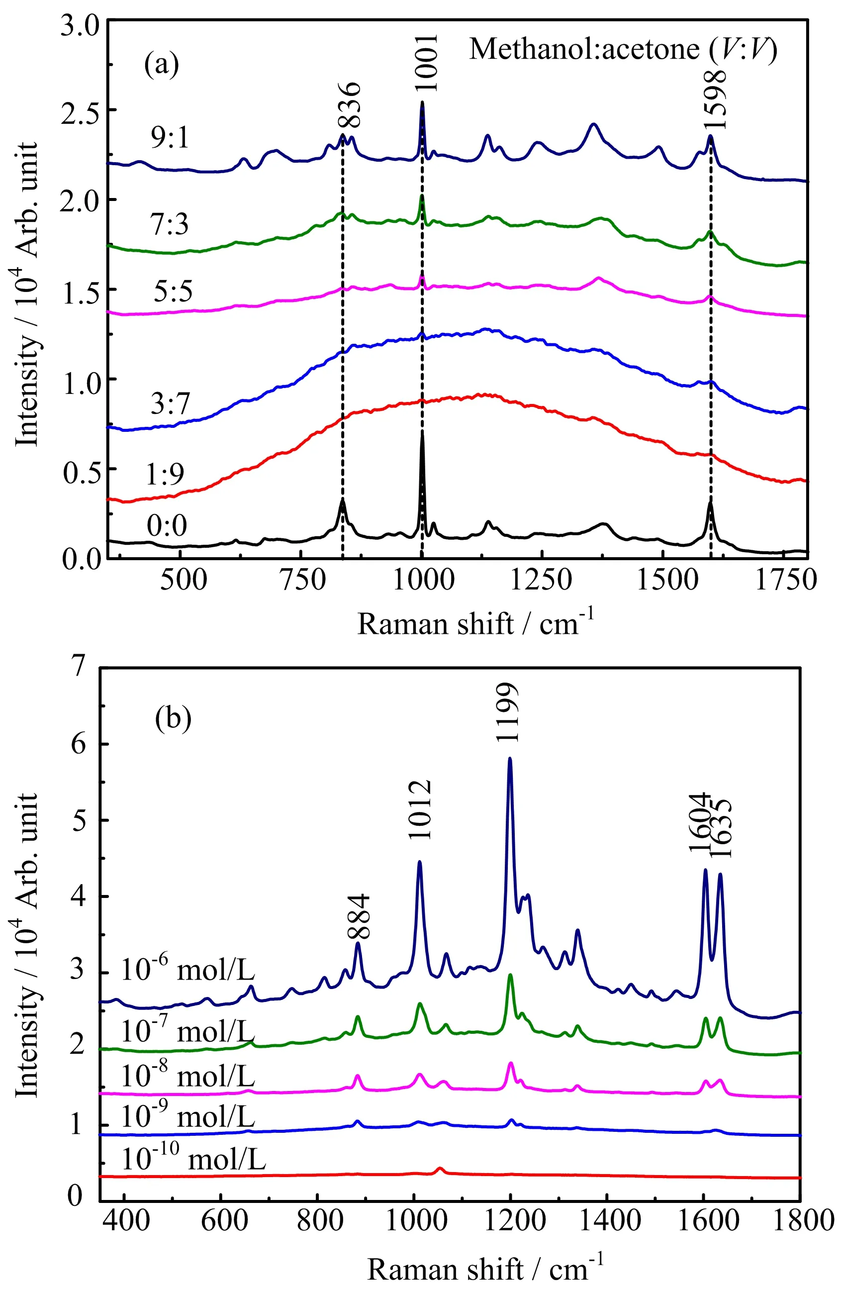
Another method to clean the SERS substrate is to remove the silver oxide with methanol,and remove the impurities on the AgNRs substrate with acetone(Text S2 in supplementary materials).After the substrate was washed with the mixed solvent of methanol and acetone in different ratios,the background signals on the substrate were recorded and displayed in FIG.4(a).The results show that the background signal of the uncleaned substrate is strong,and there are sharp peaks at 836,1001,and 1598 cm−1.When the ratio of methanol to acetone solution increased,the background peaks of the substrate gradually increased.At ratios of 1:9 and 3:7,the background signals were weakened signi ficantly and even faded,indicating that no or less impurities was on the substrate surface.
To con firm that the SERS performance of the cleaned substrate was improved,BPE with different concentrations was added to the cleaned substrate surface.As shown in FIG.4(b),the characteristic peaks intensity of BPE increased with the increasing of solution concentration.The peaks of BPE were clearly identi fied at Δν=884,1012,1199,1604,and 1635 cm−1.The limit of detection(LOD)of BPE solution was 10−6mol/L for the uncleaned substrate(FIG.S1 in supplementary material).When the ratio of methanol to acetone was 1:9,the LOD was 10−8mol/L.The characteristic peak intensities of the same concentration of BPE on the substrate after cleaning with methanol:acetone of 3:7 were much higher,which shows an LOD of 10−9mol/L and the enhancement factor(EF)of 5.7×10⁷(Text S3 and FIG.S2 in supplementary materials).Therefore,the ratio of 3:7 was better for cleaning of AgNR substrates.

FIG.4(a)The background signal of AgNR substrates after washing with the mixture of methanol and acetone in different ratios.(b)The SERS signal of BPE recorded from the AgNR substrates after washing with methanol and acetone(3:7).
From the above results,we found the SERS performances were improved signi ficantly after cleaning the substrate with two methods.However,the acid cleaning introduces pollution and increases the background signal,which may affect actual detection.Organic cleaning process is simple,and the pollution of cleaning is less than that of nitric acid cleaning.After cleaning,the substrate background signal tends to be smooth,which improves the actual detection sensitivity. Moreover,the organic cleaning achieved the LOD of 10−9mol/L,which was 1000 times greater than an uncleaned substrate.
In addition,the AgNRs substrates exhibited a high reproducibility after cleaning.The relative standard deviations(RSDs)of spot-to-spot and batch-to-batch are around 8.96%and 16.65%,respectively(FIG.S3 in supplementary materials),which is comparable to the previous reported SERS substrates[13,22,23].Furthermore,to determine the stability of the cleaned substrates,SERS signals of 10−5mol/L BPE solution were collected from the substrates with various storage time.As the result displays,the SERS intensity of BPE did not show the obvious changes with increasing storage time after cleaning,which means the AgNR substrate after cleaning is stable during the half an hour(FIG.S4 in supplementary materials).

FIG.5 (a)The optimized molecular structure of acetamiprid.(b)The Raman spectrum of acetamiprid calculated by DFT,the SERS spectrum of acetamiprid calculated by DFT and SERS spectrum of acetamiprid.Spectra were normalized by the most intensive peaks and offset for clari fication.
B.Detection of acetamiprid
To expand the applicability of the AgNRs,acetamiprid,a new neonicotinoid class of systemic insecticides,was detected using AgNRs.The stable molecular structure,Raman spectrum and SERS spectrum were initially simulated by the density function theory(DFT)calculation(FIG.5).Compared with the experimental SERS spectrum,the position of sample’s characteristic peaks and their corresponding vibration modes can be obtained.
The optimized molecular structure of acetamiprid was shown in FIG.5(a).The characteristic peak positions of acetamiprid were identi fied as Δν=745,1336,and 1535 cm−1.The experimental and calculated Raman shifts and their corresponding vibrational mode using these optimized structures were summarized in Table I.

FIG.6(a)SERS signals of acetamiprid solution with different concentrations acquired on AgNR substrates.(b)SERS intensity of acetamiprid peak at 745 cm−1with different concentrations.The black line is the detectable control line(3σ=613).The red line is the exponential fitting.
All characteristic peaks arise from different forms of molecular vibrations.For example,among the 10 identi fied peaks of the SERS spectrum shown in the Table I,the weak peak at Δν=502 cm−1is attributed to the H12-C10-H11 rocking vibration mode. The N22-C21=N17 wagging and C1-C2-C3 wagging vibration mode were found to have peaks at Δν=602 cm−1and Δν=651 cm−1.A strong peak at Δν=745 cm−1attributes to the C1−H5 wagging vibration mode and C14−C15 stretching.The characteristic peak at Δν=957 cm−1is assigned to the C4-C5-N26 scissoring mode. The N26−C5 stretching vibration correlates to Δν=1092 cm−1,and the C2−C10 stretching to Δν=1272 cm−1.The H12-C10-H11 antisymmetrical mode has a strong peak intensity at Δν=1336 cm−1,and a moderate peak intensity peak at Δν=1535 cm−1is assigned to ring breathing.
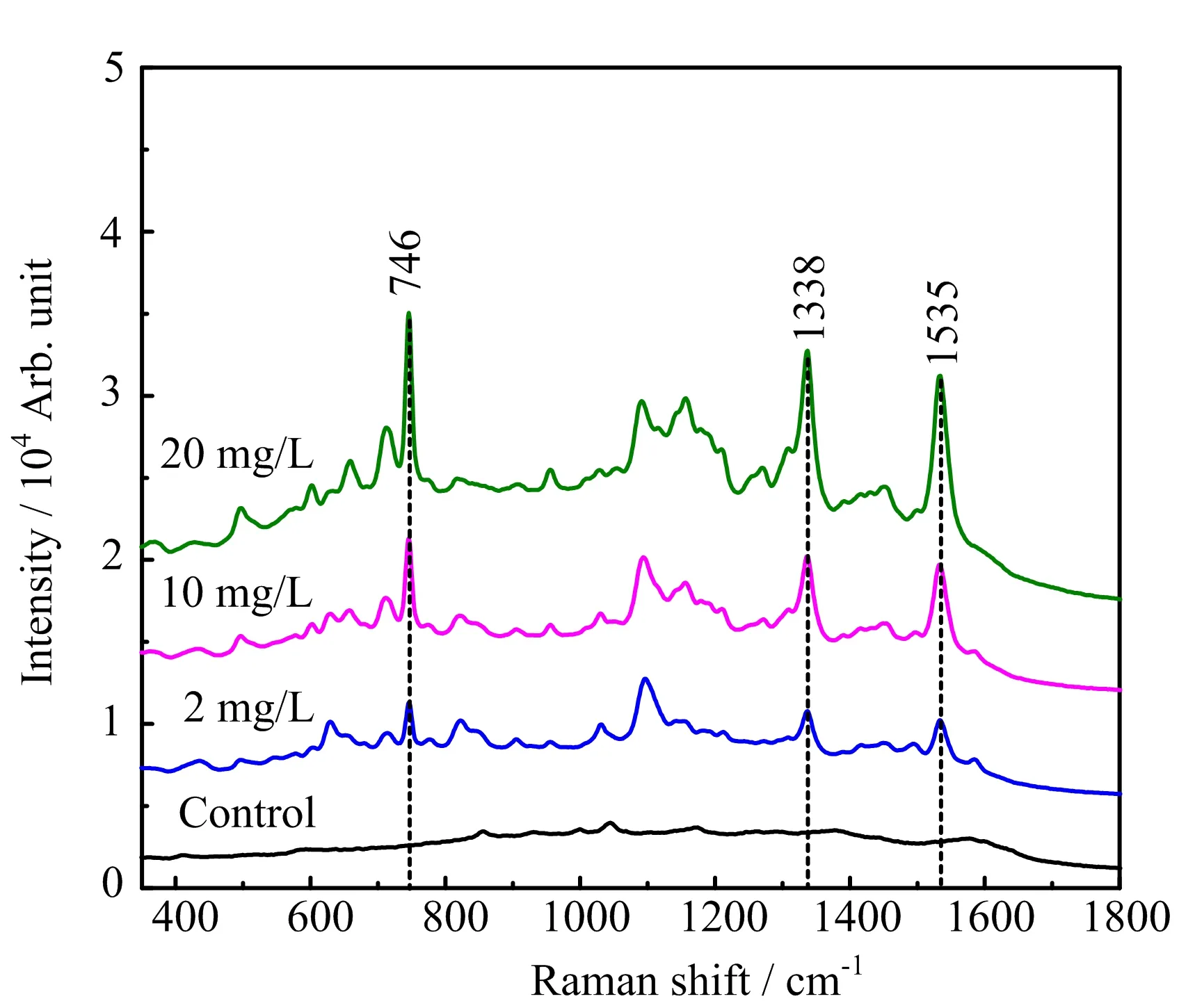
In order to determine the LOD of the acetamiprid by AgNR substrates,different concentrations acetamiprid solutions were measured. Even at the concentration of 0.1 mg/L,the relevant characteristic peaks at Δν=745,1336,and 1535 cm−1can be distinguished(FIG.6(a)).The relationship between the SERS intensity of representative peak at 745 cm−1and the concentration of acetamiprid was measured,as shown in FIG.6(b).A linear correlation between SERS intensities and logarithmic concentration was observed,I=5573.0(logC)+3505.5.The LOD and LOQ were estimated to be 0.05 mg/L(3σmethod,σis the mean square root of the noise signal,which was determined by standard deviation of the spectral intensity at a spectral region:1700−1800 cm−1)and 0.01 mg/L(10σmethod)utilizing the silver nanorod array SERS substrates as a platform,which was much lower than the China national standard 1 mg/L.This result indicates the high sensitivity of AgNRs for pesticide qualitative detection.

TABLE I The major Raman and SERS peaks of acetamiprid calculated by DFT and the experiment SERS peaks of acetamiprid,and their corresponding vibration modes.
The substrates cleaned by the organic solution were employed to test the acetamiprid from a cucumber surface.For each measurement trial,a different concentration of acetamiprid solution was add onto the cucumber surface.After extraction by water,the SERS signal of acetamiprid was obtained,as shown in FIG.7.The peaks at Δν=745,1336,and 1535 cm−1were observed clearly.Table S1 shows the recoveries of acetamiprid from cucumber by inserting the Raman intensities into the calibration curve from FIG.6(b).The recoveries of spiked acetamiprid can achieve 71.6%−115.1%.The good recovery reveals that our AgNRs is reliable in determining acetamiprid residual on the cucumber.This result showed that the real application can be achieved using the highly sensitive and reproducible AgNRs.
IV.CONCLUSION
The AgNR array substrates were fabricated by the oblique angle deposition for highly sensitive detection of acetamiprid.To remove impurities on the AgNRs substrate and improve the detection ability of the substrates,nitric acid or organic solvent cleaning,were applied to pretreat the substrate surface,respectively.The LOD of BPE after organic cleaning was determined to be 10−9mol/L and the SERS performance was improved 1000 times than those uncleaned.The acetamiprid molecule structure,the Raman spectrum and SERS spectrum were obtained by DFT simulation calculations.The characteristic peaks and their corresponding vibrational modes were also determined.In addition,the acetamiprid on cucumber was tested by the AgNR array with a LOD of 0.05 mg/L.The results suggested that the cleaned AgNR substrates can be used for SERS detection of pesticide residues on the surface of vegetables.This method provids an effective and sensitive strategy for monitoring residues on agricultural products.

FIG.7 The SERS signal of acetamiprid with different concentrations extracted from cucumber.
Supplementary material:Calculation of the mass of Ag2O on the AgNR surface(Text S1),reaction of chemicals on the AgNR surface during the cleaning(Text S2),effect of the different organic solutions and the LOD of BPE(FIG.S1),calculation of the SERS EF of the AgNR array(Text S3),SERS spectrum and Raman spectrum of BPE(FIG.S2),reproducibility of AgNR array(FIG.S3),effect of storage time on SERS signals(FIG.S4),and recovery of real sample detection(Table S1)are given.
V.ACKNOWLEDGMENTS
This work was supported by the National Natural Science Foundation of China(No.61575087,No.21505057,and No.61771227),the Natural Science Foundation of Jiangsu Province(No.BK20151164,No.BK20150227,and No.BK20170229),the Innovation Project of Jiangsu Province(No.KYLX161322),theNaturalScience Foundation of the Jiangsu Higher Education Institutions(No.17KJB140007)and Foundation of Xuzhou City(No.KC15MS030). The authors would like to thank Layne Bradley for his assistance with linguistic revision,and a Project Funded the Priority Academic Program Development of Jiangsu Higher Education Institutions.
[1]N.S.Chatterjee,S.Utture,K.Banerjee,T.P.A.Shabeer,N.Kamble,S.Mathew,and K.A.Kumar,Food Chem.196,1(2016).
[2]E.G.Amvrazi and N.G.Tsiropoulos,J.Chromatogr.A1216,2789(2009).
[3]D.Bielawski,E.Ostrea Jr.,N.Posecion Jr.,M.Corrion,and J.Seagraves,Chromatographia62,623(2005).
[4]Z.W.Xiao,M.He,B.B.Chen,and B.Hu,Talanta156/157,126(2016).
[5]M.Asensio-Ramos,J.Hernández-Borges,G.González-Hernández,and M.á.Rodrguez-Delgado,Electrophoresis33,2184(2012).
[6]J.F.Huertas-Pérez and A.M.García-Campaˆna,Anal.Chim.Acta630,194(2008).
[7]B.Liu,Y.Ge,Y.Zhang,Y.Song,Y.Y.Lv,X.X.Wang,and S.Wang,Food Agr.Immunol.23,157(2012).
[8]Z.L.Xu,H.Wang,Y.D.Shen,M.Nichkova,H.T.Lei,R.C.Beier,W.X.Zheng,J.Y.Yang,Z.G.She,and Y.M.Sun,Analyst136,2512(2011).
[9]X.Y.Zhang,Z.Y.Sun,Z.M.Cui,and H.B.Li,Sens.Actuat.B Chem.191,313(2014).
[10]Y.J.Li,C.J.Hou,J.C.Lei,B.Deng,J.Huang,and M.Yang,Anal.Sci.32,719(2016).
[11]J.Kubackova,G.Fabriciova,P.Miskovsky,D.Jancura,and S.Sanchez-Cortes,Anal.Chem.87,663(2015).
[12]L.Xue,H.X.Gu,S.Q.Yuan,and D.W.Li,RSC Adv.7,192626(2017).
[13]L.L.Qu,D.W.Li,J.Q.Xue,W.L.Zhai,J.S.Fossey,and Y.T.Long,Lab Chip12,876(2012).
[14]Y.X.Fan,K.Q.Lai,B.A.Rasco,and Y.Q.Huang,Food Control37,153(2014).
[15]C.Müller,L.David,V.Chi¸s,and S.C.Pˆınzaru,Food Chem.145,814(2014).
[16]L.Zhang,B.Wang,G.Zhu,and X.Zhou,Spectrochim.Acta A133,411(2014).
[17]J.D.Driskell,S.Shanmukh,Y.J.Liu,S.B.Chaney,X.J.Tang,Y.P.Zhao,and R.A.Dluhy,J.Phys.Chem.C112,895(2008).
[18]T.Karabacak,G.C.Wang,and T.M.Lu,J.Vac.Sci.Technol.A22,1778(2004).
[19]Y.J.Liu and Y.P.Zhao,Phys.Rev.B78,075436(2008).
[20]J.Novakovic,P.Vassiliou,and E.Georgiza,Int.J.Electrochem.Sci.8,3615(2013).
[21]T.Palomar,B.R.Barat,E.Garca,and E.Cano,J.Cult.Herit.17,20(2016).
[22]D.W.Li,W.L.Zhai,Y.T.Li,and Y.T.Long,Microchim.Acta181,23(2014).
[23]Y.T.Li,L.L.Qu,D.W.Li,Q.X.Song,F.Fathi,and Y.T.Long,Biosens.Bioelectron.43,94(2013).
 CHINESE JOURNAL OF CHEMICAL PHYSICS2018年2期
- CHINESE JOURNAL OF CHEMICAL PHYSICS的其它文章
- Investigation on Preparation and Anti-icing Performance of Superhydrophobic Surface on Aluminum Conductor
- Study of Cadmium-Doped Zinc Oxide Nanocrystals with Composition and Size Dependent Band Gaps
- Glucose Isomerization into Fructose Catalyzed by MgO/NaY Catalyst
- Analysis of Solvent Effect on Mechanical Properties of Poly(ether ether ketone)Using Nano-indentation
- Direct Synthesis of Monodisperse Hollow Molecularly Imprinted Polymers Based on Unfunctionalized SiO2for the Recognition of Bisphenol A
- Agent-Based Network Modeling Study of Immune Responses in Progression of Ulcerative Colitis
