Relationship of distraction rate with inferior alveolar nerve degeneration-regeneration shift
Ying-hua Zhao, Shi-jian Zhang, Zi-hui Yang, Xiao-chang Liu, De-lin Lei, Jing Li, Lei Wang,
1 Department of Stomatology, Huadong Hospital, Fudan University, Shanghai, China
2 Department of Oral & Maxillofacial-Head & Neck Oncology, Ninth People’s Hospital, Shanghai Jiao Tong University School of Medicine,Shanghai, China
3 Shanghai Key Laboratory of Stomatology & Shanghai Research Institute of Stomatology; National Clinical Research Center of Stomatology,Shanghai, China
4 Department of Oral Surgery, State Key Laboratory of Military Stomatology & National Clinical Research Center for Oral Diseases & Shaanxi Clinical Research Center for Oral Diseases, School of Stomatology, The Fourth Military Medical University, Xi’an, Shaanxi Province, China
5 Medical Team of 66018 PLA Troops, Beijing, China
Introduction
Distraction osteogenesis (DO) is a widely used tissue engineering technology, which promotes tissue regeneration through continuous mechanical strain. McCarthy et al.(1992) first introduced DO in a craniofacial surgery, and reported its successful application in a patient suffering from craniofacial syndromes. Since then, DO is widely used in the treatment of craniofacial bone defects and malformation(Molina et al., 1995; Corcoran et al., 1997). However, inferior alveolar nerve (IAN) injury, one of the most common complications during mandibular DO, not only causes discomfort to patients, but also hinders further improvement(Wang et al., 2002, 2009a, b).
In our previous studies, we investigated the factors that accelerated nerve recovery after DO. We found that nerve growth factor administration can accelerate the recovery of IAN in rabbits after mandibular DO (Wang et al., 2009a,b). We also applied mesenchymal stem cells modified with nerve growth factor during mandibular DO in rabbits, and found greatly improved recovery of the IANs (Wang et al.,2015). In these studies, DO was performed at a given distraction rate. Although the effects of distraction rate on bone regeneration during DO are well documented, the responses of IAN to different distraction rates remain elusive. In the present study, rabbits were subjected to DO at different distraction rates. Pin-prick test, histology and histomorphometric analysis were used to assess the relationship between distraction rate and IAN degeneration-regeneration shift during mandibular DO.
Materials and Methods
Animals
Twenty-four specific-pathogen-free male adult New Zealand rabbits aged 18–22 weeks were used in this study. All rabbits were housed individually in stainless steel cages and allowed free access to standard food and water. Animal experimental protocols (permission No. SYXK (Shaan) 2015-001) were approved by the Animal Ethics Committee of Medical Experiment at Fourth Military Medical University in China(ethics approval No. FMMU-E02318).
All the rabbits were randomly divided into four groups(n= 6 per group). The bilateral mandible of each rabbit was distracted at a rate of 1.0, 1.5 and 2.0 mm/d, separately.In the sham group, mandibular osteotomy was performed without distraction.
Surgical procedures
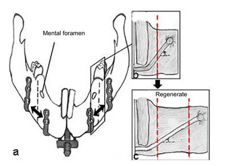
To establish the rabbit mandibular DO model, the surgical procedures were performed as described in our previous studies (Wang et al., 2009a, b, 2015). The successful criteria were new bone information and without infection. Briefly,rabbits were injected intraperitoneally with ketamine at a dose of 50 mg/kg body weight to induce anesthesia. After anesthesia, a 3 cm longitudinal incision was made in the submandibular area. Periosteum and the masseter muscle were dissected. Nerves were exposed and protected carefully. Afterwards, an osteotomy was made between premolar and mental foramen of the mandible. An external custom made distraction device (Zhongbang Titanium Biomaterials Corporation, Xi’an, China) was then closely fixed on the bone surface by six titanium screws on the forearm and four screws on the posteriorarm, and IAN and concomitant vessels were carefully protected. The structure of distraction device used in this study is shown inFigure 1, in which the figure b and c shows how the IANs distribute before and after distraction.
The incision was sutured with the distractor rod being exposed on the outside. After 3.5-day latency, rabbits in three groups were subjected to continuous distraction at a rate of 1.0, 1.5 and 2.0 mm/d, respectively. The same distance of approximately 10 mm was obtained at the end of distraction period, and a consolidation period followed thereafter.
Pin-prick test for assessing the IAN condition
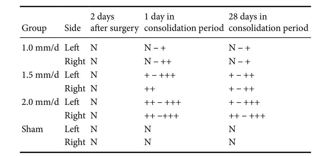
A pin-prick test was performed by the same person to assess the IAN condition of rabbits. A blunt sterile calibrated 10 g Neurotip tester pin (Owen Mumford, Brook Hill Woodstock, Oxford, UK) was used to prick the bilateral labium at the same pricking force of 10 g on day 2 post-surgery as well as day 1 and day 28 of consolidation. All the tests were repeated 20 times on each rabbit. The pin-prick test was considered positive when a rabbit started to tilt its head and struggled. The IAN condition was regarded as normal if positive results were obtained more than 17 times, slight dysfunction (+) if positive between10–16 times, medium dysfunction (++) if positive between 4–9 times, and severe dysfunction (+++) if positive less than 3 times (Wang et al.,2002).
Histological and histomorphometric analyses of nervous tissue
Rabbit IAN specimens from all groups were harvested at 41.5 days post-surgery. IAN specimens from each group were divided into two parts. One part of IAN specimens were fixed in osmium oxide for 2 hours, and embedded in epon; cross-sectional 1 μm serial sections were sliced and subjected to methylene blue staining to assess the nerve myelin sheath. Three randomly selected sections at 1-mm intervals from each IAN sample were digitized using an Olympus light microscope (BX41; Olympus, Tokyo, Japan) equipped with a 12.5 megapixel cooled CCD camera (DP71; Olympus)at a final magnification of × 400. For histomorphometric analysis, ten randomly selected fields of each sample (four fields for each section) were analyzed using a National Institutes of Health Image Analysis System (Bethesda, MD, USA).Myelinated fiber density (number per square millimeter under high magnification of a microscope) was calculated. The epon-embedded samples were further sliced into ultrathin sections with an ultramicrotome (Leica, Wetzlar, Germany).Transmission electron microscopy (Hitachi, Tokyo, Japan)was utilized to assess ultrastructural changes of IAN. The remaining IAN samples were fixed in 4% paraformaldehyde at 4°C for 4 hours, and embedded in paraffin. Longitudinal 2 μm serial sections were sliced and subjected to Bodian’s silver staining as described previously (Gambetti, 1981), Briefly,nerve fibers and nerve ending in paraffin sections were treated with silver nitrate for 10 minutes, exposed to ultraviolet and treated with sodium hyposulfite for 2 minutes.
Statistical analysis
All data were statistically analyzed with SPSS 16.0 software(SPSS, Chicago, IL, USA) and presented as the mean ±SD. Data on the myelinated fiber density passed the Shapiro-Wilk test for normality, which is well represented as measure and count data and according to normal distribution, so one-way analysis of variance was performed to identify the differences between and within groups. If the results from the comparisons between groups were significant, least significant difference multiple comparisons tests were used to explore the intergroup differences. A value ofP< 0.05 was considered statistically significant.
Results
Pin-prick test
All rabbits recovered well. As shown inTable 1, the pinprick test results showed that IAN condition was normal 2 days after surgery, indicating that IAN was not injured during osteotomy and the implantation of distraction device. Nearly all rabbits in the 1.0 mm/d, 1.5 mm/d and 2.0 mm/d groups that underwent mandible distraction suffered from varying degrees of IAN dysfunction, with identical degrees between left and right sides in the same animal. IAN condition of rabbits in the sham group, which received osteotomy but without distraction, appeared normal during consolidation period. We compared pin-prick results of the 1.0 mm/d, 1.5 mm/d and 2.0 mm/d groups, and found that IAN functioned poorly at a high distraction rate. Notably,at 28 days of consolidation, only the rabbits in the 1.0 mm/d group had recovered IAN function amongst all the rabbits that underwent mandible distraction. The pin-prick test suggested that mechanical strain resulted in IAN dysfunction at a high distraction rate.
Histological analysis of nervous tissue
IAN specimens of the 1.0 mm/d and 1.5 mm/d groups were much finer than in the sham group, and shared a relatively similar diameter in the 1.0 mm/d and 1.5 mm/d groups.However, the IAN diameters in the 2.0 mm/d group were more different from each other, and were finer than in the normal group (data not shown).
Toluidine blue staining results showed that each IAN in the sham group constituted of 4–8 nerve tracts. Myelin sheaths of myelinated nerve fiber appeared dense, thick, and uniform in thickness, and were stained deep blue. However,the cable was not stained, and no obvious degeneration or regeneration was found in the myelinated fiber (Figure 2D).The myelin sheaths in the 1.0 mm/d group were sparser than in the sham group. We observed mild to moderate neurodegeneration, such as demyelination of myelinated fibers,plate separation and axonal thickening (Figure 2A). Also,some immature nerve fibers and active Schwann cells were present, indicating that nerve regeneration had been initiated. In comparison to the sham group, the myelin sheaths of rabbits in the 1.5 mm/d group were sparse and thin. More neurodegeneration and clusters of axons were observed, and interstitial nerve bundles increased (Figure 2B). The diameter of nerve fibers decreased sharply in the 2.0 mm/d group.The myelin sheaths of rabbits were finest in the 2.0 mm/d group than in the 1.0 and 1.5 mm/d groups. Serious disorder in the structure of the nerve fiber was observed in the 2.0 mm/d group, with little nerve regeneration (Figure 2C). The myelinated fiber density in the 2.0 mm/d group was signif icantly lower compared with the 1.0 mm/d, 1.5 mm/d and sham groups (Figures 2E;P< 0.05).
Using transmission electron microscopy, myelinated nerve fibers in the sham group were uniform in thickness,without obvious degeneration. Moreover, the axonal electron density was normal with few unmyelinated nerve fibers(Figure 3D,H). Both myelinated and unmyelinated nerve fibers in the 1.0 mm/d group had mild to moderate neurodegeneration, which was consistent with the toluidine blue staining results (Figure 3A, E). Furthermore, there were immature nerve fibers and active Schwann cells, indicating that the damaged nerve fibers had entered the regeneration stage(Figure 3A, E). Many myelinated and unmyelinated nerve fibers in the 1.5 mm/d group underwent moderate to severe neurodegeneration, and also contained some macrophages(Figure 3B, F). More number of myelinated and unmyelinated nerve fibers in the 2.0 mm/d group suffered from neurodegeneration, and also contained more macrophages than in the 1.0 mm/d and 1.5 mm/d groups (Figure 3C, G).
Bodian’s silver staining results showed that the nerve axons in the 1.0 mm/d group were uniform in thickness and arranged parallel to the long axis of nerve, and had a few disconnecting axons. In the 1.5 mm/d group, nerve axons were uneven in thickness and had more disconnecting axons than in the 1.0 mm/d group. In the 2.0 mm/d group, nerve axons were disordered, uneven in thickness and contained more disconnecting axons and tumor-like enlargement at the broken ends. Enormous axon fragments were visible in the 2.0 mm/d group (Figure 4A–C).

Figure 1 Schematic diagram of the structure of distractor and how the inferior alveolar nerves distribute before and after distraction.

Figure 2 Histological and histomorphometric analyses of the inferior alveolar nerves under different distraction rates (toluidine blue staining).

Figure 3 Transmission electron microscopy of the inferior alveolar nerves under different distraction rates after 28 days of consolidation.

Figure 4 Bodian’s silver staining for axial sections of the inferior alveolar nerves under different distraction rates after 28 days of consolidation.

Table 1 Pin-prick test results on the inferior alveolar nerves in rabbits undergoing mandibular distraction osteogenesis
Discussion
IAN injury not only causes discomfort to patients, but also hinders further improvement as a common complication in mandibular DO (Wang et al., 2002, 2009a, b). It is unclear how the injury and regeneration of IAN occurs. Therefore, it is important to clarify the relationship of the distraction rate with the regeneration of IAN during mandibular DO. Various animals, including rats, dogs, monkeys and goats, have been employed in the investigation of DO (Al-Ruhaimi et al.,2001; Wang et al., 2002; Long et al., 2009). Due to various advantages, such as surgery tolerance, convenient management,and relatively low costs, rabbits are widely accepted animal models of DO (Aida et al., 2003; Al-Sebaei et al., 2005).
In accordance with our previous work (Wang et al., 2009a,b), a rabbit mandibular DO model was used in the present study. The recovery of rabbits is slightly faster than that of humans, and it has been documented that the most suitable latency period for rabbit mandibular DO was 3 days or 5 days (Aida et al., 2003; Al-Sebaei et al., 2005). Furthermore,a proper consolidation period is also important to a successful DO. In the present study, the rabbits adapted well to the distractor, and almost recovered from the surgery in 3 days.Therefore, it was appropriate to leave 3.5 days for the latency period. It has been previously reported that distraction at a rate of 1.0 mm/d was most suitable for DO. A higher rate(> 1.3 mm/d) can result in poor bone formation (Al-Ruhaimi et al. 2001). However, some researchers deliberately adopted a high rate distraction (1.5 and even 2.0 mm/d) in experiments to accelerate callus maturation (Sakurakichi et al. 2004). To study the effect of distraction rates on the IANs, we used nerve tissue functional and pathological methods to analyze IAN conditions under commonly used distraction rates of 1.0, 1.5 and 2.0 mm/d. Previous studies have established different sham groups to investigate DO,including no operation side of the same animal, fixation of the bone stump using Kirschner wire after osteotomy and undergoing skin incision without osteotomy (Hu et al. 2001;Wang et al. 2002). However, these studies failed to account for the effect of the distraction device inserted on the nerve.Thus, a self-sham group is essential. Although Al-Sebaei et al. (2005) performed both surgical incision and distraction device placement on each side of the mandible, it was difficult to be consistent in the distraction of each side due to distinctions in the distraction device, operation, and force loaded on the mandible. In the present study, the distraction device was designed to be symmetrical and both sides of the mandible were able to be distracted simultaneously. Indeed,each rabbit suffered from identical degrees of functional and histological changes in the IANs between left and right sides,enabling the establishment of a good self-control in future manipulation studies.
During DO, IAN may be damaged from both direct injures and distraction strain. IAN is located in the narrow submandibular nerve tube. Mandible osteotomy, distractor installation, and displacement of bone stump all can cause direct injuries to IAN during DO. To avoid direct injuries to IAN during the operation, we have studied how IAN runs in the mandible of rabbits before the operation, and practiced mandible osteotomy and distractor installation repeatedly in thein vitromodels. Additionally, the results obtained in the sham group proved that IAN was not injured during the operation. In this study, these results are in line with previously reported studies on the mandibular DO of goats (Hu et al., 2001). The data suggest the existence of a physiological limit of the nerve tissue to endure stress during DO. Higher stress load may cause serious influence on the synthesis and proliferation of nerve cells, which can increase the neural degeneration. Using a rabbit femur DO model, Yokota et al. (2003) found that the sciatic nerve did not degenerate at the rate of 1 mm/d, and only showed mild degeneration of a small number of unmyelinated fibers at the rate of 2 mm/d, with no degeneration of the myelinated fibers. In contrast, the IAN in this study was more susceptible to the distraction stress. The possible reasons may be restriction of the nerve branches in the mandible, and the lateral force of IAN during the distraction. According to the sample measurement, we found that the angle between the IAN and the horizontal axis was about 50 degrees. After the anterior mandible moves forward, IAN between the mental foramen and the first premolar still remains straight; moreover, the angle of the horizontal axis is reduced to 35 degrees. This characteristic indicated that the lateral force of the mandible to IAN was inevitable during the distraction and the actual speed of IAN elongation was higher than that of distraction loaded. Therefore, more neurodegeneration was observed in this study.
Axons and myelin degeneration disruption products,macrophages and Schwann cells were observed in all the experimental groups, along with different degrees of nerve regeneration with activated Schwann cells. Schwann cells are known to play a critical role in the nerve repair. Hyperplastic and activated Schwann cells can not only phagocytize the disruption products, but also guide the growth of regenerat-ed nerves. We found that regeneration was not proportional to the degree of degeneration, as the 1.0 mm/d group did not show much degeneration. This indicated that it takes time for the nerve to repair from damage, and it may be as much as 12 weeks (Wang et al., 2002). Importantly, histological data indicated that the nerve regeneration in the 1.0 and 1.5 mm/d groups was more abundant than in the 2.0 mm/d group. Thus, the distraction rate was strongly associated with the IAN functions. The distraction rates of 1.0 and 1.5 mm/d had regenerative effects on the IAN, whereas higher rates such as 2.0 mm/d may create a demanding situation for the regeneration capability of IANs.
Our study has limitations. Because the speed of nerve repair depended on the number and activity of Schwann cells,and the proliferation of Schwann cells is regulated by nerve growth factor and other neurotrophic factors, application of neurotrophic factors may promote the repair of IAN injuries in mandibular DO (Wang et al., 2009a, b, 2015). It is still unclear how the tension stress regulates axons and Schwann cells. More detailed studies are needed to clarify the exact molecular mechanisms which contribute to the degeneration-regeneration shift during mandibular DO.
To conclude, the IAN was minimally injured at lower rates of distraction in rabbit mandibular DO. The present study provides an experimental basis for the relationship of distraction rate with degeneration-regeneration shift during the clinical application of DO, and may facilitate reducing nerve complications during DO treatments.
Author contributions:YHZ, SJZ and XCL participated in practical achievement of the experiments. SJZ, YHZ, ZHY and LW collected, analyzed and interpreted the data. LW, JL and YHZ wrote the paper. JL and LW designed the study and provided financial and administrative support. All authors approved the final version of the paper.
Conflicts of interest:None declared.
Financial support:This study was supported by the National Natural Science Foundation of China, No. 81270015 and 81771046. Funders had no involvement in the study design; data collection, management, analysis, and interpretation; paper writing; or decision to submit the paper for publication.
Research ethics:The study protocol was approved by the Animal Ethics Committee of Medical Experiment at Fourth Military Medical University of China (approval number: FMMU-E02318). The experimental procedure followed the United States National Institutes of Health Guide for the Care and Use of Laboratory Animals (NIH Publication No. 85-23, revised 1985).
Data sharing statement:Datasets analyzed during the current study are available from the corresponding author on reasonable request.
Plagiarism check:Checked twice by iThenticate.
Peer review:Externally peer reviewed.
Open access statement:This is an open access article distributed under the terms of the Creative Commons Attribution-NonCommercial-ShareAlike 3.0 License, which allows others to remix, tweak, and build upon the work non-commercially, as long as the author is credited and the new creations are licensed under identical terms.
Al-Ruhaimi KA (2001) Comparison of different distraction rates in the mandible: an experimental investigation. Int J Oral Maxillofac Surg 30:220-227.
Al-Sebaei M, Gagari E, Papageorge M (2005) Mandibular distraction osteogenesis: a rabbit model using a novel experimental design. J Oral Maxillofac Surg 63:664-672.
Corcoran J, Hubli EH, Salyer KE (1997) Distraction osteogenesis of costochondral neomandibles: a clinical experience. Plast Reconstr Surg 100:311-315.
Gambetti P, Autilio Gambetti L, Papasozomenos SC (1981) Bodian’s silver method stains neurofilament polypeptides. Science. 213:1521-1522.
Hu J, Tang Z, Wang D, Buckley MJ (2001) Changes in inferior alveolar nerve after mandibular lengthening with different rates of distration.J Oral Maxillofac Surg 59:1041-1045.
Long J, Tang W, Fan YB, Tian WD, Feng F, Liu L, Zheng XH, Jing W,Wu L (2009) Effects of rapid distraction rate on new bone formation during mandibular distraction osteogenesis in goats. Injury 40:831-834.
McCarthy JG, Schreiber J, Karp N, Thorne CH, Grayson BH (1992)Lengthening the human mandible by gradual distraction. Plast Reconstr Surg 89:1-8.
Molina F, Ortiz Monasterio F (1995) Mandibular elongation and remodeling by distraction: a farewell to major osteotomies. Plast Reconstr Surg 96:825-840.
Sakurakichi K, Tsuchiya H (2004) Effects of timing of low-intensity pulsed ultrasound on distraction osteogenesis. J Orthop Res 22:395-403.
Wang L, Cao J, Lei DL, Cheng XB, Yang YW, Hou R, Zhao YH, Cui FZ(2009a) Effects of nerve growth factor delivery via a gel to inferior alveolar nerve in mandibular distraction osteogenesis. J Craniofac Surg 20:2188-2192.
Wang L, Zhao YH, Cheng XB, Yang YW, Liu GC, Ma Q, Shang HT,Tian L, Lei DL (2009sb) Effects of locally applied nerve growth factor to the inferior alveolar nerve histology in a rabbit model of mandibular distraction osteogenesis. Int J Oral Maxillofac Surg 38:64-69.
Wang L, Zhao YH, Cao J, Yang XJ, Lei DL (2015) Mesenchymal stem cells modified with nerve growth factor improve recovery of the inferior alveolar nerve after mandibular distraction osteogenesis in rabbits. Br J Oral Maxillofac Surg 53:279-284.
Wang XX, Wang X, Zi L (2002) Effects of mandibular distraction osteogenesis on the inferior alveolar nerve: an experimental study in monkeys. Plast Reconstr Surg 109:2373-2383.
Yokota A, Doi M, Ohtsuka H, Abe M (2003) Nerve conduction and microanatomy in the rabbit sciatic nerve after gradual limb lengthening distraction neurogenesis. J Orthop Res 21:36-43.
- 中国神经再生研究(英文版)的其它文章
- Neuroprotective effects of statins against amyloid βinduced neurotoxicity
- Detection of thinned corticospinal tract and corticoreticular pathway in a patient with a calf circumference discrepancy
- Dyslipidemia modulates Müller glial sensing and transduction of ambient information
- Mitochondrial transplantation strategies as potential therapeutics for central nervous system trauma
- A new direction for Alzheimer’s research
- DNA plasticity and damage in amyotrophic lateral sclerosis

