乳头状肾细胞癌的临床特征及预后分析
陆金金 陈璐 宋晓东 胡志全 刘继红 陈志强 王少刚 许盛飞 杨为民
乳头状肾细胞癌的临床特征及预后分析
陆金金 陈璐 宋晓东 胡志全 刘继红 陈志强 王少刚 许盛飞 杨为民
目的提高对乳头状肾细胞癌(PRCC)的诊治水平,评估PRCC手术后的预后情况。方法分析我院2004年5月~2012年12月期间收治的52例PRCC癌患者的病例资料,并进行术后的随访。结果52例PRCC患者(男39例,女13例,中位年龄50.3岁)中,10例因血尿就诊,18例因腰痛就诊,既有血尿又有腰痛者为4例,其余20例为体检时偶然发现。肿瘤直径平均5.9 cm(1.4~15 cm)。其中10例病人(肿瘤直径<5 cm,无转移征象且位于肾的上极或者下极)行保留肾单位手术,42例行肾肿瘤根治性切除术。TNM分期分别为:Ⅰ期有37例,Ⅱ期有9例,Ⅲ期有2例,Ⅳ期有4例。共有44例获得术后随访,随访时间10~122个月,平均随访46.9个月。10例行保留肾单位手术的病人均存活,平均随访时间50.9个月(19~81个月)。有4例分别于术后10、16、16、30个月死亡。术后1年存活率为97.7%(43/44),3年存活率为87.5%(28/32),5年存活率为77.8%(14/18)。结论乳头状肾细胞癌的临床表现无特异性,病人来医院就诊时肿瘤大多数处于Ⅰ期,治疗上以手术切除治疗为主,术后给予免疫制剂治疗,病人预后较好。
乳头状肾细胞癌; 病理学; 免疫组织化学; 预后
乳头状肾细胞癌(papillary renal cell carcinoma,PRCC)占所有肾细胞癌总数的7.0%~14.0%[1],生长速度较慢,大多数(本组病例中约71.1%)病人在手术时处于I期,预后比其他类型肾细胞癌好。我们对52例PRCC病人的临床资料进行分析。
对象与方法
一、对象
2004年5月~2012年12月,我院共收治52例PRCC病人,占同期收治肾癌病人总数的7.9%(52/662)。52例PRCC病人中男性共39例,女性13例,年龄13~81岁,平均(50.3±15.1)岁,高发年龄段在50~60岁之间,男女病人的比例大约是3∶1(图1)。
除20例(38.5%)病人为体检时偶然发现,其余病人10例(19.2%)表现为血尿,18例(34.6%)腰痛,4例(7.7%)既有血尿又有腰痛。此外,还有2例病人伴有发热症状。
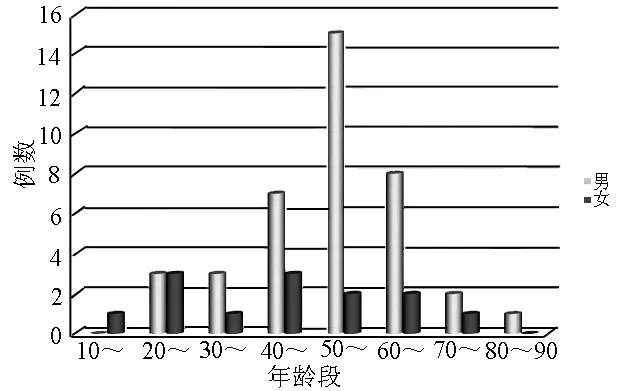
图1 各年龄段男女发病数分布图
二、方法
本研究所有病人PRCC均位于一侧肾脏,右侧35例,左侧17例。肿瘤最大直径为1.4~15 cm,平均(5.9±2.9)cm,本组病例术前诊断主要依据CT平扫+增强。本组病例中CT平扫23例呈低密度影,2例高密度影,3例等密度影,1例高低混杂信号影;CT增强27例呈轻度地不均匀强化,2例有明显强化。病人典型CT表现见图2、图3。10例PRCC病人行MRI检查,呈等T1长T2信号。另有部分病人CT、MRI于外院完成。10例病人(肿瘤<5cm,无转移征象且位于肾的上极或者下极)行保留肾单位手术,42例行肾肿瘤根治性切除术。术后病检显示,TNM分期:Ⅰ期37例(71.2%),Ⅱ期9例(17.3%),Ⅲ期2例(3.8%),Ⅳ期4例(7.7%)。
三、统计学处理
应用SPSS 19.0软件进行统计学分析,连续变量使用t检验,定性变量使用χ2检验。生存曲线以Kaplan-Meier法来进行绘制。通过应用Cox比例风险回归模型分析本研究中的各因素对病人生存率的影响。P<0.05为差异具有统计学意义。
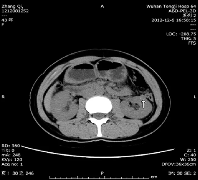
稍低密度肿块影,其内密度不均,可见钙化影,突出于肾脏边界
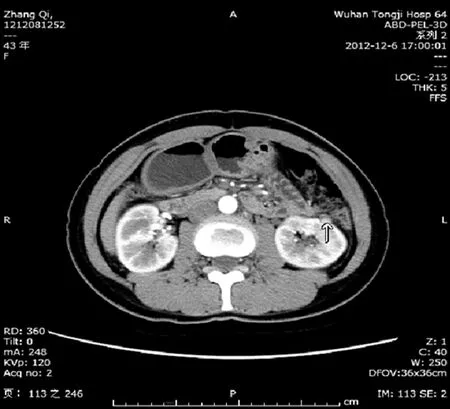
轻度不均匀强化
结 果
术后共44例病人取得随访,随访时间10~122个月,平均46.9个月。其中Ⅰ、Ⅱ、Ⅲ、Ⅳ期PRCC病人生存率分别为96.9%、85.7%、50.0%和66.7%;随访至今各期PRCC病人的平均生存时间分别为52.9个月、31.0个月、28.5个月和32.0个月。本研究中有2例PRCC病人病检提示广泛的出血坏死,但与病人的预后情况无相关性(P>0.05)。10例肾部分切除术的病人都存活至最后一次随访,随访19~81个月,平均随访50.9个月。在随访中4例PRCC病人分别于术后10、16、16、30个月死亡。死于肿瘤转移或者腹膜后肿瘤复发。生存分析见图4。本研究中PRCC病人术后1年存活率为97.7%(43/44),3年存活率为87.5%(28/32),5年存活率为77.8%(14/18)。影响病人预后的主要因素为肿瘤的分期(Ⅱ/Ⅲ/Ⅳ期vsⅠ期:HR=10.281,P=0.044<0.05)。见表1。本组病例中2例行根治性切除术的PRCC病人分别于手术后1年、2年出现原位或者肾区肿瘤复发,再次行手术,术后恢复情况可。
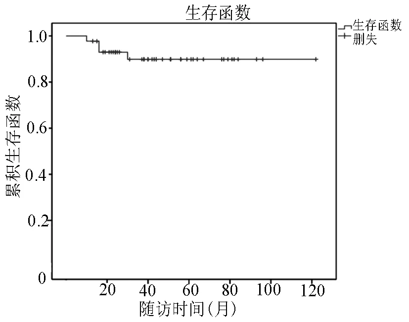
图4 随访病人的肿瘤特异性生存函数

表1 预后相关的单因素、多因素分析
讨 论
PRCC临床罕见,最早是在1976年由Mancilla-Jimenez等[2]作为肾细胞癌(renal cell carcinoma,RCC)的一种临床病理亚型而提出。国外文献显示PRCC占RCC的10.0%~15.0%[1],国内报道其所占比例较低,约为1.9%~7.5%[3-4],本研究中大约为7.9%(52/662),低于国外报道。本研究中PRCC病人的高发年龄为:50~60岁,最小年龄仅为13岁,男女比例3∶1。PRCC的临床表现变化较大,部分病人以腰痛、血尿、发热为首发症状,其余无明显的临床表现,偶然在体检时发现。肿瘤多位于肾脏组织的上极或下极,其直径一般>2 cm(本组最小者仅为1.4 cm)。本研究发现PRCC的CT影像学特点为:CT平扫肿瘤密度较低;CT增强主要表现为轻度不均匀强化,其增强CT值较正常肾组织的增强CT值低。多灶性是PRCC的一个特点,约占所有PRCC的20.0%~41.0%[5-6],但在本研究中仅有1例病人肿瘤表现为多灶性。根据病理检查结果PRCC又可以细分为两个亚型:1型PRCC细胞较小、胞质淡染嗜碱性、核圆形或者椭圆形、单层排列;2型PRCC细胞较大、胞浆嗜酸性、核大、核仁明显、细胞呈假复层排列。有文献表明1型PRCC的预后比2型的好[7-9]。免疫组化检查中PRCC有其独特性,表现为CK7(+)、CK34betaE12(+)、Vimentin(+);RCC(-)及P504s/AMACR(-)[10-11]。Williamson等[12]对395例行手术治疗后的PRCC病人的生存情况进行了长期随访,结果表明,5、10年生存率可达到91.8%和87.7%,PRCC病人预后较好。Patard等[13]的研究显示,PRCC、透明细胞癌5年生存率分别为79.4%和73.2%。PRCC预后较后者好,术后免疫制剂治疗可使部分病人受益。PRCC的预后主要与肿瘤的亚型、TNM分期、有无远处转移等有关[6,12,14]。除了医学上的治疗外,对肿瘤病人的心理上的诊治也逐渐受到关注,它不仅能改善肿瘤病人的生活质量,同时还能对病人的医学治疗产生积极的影响[15]。
总而言之,PRCC临床表现无特异性;影像学上以CT检查为主,平扫呈低密度影,增强显示轻度不均匀强化;确诊依据术后病理检查和免疫组化;肿瘤生长缓慢,就诊时多数病人处于Ⅰ期,治疗方法主要为肾根治性切除术和保留肾单位手术,术后给予免疫制剂,病人预后较好。
[1] Störkel S,Eble JN,Adlakha K,et al.Classification of renal cell carcinoma:Workgroup No.1.Union Internationale Contre le Cancer(UICC)and the American Joint Committee on Cancer(AJCC)[J].Cancer,1997,80(5):987-989.
[2] Mancilla-Jimenez R,Stanley RJ,Blath RA.Papillary renal cell carcinoma:a clinical,radiologic,and pathologic study of 34 cases[J].Cancer,1976,38(6):2469-2480.
[3] 张磊,石怀银,洪宝发.乳头状肾细胞癌临床病理分析[J].解放军医学杂志,2005,30(4):303-305.
[4] 孔祥田,曾荔,宓培,等.乳头状肾细胞癌的临床病理表现[J].中华泌尿外科杂志,2001,22(2):77-80.
[5] Richstone L,Scherr DS,Reuter VR,et al.Multifocal renal cortical tumors:frequency,associated clinicopathological features and impact on survival[J].J Urol,2004,171(2 Pt 1):615-620.
[6] Mejean A,Hopirtean V,Bazin JP,et al.Prognostic factors for the survival of patients with papillary renal cell carcinoma:meaning of histological typing and multifocality[J].J Urol,2003,170(3):764-767.
[7] Steffens S,Janssen M,Roos FC,et al.Incidence and long-term prognosis of papillary compared to clear cell renal cell carcinoma-a multicentre study[J].Eur J Cancer,2012,48(15):2347-2352.
[8] Ledezma RA,Negron E,Paner GP,et al.Clinically localized type 1 and 2 papillary renal cell carcinomas have similar survival outcomes following surgery[J].World J Urol,2015,34(5):1-7.
[9] Herrmann E,Trojan L,Becker F,et al.Prognostic factors of papillary renal cell carcinoma:results from a multi-institutional series after pathological review[J].J Urol,2010,183(2):460-466.
[10] Tickoo SK,Reuter VE.Differential diagnosis of renal tumors with papillary architecture[J].Adv Anat Pathol,2011,18(2):120-132.
[11] Gobbo S,Eble JN,Maclennan GT,et al.Renal cell carcinomas with papillary architecture and clear cell components:the utility of immunohistochemical and cytogenetical analyses in differential diagnosis[J].Am J Surg Pathol,2008,32(12):1780-1786.
[12] Williamson SR,Eble JN,Cheng L,et al.Clear cell papillary renal cell carcinoma:differential diagnosis and extended immunohistochemical profile[J].Mod Pathol,2013,26(5):697-708.
[13] Patard JJ,Leray E,Rioux-Leclercq N,et al.Prognostic value of histologic subtypes in renal cell carcinoma:a multicenter experience[J].J Clin Oncol,2005,23(12):2763-2771.
[14] Pignot G,Elie C,Conquy S,et al.Survival analysis of 130 patients with papillary renal cell carcinoma:prognostic utility of type 1 and type 2 subclassification[J].Urology,2007,69(2):230-235.
[15] 罗莎莉,隋海洋,侯公林.癌症患者心理压力量表的编制和测量学属性的研究[J].华中科技大学学报(医学版),2013,42(1):94-98.
Theclinicalfeaturesandprognosisofpapillaryrenalcellcarcinoma
LUJinjin,CHENLu,SONGXiaodong,etal.
(DepartmentofUrology,TongjiHospital,TongjiMedicalCollegeofHuazhongUniversityofScienceandTechnology,Wuhan430030,China)
ObjectiveTo improve the level of diagnosis and treatment of papillary renal cell carcinoma(PRCC),and to evaluate the prognosis of it.MethodsThe clinical data and survival status of 52 PRCC patients underwent surgery from May 2004 to December 2012 were collected.ResultsAmong the 52 patients(39 men and 13 women,mean age 50.3 years),10 cases presented with hematuria,18 with flank pain and 4 with both hematuria and flank pain.The remaining 20 cases were discovered incidentally.The size of tumor ranged from 1.4cm to 15cm(mean diameter 5.9cm).Forty-two patients underwent radical nephrectomy(80.8%)and 10 patients(tumor size less than 5cm,no signs of metastasis and the tumor located at the upper or lower pole)underwent partial nephrectomy(19.2%).The postoperative pathological examination showed that 37 cases(71.2%)were at the stageⅠ,9(17.3%)at the stageⅡ,2(3.8%)at the stageⅢ,4(7.7%)at the stageⅣ.44 patients were followed up,the median follow-up period after surgery was 46.9 months(range 10 to 122).10 patients,who underwent partial nephrectomy,survived so far.The median follow-up time is 50.9 months(range 19 to 81).4 patients died respectively in 10,16,16,30 months after surgery.The 1-year,3-year,5-year survival rate of PRCC 97.7%(43/44),87.5%(28/32),77.8%(14/18)respectively.ConclusionsPRCC has a nonspecific appearance; most of patients are often at the stage I of the tumor at the time of diagnosis.Surgery is the gold standard treatment of PRCC,including radical and partial nephrectomy.Postoperatively,patients who
an immunosuppressive therapy routinely,have a better prognosis comparing with other types of renal carcinoma.
Papillary renal cell carcinoma; Pathology; Immunohistochemistry; Prognosis
2016-09-12)
(本文编辑:徐文聃)
10.3969/j.issn.1005-6483.2017.10.018
430030 武汉,华中科技大学同济医学院附属同济医院泌尿外科
杨为民,Email:wmyang@tjh.tjmu.edu.cn

