Effect of tourniquet use during total knee arthroplasty on global inflammatory cytokine changes associated with ischemiareperfusion injury
Brian Wu, Ryan Graff, Ido Badash, Joseph G. Skeate,, Christianne Lane, Ibrahim Mansour, Ravi Rao, Anthony Yi,Jarrad Merriman, C. Thomas Vangsness, George Rick Hatch III, Larry Dorr, Paul Gilbert, E. Todd Schroeder, *
1 Keck School of Medicine of the University of Southern California, Los Angeles, CA, USA
2 Department of Molecular Microbiology & Immunology, University of Southern California, Los Angeles, CA, USA
Background
Total knee arthroplasty (TKA) is the most common surgical remediation for patients with long-standing osteoarthritis (OA). A major clinical barrier that has been noted following TKA surgery is persistent muscle atrophy and weakness (Walsh et al., 1998; Mizner et al., 2005;Valderrabano et al., 2007; Halinen et al., 2009). After TKA, ambulation and stair climbing speeds have been shown to be half that of age-matched controls one-year post-surgery (Walsh et al., 1998). Additionally, it has been reported that the gait speed of patients who have undergone TKA is 16-22% less than that of healthy patients (McClelland et al., 2009). Further investigation has suggested that atrophy and failure of voluntary activation contributes to the majority (~85%) of strength lost in leg muscle tissue, and that the quadriceps of patients undergoing TKA are as much as 40% weaker 1 year after surgery when compared to healthy controls (Ouellet et al., 2002; Mizner et al., 2005). Limitations to successful rehabilitation of these patients may include the pre-surgical decrease in voluntary muscle activation and force production, but recent studies point to persistent quadriceps and plantar flexor atrophy as the main contributors to decreased strength and marginal return of physical function even after 1 year of recovery(Valderrabano et al., 2007; Meier et al., 2009). Thus, it is clear that long-term de ficits are demonstrated by patients undergoing TKA surgeries, warranting investigation into the mechanisms preventing recovery or inducing atrophy after these operations (Snyder-Mackler et al., 1994; Walsh et al., 1998; Antolic et al., 1999; Eastlack et al., 1999; Valderrabano et al., 2007; Palmieri-Smith et al., 2008).
During TKA surgeries, a tourniquet is often applied to the proximal thigh to stop blood flow and maintain a clear surgical field. Muscle cells have been found to be particularly sensitive to tourniquet use, as ischemic conditions cause damage to tissue beds that increases relative to the duration of ischemia (Hammers et al., 2008). This phenomenon is caused by biochemical processes that drive metabolism in myocytes. Between meals, approximately 80% of skeletal muscle energy is generated by a continuous supply of free fatty acids being completely oxidized to produce adenosine triphosphate (ATP), a process that represents 95% of the total metabolism in the lower extremities (Ruderman et al.,1977). During ischemia, this supply of circulating free fatty acids as well as oxygen is cut off, forcing ATP demand to be met solely by burning up endogenous phosphocreatine (PCr)and glycogen stores. These reserves are rapidly depleted, and their usage creates localized regions of low pH conditions within myocytes, ultimately inhibiting glycolysisviaa negative feedback loop (Bangsbo et al., 1996). Following surgery,the tourniquet is released and blood flow is re-established to the surgical site (reperfusion event). The initial ischemic stress placed on myocytes is exacerbated during reperfusion, catalyzing a second phase of injury through the rapid production of reactive oxygen species (ROS) that are in signi ficant excess of the endogenous buffering capacity of muscle cells. At the cellular level, these free radicals induce oxidative damage to mitochondria, proteins, lipids, and DNA.As a result, both tissue swelling and vascular endothelial cell dysfunction result in local in flammation as well as microvascular dysfunction, and can render areas of muscle that have undergone the most damage necrotic (Kalogeris et al., 2012).One mediator exacerbating muscle damage may be the in flammatory pathway stimulated by ischemia-reperfusion injury (IRI). Studies investigating the relationship between tourniquet use and IRI have indicated that there is an increase in the levels of IRI-associated in flammatory cytokines following tourniquet use (Tran et al., 2011). Additionally, research has demonstrated increased circulating levels of the in flammatory cytokines interleukin-1 (IL-1),IL-6, and tumor necrosis factor-α (TNF-α) in the plasma of rats within 60 minutes of reperfusion following ischemia.The effects of IRI in in this model were signi ficantly attenuated when treated with polyclonal antibodies against TNF-α or an IL-1 receptor agonist, suggesting that these cytokines play a key role in IRI (Seekamp et al., 1993).Presumably, ROS-mediated damage to cells resulting from reperfusion following ischemia initiates an in flammatory response characterized primarily by the production of these cytokines, as shown by their upregulated presence in plasma samples. Muscle catabolism is associated with an increase in the production of in flammatory cytokines such as IL-1,IL-6, and TNF-α, and it has been shown that IL-6 signaling through the JAK/STAT3 pathway is linked to acute muscle atrophy events (Bonetto et al., 2011; Costamagna et al.,2015). Furthermore, IL-1, IL-6 and TNF-α are present in elevated amounts in the plasma of patients experiencing myocardial infarctions as well as renal ischemia-reperfusion injury, suggesting a role of these cytokines in muscle and tissue atrophy (Nian et al., 2004; Li et al., 2011; Malek et al., 2015). Overall, these previous studies provide some foundational evidence that tourniquet use during TKA may increase the levels of IRI-related cytokines and induce in flammatory signaling that plays a role in muscle catabolism and atrophy (Seekamp et al., 1993; Nian et al.,2004; Bonetto et al., 2011; Li et al., 2011; Tran et al., 2011;Costamagna et al., 2015; Malek et al., 2015).
The goal of this experimental pilot study is to examine the in fluence that tourniquet use during TKA may have on inducing IRI in muscle tissues, as measured by the changes in circulating in flammatory cytokines following tourniquet use during these surgeries. Through establishing a measurable link between tourniquet use during TKA and induction of IRI, we aim to provide guidance for future therapeutics which may be used to counteract the effects of TKA-induced IRI, in the hopes of reducing patient recovery time as well as limiting initial muscle atrophy events after surgery.
Subjects and Methods
Subjects
This study includedmen and women (18 years and older,n= 50) who were scheduled to undergo TKA surgery with an orthopedic surgeon at the orthopedic medicine clinic at the Keck School of Medicine of the University of Southern California (USC), USA. An insitutional review board ap-proval was obtained from the USC Keck School of Medicine(HS-12-00534). Subjects were recruited by an orthopedic surgeon or study team member during a pre-surgical of fice visit. Randomization of study subjects was not performed, as use of a tourniquet was determined by the orthopedic surgeon;the surgeon determined if a tourniquet was required based on the necessity for a clear field and whether it was necessary to ensure a rigid bone-implant cementation.
Three groups were established: no tourniquet, operative tourniquet, and tourniquet during implant cementation groups. Eighteen subjects were enrolled in each group.
Exclusion criteria were pregnancy, peripheral vascular disease, or any uncontrolled medical conditions such as heart, liver, kidney, blood, or respiratory disease. In situations where participants sorted into the ‘no tourniquet’ group were later determined to require a tourniquet, these participants were considered part of the control, non-tourniquet group under an intention-to-treat (ITT) analysis.
Surgical and tourniquet procedures
All subjects had TKA surgery as per standard surgical protocol established at USC. The TKA procedures followed were in accordance with the ethical standards of the responsible committee on human experimentation (institutional and national) and with theHelsinki Declarationof 1975, as revised in 2000 and 2008. All patients underwent primary total knee arthroplasty with minimally invasive techniques and cemented implant prostheses (Zimmer Inc., Warsaw, IN,USA). All of the operations were performed by the senior surgeons, and involved a midline skin incision and trivector medial parapatellar approach.
Subjects in the operative tourniquet group had the tourniquet in flated from initial incision to completion of cementation while patients in the tourniquet during cementation group had the tourniquet in flated only during cementation of the implant. Tourniquets in both groups were in flated to 275 mmHg (1 mmHg = 0.133 kPa) or greater and were de flated after cementation. Cuff time ranged from 8 minutes to 90 minutes across all groups. Tourniquets were released at the completion of patella cementation, and the knees were subsequently held in extension with compression of the patella for at least 10 minutes as the cement completed polymerization.Surgical anesthesia was administered with an epidural, spinal,or general anesthetic, along with a preoperative femoral nerve block used for analgesia.
Inflammatory cytokine/chemokine analysis
For in flammatory cytokine analysis, 8 mL of blood was collected at baseline from the median cubital vein, just prior to cuff in flation (tourniquet groups) and 8 mL of blood was again drawn at the end of surgery, after cuff de flation (tourniquet groups). Total blood drawn did not exceed 16 mL. Venipuncture site was determined by the anesthesiologist, but in general was an antecubital vein. Blood samples were collected within 20 minutes following cuff de flation. Serum was then extracted from patient whole blood through high-speed centrifugation and analyzed with the Bio-Plex Pro™ Human Cytokine 27-plex Assay (Bio-Rad Laboratories, Hercules, CA, USA) measuring FGF basic, Eotaxin, G-CSF, GM-CSF, IFN-γ, IL-1β,IL-1ra, IL-2, IL-4, IL-5, IL-6, IL-7, IL-8, IL-9, IL-10, IL-12(p70), IL-13, IL-15, IL-17, IP-10, MCP-1(MCAF), MIP-1α,MIP-1β, PDGF-BB, RANTES, TNF-α, and VEG. If results were out of range, they were dropped from the analysis.
Statistical analysis
Sample characteristics were reported for each group, comparing patient characteristics for the total sample, as well as by initial and actual treatment. ITT analyses were performed comparing the change from pre-to-post values in cytokines across initial treatment assignment using the Kruskal-WallisH-test, a non-parametric corollary to an variance analysis test.This was due to the preponderance of variables with unequal variances and non-normal distributions. Additionally, Cohen’s d was calculated to estimate the effect size of the mean difference between groups. As a general note, Cohen’s d represents how many standard deviations apart the means are: in general,0.2 is considered small, 0.5 is moderate, 0.7 is large. Further analyses were performed using the same methods on the actual treatment received. Analyses were performed using SPSS 21.0 software (IBM, Armonk, NY, USA).
Results
Baseline data
A total of 54 patients undergoing TKA surgeries were enrolled in this study and separated into three equal groups: no tourniquet (No T), operative tourniquet (OT), and tourniquet during cementation (TDC) (Figure 1). The average length of operative tourniquet in flation was 45.8 minutes (+/- 8.5 minutes) compared to 9.3 minutes (+/- 1.5 minutes) for the tourniquet during cementation group. In four instances, the surgeon was unable to collect blood and tissue samples prior to the start of surgery. These four patients were thusdropped from the study and subsequently removed from analyses.The demographics of the 50 patients included in the study are shown in Table 1.
Effect of tourniquet use during TKA on global inflammatory cytokine changes associated with IRI

Figure 1: Flow chart of study procedures.
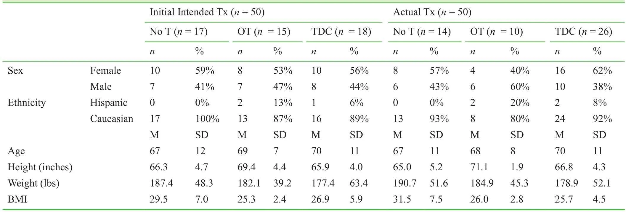
Table 1: Demographic information of patients enrolled in the study
Under the intent to treat analysis, the results generally showed constant or decreasing levels of cytokines in the operative tourniquet and no tourniquet groups and constant or increasing levels in the tourniquet during cementation group (Figure 2). IL-4, IL-5, and TNF-α were the only cytokines to exhibit a statistically signi ficant difference between groups, with decreased cytokines following surgery in the no tourniquet and tourniquet groups and increased cytokines following surgery in the tourniquet during cementation group (Table 2). IL-1β exhibited similar differences between the three groups that trended towards signi ficance (P= 0.06), decreasing in the operative tourniquet and no tourniquet groups and increasing in the tourniquet during cementation group (Table 2).
When the analysis was performed with subjects grouped according to the actual treatment they received, the data is consistent with the ITT analysis in that levels of cytokines generally decreased after surgery in the no tourniquet and operative tourniquet groups, while cytokine increases after surgery were noted in the tourniquet during cementation group (Figure 3). Statistically signi ficant differences were found for IL-12, with the no tourniquet and operative tourniquet groups exhibiting decreases after surgery, and the tourniquet during cementation group demonstrating an increase in IL-12. MCP-1 and IL-1β exhibited similar patterns, which trended towards signi ficance, of decreasing postoperatively in the no tourniquet and operative tourniquet groups and increasing after surgery in the tourniquet during cementation group (Pvalues of 0.05 and 0.06, respectively)(Table 3).
Discussion
The data from this prospective study demonstrated that induction of IRI, based on global in flammatory cytokine changes, shows either a statistically signi ficant difference or one that is trending towards signi ficance with altered tourniquet use in TKA surgeries. Based on our data, there is no signi ficant increase in the pro-in flammatory cytokine pro file of patients who received no tourniquet during surgical procedure and those receiving tourniquet application throughout the procedure. However, the use of a tourniquet when applied solely during implant cementation did result in statistically signi ficant, or trending toward signi ficant,increases in the expression of speci fic in flammatory cytokines (TNF-α, MCP-1 and IL-1β) as compared to full-time tourniquet use or lack-of tourniquet use.This difference potentially outlines differences in the relative risk of ischemia-reperfusion injury to patients depending on the time at which a tourniquet is applied during TKA surgery.In flammation occurring in IRI is shown to have a profound effect on immune responses. A recent study of renal IRI indicates that Th1 cells may exacerbate IRI while Th2 cells may attenuate the effects IRI. Th1 cells are known to produce TNF-α and IL-1β, both well-characterized inducers of IRI,while Th2 cells are characteristic in their release of IL-4, a cytokine associated with attenuation of IRI (Yokota et al.,2003; Boros et al., 2006). Interestingly, in the intent-to-treat analysis, TNF-α and IL-4 were two of the three cytokines found to be signi ficantly differentially expressed between groups with tourniquet during cementation as opposed to operative tourniquet or no tourniquet. The third, IL-5, is another cytokine characteristic of a Th2 in flammatory response. Through this intent-to-treat analysis, both pro-IRI(TNF-α and IL-1β) and anti-IRI (IL-4 and IL-5) cytokines were upregulated in response to tourniquet application during cementation, preventing de finitive conclusions as to the effects of tourniquet use during cementation on potential induction of IRI in total knee arthroplasty.
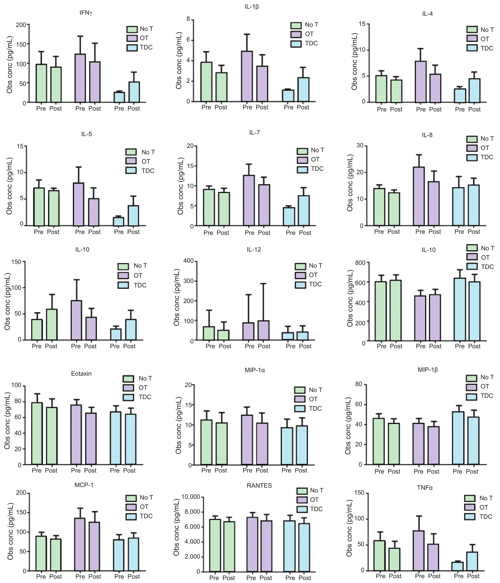
Figure 2: Preoperative and postoperative serum concentrations (conc) of in flammatory cytokines (pg/mL) based on an intention-to-treat analysis.
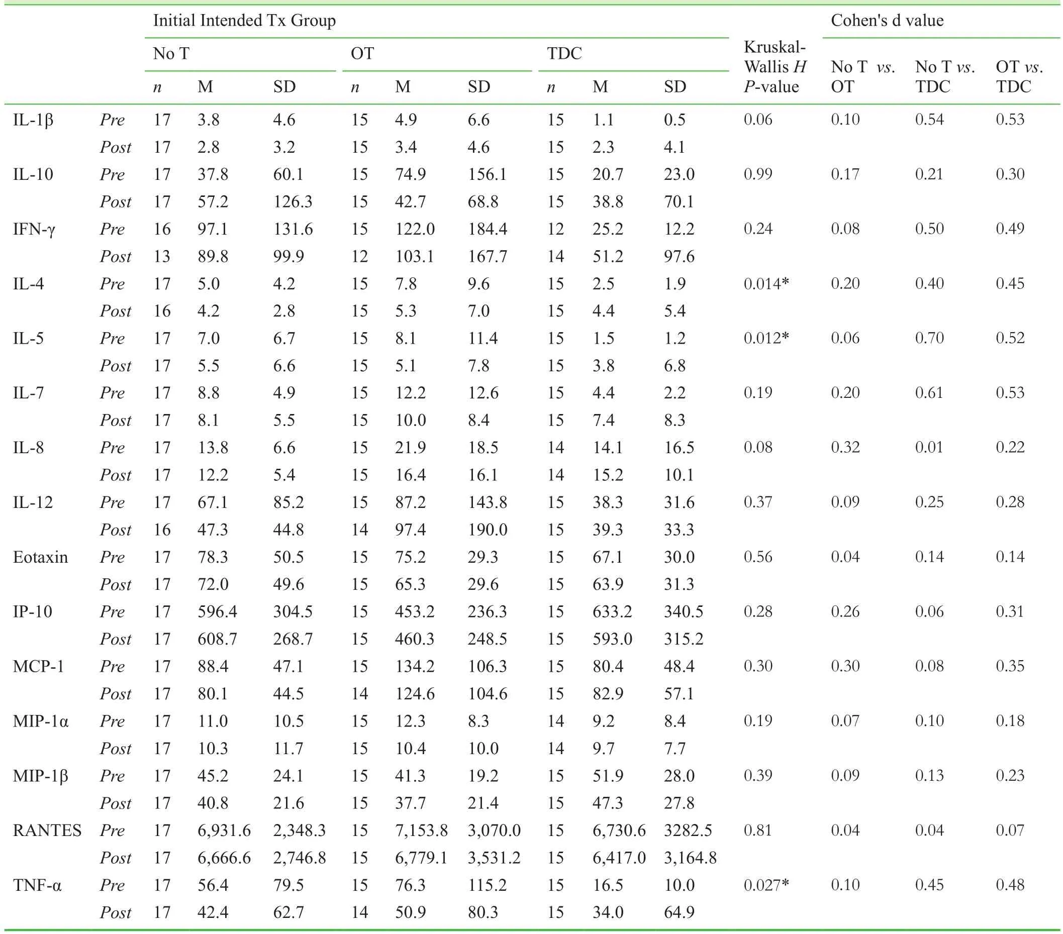
Table 2: Preoperative and postoperative serum cytokine concentrations (pg/mL) based on an intention-to-treat analysis
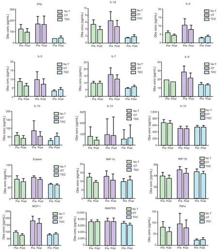
Figure 3: Preoperative and postoperative serum concentrations (conc) of in flammatory cytokines (pg/mL) based on actual treatment groups.
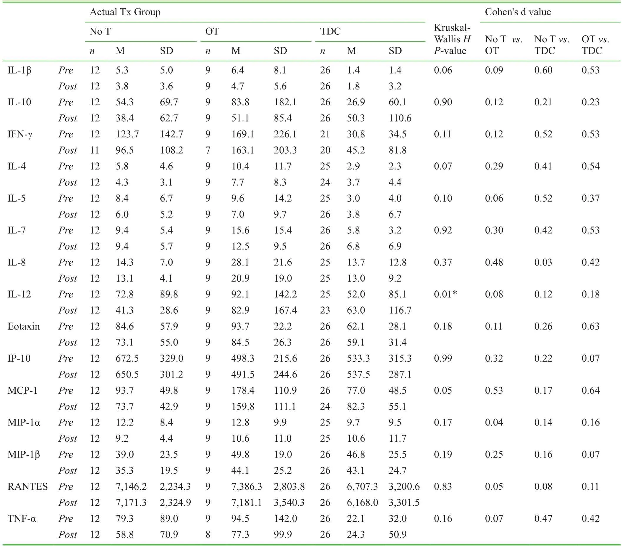
Table 3: Preoperative and postoperative serum cytokine concentrations (pg/mL) based on actual treatment groups
In the analysis of groups according to the actual treatments received by patients, IL-12 was the only cytokine found to be differentially upregulated in patients receiving tourniquets during implant cementation and downregulated in patients with operative tourniquets or no tourniquet application. IL-12 promotes the growth of Th1 cells and inhibits Th2 cell expansion, indicating a potential role for this cytokine in the induction of IRI when tourniquets are applied during cementation (Hamza et al., 2010). Our analysis revealed similar increases in the concentrations of IL-1β(trending towards signi ficance,P= 0.06) and MCP-1 (P=0.05) in patients receiving tourniquet only during implant cementation, which are noteworthy due to the previously described role of IL-1β in IRI, as well as the involvement of MCP-1 in reperfusion injuries (Gourmala et al., 1997;Sung et al., 2002). Therefore, through the actual treatment analysis, there is some indication that tourniquet during cementation in TKA may produce an in flammatory response indicative of IRI. However, it is unclear why tourniquet use throughout the procedure did not show similar changes in in flammatory cytokines, or why both anti-in flammatory and pro-in flammatory cytokines were found to increase with use of tourniquet during cementation in the ITT group.
One potential cause for the lack of signi ficant directional cytokine changes found in this study is the complexity of the human immune response to knee injury. The cytokine pro file of patients may vary according to several factors: time of initial injury, extent of injury, the patient's comorbidities, BMI,immune status, and any other acute or chronic injuries that were not addressed in this study (Furman et al., 2015). Moreover, the interplay of cytokines and the variability of baseline levels of in flammatory markers presents another challenge to analyzing cytokine pro files. For example, patients with chronic osteoarthritis, such as those undergoing TKA, have been identi fied as having high levels of IL-6, TNF-α, IL-17,and IL-18 that could confound the measurement of in flammatory cytokine changes from tourniquet use. On the other hand, a protective effect from the anti-in flammatory IL-10 has been noted in patients who had osteoarthritis and exercised regularly (Wang et al., 2015). It is thus clear that variations in in flammatory cytokines in patients undergoing TKA may be caused by a number of factors beyond the variable use of a tourniquet during these surgeries.
While our study analyzing global in flammatory cytokine changes did not directly indicate that IRI is more strongly induced with use of a tourniquet during TKA surgeries,previous studies analyzing local in flammatory cytokines do demonstrate differences in the in flammatory response in relation to knee surgery requiring tourniquet use. In one previous study that examined the effectiveness of C-reactive protein (CRP), IL-6, creatine kinase (CK), and myoglobin as markers of in flammation and muscle injury due to tourniquet use in TKA, researchers found increased levels of CRP, IL-6, and CK in groups of patients requiring tourniquets as compared to those not requiring tourniquets(Huang et al., 2014). Therefore, the most likely cause for the different cytokine patterns found in this study was the sole use of the antecubital vein, accessed above the tourniquet placement site, for the collection of plasma cytokine samples. If blood were also collected from the ischemic site below the tourniquet zone, such as from the vena saphena magna, a different cytokine pro file, indicating in flammatory changes associated with tourniquet use,may have been obtained. Our results therefore suggest that systemic in flammatory changes may not be as signi ficant as local ones following tourniquet use, and therefore should not be used as the sole outcome marker of IRI caused by tourniquet application
Still, there is value to this study’s method of analyzing systemic changes in in flammatory cytokines as a means of studying IRI. For example, systemic measurements of cytokine changes may be a useful indicator for evaluating the severity of IRI in patients, which can then inform treating physicians of potential interventions to reverse severe incidences of IRI in an effort to reduce recovery times.In the tourniquet during cementation group, there were signi ficant increases, and increases that trended towards signi ficance, of in flammatory cytokines associated with IRI and muscle atrophy. These preliminary findings are encouraging, and should be explored in future studies with larger sample sizes, randomization of patients to treatment groups, and normalization of cytokine changes (adjusting for endogenous baseline levels of each patient) to determine if more signi ficant results in global cytokine changes can be found. Furthermore, measuring ischemic metabolites before and after surgery would enhance the data by suggesting the ischemic states of patients at the time of blood sample collection. It has been shown that ischemic metabolite levels, including glucose, lactate, pyruvate and glycerol,differ with the use of a tourniquet during TKA (Ejaz et al.,2015). Through determining the ischemic states of patients,as well as in flammatory reactions following tourniquet use,future studies will be able to establish a more conclusive link between the use of tourniquets during TKA, ischemia and in flammation-mediated reperfusion injury.
Overall, this study has not been able to demonstrate a statistically signi ficant increase in global in flammatory cytokine response after TKA surgery when comparing patients who required the use of a tourniquet and those who did not. If changes in in flammatory cytokines are an accurate indicator of IRI induction, it is possible that tourniquet use during TKA has no signi ficant in fluence on the occurrence of reperfusion injury and muscle atrophy in patients. Given the findings of other studies on this topic, it is more likely that the measurement of cytokine changes globally is not as effective in detecting an in flammatory reaction as measurements made from the local ischemic zone below the tourniquet.Therefore, additional studies with larger sample sizes and plasma cytokine collection localized to the ischemic site are necessary to conclusively determine if tourniquets can indeed be used in these surgeries without inducing IRI and increasing the risk of muscle atrophy. Furthermore, if global cytokine analysis is to be performed in a patient population,it should be combined with other measurements, such as the level of ischemic metabolites, to more accurately indicate the severity of IRI induction and better inform treating physicians about which interventions should be utilized to reduce recovery times.
Declaration of patient consent
The authors certify that they have obtained all appropriate patient consent forms. In the form the patient(s) has/have given his/her/their consent for his/her/their images and other clinical information to be reported in the journal. The patients understand that their names and initials will not be published and due efforts will be made to conceal their identity, but anonymity cannot be guaranteed.
Conflicts of interest
None declared.
Author contributions
BW, RH, LD, PG, and TS provided conception, design, trial protocol and initiation of the project; BW was the study coordinator;IM, RR, AY, JM, and BW were involved in patient recruitment,data collection and entry; CL, JS, BW were responsible for data analysis and entry; RG, IB, BW, CL drafted and finalized the manuscript. All authors have read and approved the final manuscript.
Plagiarism check
This paper was screened twice using CrossCheck to verify originality before publication.
Peer review
This paper was double-blinded and stringently reviewed by international expert reviewers.
Antolic V, Strazar K, Pompe B, Pavlovcic V, Vengust R, Stanic U,Jeraj J (1999) Increased muscle stiffness after anterior cruciate ligament reconstruction-memory on injury? Int Orthop 23:268-270.
Bangsbo J, Madsen K, Kiens B, Richter EA (1996) Effect of muscle acidity on muscle metabolism and fatigue during intense exercise in man. J Physiol 495:587-596.
Bonetto A, Aydogdu T, Kunzevitzky N, Guttridge DC, Khuri S, Koniaris LG, Zimmers A (2011) STAT3 activation in skeletal muscle links muscle wasting and the acute phase response in cancer cachexia. PLoS One 6:e22538.
Boros P, Bromberg JS (2006) New cellular and molecular immune pathways in ischemia/reperfusion injury. Am J Transplant 6:652-658.
Costamagna D, Costelli P, Sampaolesi M, Penna F (2015) Role of in flammation in muscle homeostasis and myogenesis. Mediators In flamm 2015:805172.
Eastlack ME, Axe MJ, Snyder-Mackler L (1999) Laxity, instability,and functional outcome after ACL injury: copers versus noncopers. Med Sci Sports Exerc 31:210-215.
Ejaz A, Laursen AC, Kappel A, Jakobsen T, Nielsen PT, Rasmussen S (2015) Tourniquet induced ischemia and changes in metabolism during TKA: a randomized study using microdialysis. BMC Musculoskelet Disord 16:326.
Furman BD, Kimmerling KA, Zura RD, Reilly RM, Zlowodzki MP, Huebner JL, Kraus VB, Guilak F, Olson SA (2015) Articular ankle fracture results in increased synovitis, synovial macrophage in filtration, and synovial fluid in flammatory cytokines and chemokines. Arthritis Rheumatol 67:1234-1239.
Gourmala NG, Buttini M, Limonta S, Sauter A, Boddeke HW(1997) Differential and time-dependent expression of monocyte chemoattractant protein-1 mRNA by astrocytes and macrophages in rat brain: effects of ischemia and peripheral lipopolysaccharide administration. J Neuroimmunol 74:35-44.
Halinen J, Lindahl J, Hirvensalo (2009) Range of motion and quadriceps muscle power after early surgical treatment of acute combined anterior cruciate and grade-III medial collateral ligament injuries. A prospective randomized study. J Bone Joint Surg Am 91:1305-1312.
Hammers DW, Merritt EK, Matheny RW Jr, Adamo ML, Walters TJ, Estep JS, Farrar RP (2008) Functional de ficits and insulin-like growth factor-I gene expression following tourniquet-induced injury of skeletal muscle in young and old rats. J Appl Physiol 105:1274-1281.
Hamza T, Barnett JB, Li B (2010) Interleukin 12 a key immunoregulatory cytokine in infection applications. Int J Mol Sci 11:789-806.
Huang ZY, Pei FX, Ma J, Yang J, Zhou ZK, Kang PD, Shen B(2014) Comparison of three different tourniquet application strategies for minimally invasive total knee arthroplasty: a prospective non-4randomized clinical trial. Arch Orthop Trauma Surg 134:561-570.
Kalogeris T, Baines CP, Krenz M, Korthuis RJ (2012) Cell biology of ischemia/reperfusion injury. Int Rev Cell Mol Biol 298:229-317.
Li JP, Lu L, Wang LJ, Zhang FR, Shen WF (2011) Increased serum levels of S100B are related to the severity of cardiac dysfunction, renal insuf ficiency and major cardiac events in patients with chronic heart failure. Clin Biochem 44:984-988.
Malek M, Nematbakhsh M (2015) Renal ischemia/reperfusion injury; from pathophysiology to treatment. J Renal Inj Prev 4:20-27.
McClelland JA, Webster KE, Feller JA (2009) Variability of walking and other daily activities in patients with total knee replacement. Gait Posture 30:288-295.
Meier WA, Marcus RL, Dibble LE, Foreman KM, Peters CL, Mizner RL, LeStayo PC (2009) The long-term contribution of muscle activation and muscle size to quadriceps weakness following total knee arthroplasty. J Geriatr Phys Ther 32:79-82.
Mizner RL, Petterson SC, Snyder-Mackler L (2005) Quadriceps strength and the time course of functional recovery after total knee arthroplasty. J Orthop Sports Phys Ther 35:424-436.
Mizner RL, Petterson SC, Stevens JE, Vandenborne K, Snyder-Mackler L (2005) Early quadriceps strength loss after total knee arthroplasty. The contributions of muscle atrophy and failure of voluntary muscle activation. J Bone Joint Surg Am 87:1047-1053.
Nian M, Lee P, Khaper N, Liu P (2004) In flammatory cytokines and postmyocardial infarction remodeling. Circulation 94:1543-1553.
Ouellet D, Moffet H (2002) Locomotor de ficits before and two months after knee arthroplasty. Arthritis Rheum 47:484-493.
Palmieri-Smith RM, Thomas AC, Wojtys EM (2008) Maximizing quadriceps strength after ACL reconstruction. Clin in Sports Med 27:405-424.
Ruderman NB, Goodman MN, Berger M, Hagg S (1977) Effect of starvation on muscle glucose metabolism: studies with the isolated perfused rat hindquarter. Fed Proc 36:171-176.
Seekamp A, Warren JS, Remick DG, Till GO, Ward PA (1993) Requirements for tumor necrosis factor-alpha and interleukin-1 in limb ischemia/reperfusion injury and associated lung injury. Am J Pathol 143:453-463.
Snyder-Mackler L, De Luca PF, Williams PR, Eastlack ME, Bartolozzi AR 3rd(1994) Re flex inhibition of the quadriceps femoris muscle after injury or reconstruction of the anterior cruciate ligament. J Bone Joint Surg Am 76:555-560.
Sung FL, Zhu TY, Au-Yeung KK, Siow YL, O K (2002) Enhanced MCP-1 expression during ischemia/reperfusion injury is mediated by oxidative stress and NF-kappaB. Kidney Int 62:1160-1170.
Tran TP, Tu H, Pipinos II, Muelleman RL, Albadawi H, Li Y-L(2011) Tourniquet-induced acute ischemia-reperfusion injury in mouse skeletal muscles: involvement of superoxide. Eur J Pharmacol 650:328-334.
Valderrabano V, Nigg BM, von Tscharner V, Frank CB, Hintermann BJ (2007) Leonard goldner award 2006. Total ankle replacement in ankle osteoarthritis: an analysis of muscle rehabilitation. Foot Ankle Int 28:281-291.
Walsh M, Woodhouse LJ, Thomas SG, Finch E (1998) Physical impairments and functional limitations: a comparison of individuals 1 year after total knee arthroplasty with control subjects. Phys Ther 78:248-258.
Wang X, Hunter D, Xu J, Ding C (2015) Metabolic triggered in flammation in osteoarthritis. Osteoarthritis Cartilage 23:22-30.
Yokota N, Burne-Taney M, Racusen L, Rabb H (2003) Contrasting roles for STAT4 and STAT6 signal transduction pathways in murine renal ischemia-reperfusion injury. Am J Physiol Renal Physiol 285:F319-325.
 Clinical Trials in Orthopedic Disorder2017年1期
- Clinical Trials in Orthopedic Disorder的其它文章
- Information for Authors -Clinical Trials in Orthopedic Disorders
- Genetic risk factors of degenerative intervertebral disc disease:a case-control study
- MRI appearance of injured ligaments and/or tendons of the ankle in different positions: study protocol for a single-center,diagnostic clinical trial
- Effect of periprosthetic fracture on hip function after femoral neck-preserving total hip arthroplasty: study protocol for a prospective, single-center, self-controlled trial with 2-year follow-up
- New bone fixation plate for the repair of avulsion fracture of the tibial attachment of the posterior cruciate ligament: study protocol for a prospective, open-label, self-controlled, clinical trial
- Risk factors for ligamentum flavum hypertrophy in lumbar spinal stenosis patients from the Xinjiang Uygur Autonomous Region, China: protocol for a retrospective, single-center study
