
Development of tissue-selective gene delivery system with ultrasound
2017-01-19 11:37:441MutsumiSugiiHitoshiUrugJohnUngYoichiNegishiSneOdKzuoMruym
1Mutsumi Sugii Hitoshi UrugJohn UngYoichi Negishi,Sne Od Kzuo Mruym
aFaculty of Pharma-Sciences,Teikyo University,Tokyo,Japan
bSchool of Pharmacy,Tokyo University of Pharmacy and Life Science,Tokyo,Japan
Development of tissue-selective gene delivery system with ultrasound
Ryo Suzukia,*,Daiki Omataa,1,Yusuke Odaa,Mutsumi Sugiia, Hitoshi Urugaa,Johan Ungaa,Yoichi Negishib,Sanae Odaa, Kazuo Maruyamaa
aFaculty of Pharma-Sciences,Teikyo University,Tokyo,Japan
bSchool of Pharmacy,Tokyo University of Pharmacy and Life Science,Tokyo,Japan
A R T I C L E I N F O
Article history:
Available online 25 November 2015
Ultrasound
Nanobubbles
Gene delivery
Gene therapy is applied into cardiovascular diseases,cancer and diseases that are due to genomic causes.Viral vectors are effcient carriers of genes for transduction,but some problems have become evident.Delivery vectors that are highly potent in terms of gene transduction effciency should also be safe and easy to apply.Non-viral vectors have recently received focus as gene carriers,but their transduction effciency is very low and not suitable for in vivo gene delivery.In addition,it is important to develop tissue-specifc or selective gene delivery system to avoid side effects in gene therapy.However, the gene delivery system which can easily change a transfection site has not been developed.Gene delivery with ultrasound is expected to be an attractive method for controlling gene delivery site due to induced driving force of gene transfection at the limited area where it is insonated.In this study,we assessed the feasibility of tissue selective gene delivery with nanobubbles and ultrasound exposure.
Gene delivery into liver or brain–Luciferase coded plasmid DNA(pCMV-Luc)(100 μg)and nanobubbles(500 μg)suspension was injected into the tail vein of mice.Then,US was transdermally exposed to liver(frequency:1 MHz,1 W/cm2, 1 min)or transcranially exposed to brain(frequency:1 MHz, 1.2 W/cm2,1 min).After 1 day of injection,the luciferase expressions were measured.When ultrasound was exposed to liver,luciferase expression in the liver was higher than that in other tissues(Fig.1).On the other hand,when ultrasound was exposed to brain,luciferase expression was observed in the brain.From these results,it was suggested that the tissue of gene delivery was controllable by changing the site of ultrasound exposure.
In addition,we confrmed the gene expression cells in gene delivery for liver.In this case,gene expression was observed in parenchymal cells.Moreover,we also confrmed the parts of gene expression in the brain after gene delivery.Gene expression was observed at wide area in the brain.From these results,it is guessed that plasmid DNA might be extravasated with jet stream induced by cavitation of nanobubbles and delivered into parenchymal cells in the liver and brain.Therefore,the combination of nanobubbles and ultrasound exposure would be a noninvasive and tissue selective gene delivery system.
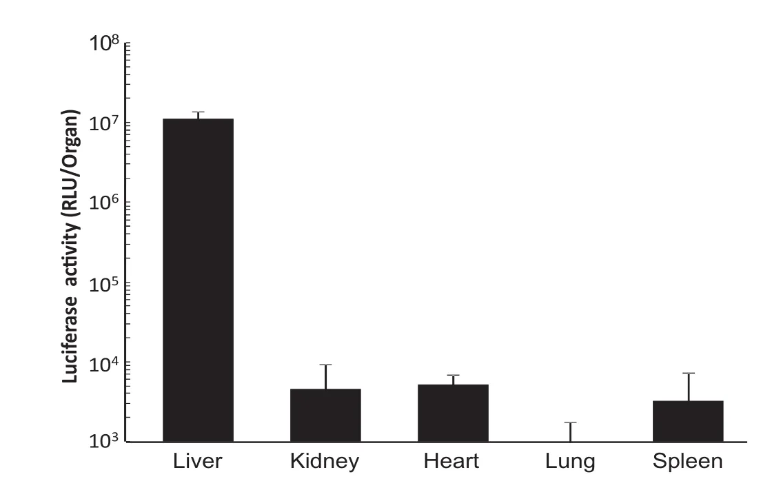
Fig.1–Luciferase expression with nanobubble and ultrasound exposure for liver.
Acknowledgement
This study was supported by MEXT-supported Program for the Strategic Research Foundation at Private Universities,2013–2017.
*E-mail address:r-suzuki@pharm.teikyo-u.ac.jp.
1JSPS Research Fellow.
Peer review under responsibility of Shenyang Pharmaceutical University.
http://dx.doi.org/10.1016/j.ajps.2015.11.067
1818-0876/©2016 Production and hosting by Elsevier B.V.on behalf of Shenyang Pharmaceutical University.This is an open access article under the CC BY-NC-ND license(http://creativecommons.org/licenses/by-nc-nd/4.0/).
 Asian Journal of Pharmacentical Sciences2016年1期
- Asian Journal of Pharmacentical Sciences的其它文章
- Determination of the antidepressant effect of mirtazapine augmented with caffeine using Swiss-albino mice
- Photosafety testing of dermally-applied chemicals based on photochemical and cassette-dosing pharmacokinetic data
- Biopharmaceutics classifcation system(BCS)-based biowaiver for immediate release solid oral dosage forms of moxifoxacin hydrochloride (Moxifox GPO)manufactured by the Government Pharmaceutical Organization(GPO)
- Bioequivalence study of abacavir/lamivudine (600/300-mg)tablets in healthy Thai volunteers under fasting conditions
- Evaluation of cytotoxic and infammatory properties of clove oil microemulsion in mice
- Analytical method development of pregabalin and related substances in extended release tablets containing polyethylene oxide