
Morphological changes of doxorubicin-loaded liposomes observed by atomic force microscopy
2017-01-19 11:37:40NaokiTakahashiKeisukeUedaKenjirouHigashiKeijiYamamotoKunikazuMoribe
Naoki Takahashi,Keisuke Ueda,Kenjirou Higashi,Keiji Yamamoto, Kunikazu Moribe
Graduate School of Pharmaceutical Sciences,Chiba University,1-8-1 Inohana,Chuo-ku,Chiba 260-8675,Japan
Morphological changes of doxorubicin-loaded liposomes observed by atomic force microscopy
Naoki Takahashi*,Keisuke Ueda,Kenjirou Higashi,Keiji Yamamoto, Kunikazu Moribe
Graduate School of Pharmaceutical Sciences,Chiba University,1-8-1 Inohana,Chuo-ku,Chiba 260-8675,Japan
A R T I C L E I N F O
Article history:
Available online 23 November 2015
Liposome
Doxorubicin
Atomic force microscopy
Liposomes,closed vesicles composed of lipid bilayers,have been widely used as pharmaceutical carriers.Liposomal formulations containing doxorubicin(DOX)of an anticancer drug are developed to reduce toxic side effects and to improve drug accumulation at tumor tissues.An encapsulation of DOX into the inner water phase of liposomes results in the formations of fbrous DOX bundles and the elongation of the liposomes[1]. In this study,atomic force microscopy(AFM),which allows nanoscale imaging directly in an aqueous environment,was utilized to observe the morphological changes of the liposomes induced by the DOX encapsulation.Furthermore,the effect of storage on liposomal morphology was also evaluated.
1,2-Dimyristoyl-sn-glycero-3-phosphocholine(DMPC)/ cholesterol(60:40,mol%)liposomes were prepared by the hydration and extrusion method.DOX was loaded into the inner water phase of liposomes by the ion gradient method at the molar ratio of DOX from 20%to 40%against total lipid components.The liposomes were immobilized on(3-Aminopropyl)triethoxysilane(APTES)-modifed mica for AFM measurements.
DOX-unloaded liposomes represented circular shape in AFM images(Fig.1A).The encapsulation of 20%DOX in the liposomes changed the liposomal morphology from sphere to rod shape(Fig.1B).Furthermore,the ratio of rod-shaped liposomes signifcantly increased with the enhanced DOX loading from 20%to 40%(Fig.1C).This AFM result in an aqueous environment is consistent with the previous cryogenic transmission electron microscopy studies;the liposomal morphology is changed to rod shape with the increase in DOX concentrations,accompanied with the linearly expansion of fbrous DOX bundles.Interestingly,several circular particles in theAFM image showed concave shape(Fig.1D).The thickness at the center of concave particles was determined as 11±1 nm,corresponding to about twice the theoretical height of the lipid bilayers. In contrast,the peripheral height of the concave particles was over 20 nm,indicating the existence of curved bundles of DOX fbers such as the ring and‘U’shape.The stability of 40%DOX-loaded liposomes was evaluated by storing them for 7 days at 4°C.In comparison with the freshly prepared liposomes,the rod-shaped particles were decreased with the increase of the concave particles at liposomes stored for 7 days(Fig.1E).Hence, after the storage,the rod-shaped liposomes containing linear bundles of DOX fbers transformed into the spherical liposomes containing curved bundles.
Spherical liposomes were deformed to rod shape due to the formation of linear bundles of DOX fbers depending on DOXloading.The storage of 40%DOX-loaded liposomes induced the transformation of rod-shaped liposomes into spherical ones, accompanied with the transformation of fbrous DOX bundles from linear to curve.AFM measurements provide detail information on the changes of liposomal morphology directly in an aqueous environment.
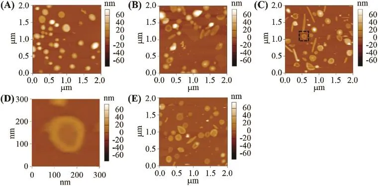
Fig.1–AFM images of(A)unloaded liposomes,(B)20%DOX-loaded liposomes(freshly prepared),(C)40%DOX-loaded liposomes(freshly prepared),(D)expanded concave particle in the dotted square of the image(C),and(E)40%DOX-loaded liposomes(stored for 7 days).
R E F E R E N C E
[1]Li X,Hirsh DJ,Cabral-Lilly D,et al.Doxorubicin physical state in solution and inside liposomes loaded via a pH gradient. Biochim Biophys Acta 1998;1415:23–40.
*E-mail address:naoki.t.1227@chiba-u.jp.
Peer review under responsibility of Shenyang Pharmaceutical University.
http://dx.doi.org/10.1016/j.ajps.2015.10.044
1818-0876/©2016 The Authors.Production and hosting by Elsevier B.V.on behalf of Shenyang Pharmaceutical University.This is an open access article under the CC BY-NC-ND license(http://creativecommons.org/licenses/by-nc-nd/4.0/).
 Asian Journal of Pharmacentical Sciences2016年1期
- Asian Journal of Pharmacentical Sciences的其它文章
- Determination of the antidepressant effect of mirtazapine augmented with caffeine using Swiss-albino mice
- Photosafety testing of dermally-applied chemicals based on photochemical and cassette-dosing pharmacokinetic data
- Biopharmaceutics classifcation system(BCS)-based biowaiver for immediate release solid oral dosage forms of moxifoxacin hydrochloride (Moxifox GPO)manufactured by the Government Pharmaceutical Organization(GPO)
- Bioequivalence study of abacavir/lamivudine (600/300-mg)tablets in healthy Thai volunteers under fasting conditions
- Evaluation of cytotoxic and infammatory properties of clove oil microemulsion in mice
- Analytical method development of pregabalin and related substances in extended release tablets containing polyethylene oxide