
Life cycle and morphology of development stages of Physocephalus dromedarii(Nematoda∶Spirocercidae)
2016-10-22 01:08:22RolfKarlSchusterSarithaSivakumarorgKinne
Rolf Karl Schuster,Saritha Sivakumar,J¨org Kinne
Central Veterinary Research Laboratory,PO Box 597,Dubai,United Arab Emirates
Life cycle and morphology of development stages of Physocephalus dromedarii(Nematoda∶Spirocercidae)
Rolf Karl Schuster*,Saritha Sivakumar,J¨org Kinne
Central Veterinary Research Laboratory,PO Box 597,Dubai,United Arab Emirates
ARTICLE INFO
Article history:
in revised form 30 Nov 2015,2nd revised form 1 Feb,3rd revised form 19 Feb 2016
Accepted 12 Jun 2016
Available online 25 Aug 2016
Physocephalus dromedarii Nematoda
Life cycle
Dromedary
Abomasum
Objective:To study the development of Physocephalus dromedarii(P.dromedarii)in the final host.
Methods:For this,5 adult dromedaries were orally infected with third larval stages of P.dromedarii obtained from naturally infected scarab beetles(Scarabaeus cristatus).The camels were necropsied 14,42,70,84 and 280 days after infection and their abomasi were examined for the presence of nematodes.
Results:Early 4th stage larva occurred already 2 weeks after infection.They were still in the sheet of the 3rd stage larva.Six weeks after infection,the nematodes became juvenile male and female adults measuring 9 and 10 mm,respectively.Their size doubled at 10 weeks post infection and patency was reached at 12 weeks.P.dromedarii was still present in the camel that was examined 40 weeks after infection.
Conclusions:As a result of experimental infection of the natural host,the determined prepatent period of P.dromedarii equalled 12 weeks.
Original articlehttp://dx.doi.org/10.1016/j.apjtb.2016.08.001
1.Introduction
Nematodes of the genus Physocephalus in dromedaries were reported for the first time from Algeria[1]and Turkmenistan[2]in 1912 and 1924,respectively.In his thesis,Badanin[3]drew attention that Physocephalus specimens found in the abomasi of dromedaries in Turkmenistan differ from those in pigs in larger dimensions and marked swelling in the midbody of females. These features in connection with a different intermediate host spectrum and bigger measurements of 3rd stage larvae found in scarab beetles were reasons to divide Physocephalus sexalatus(P.sexalatus)into two subspecies,Physocephalus sexalatus sexalatus found in pigs and Physocephalus sexalatus dromedarii occurring in dromedaries[4].The first cases of Physocephalus infection of dromedaries in Dubai were diagnosed in July 2011 and October 2012.Both positive animals originated from a camel dairy farm that previously had imported dromedaries from abroad.A closer look at these parasites revealed further peculiarities that allowed upgrading the former subspecies to species level[5].Both P.sexalatus from pigs and Physocephalus dromedarii(P.dromedarii)from camels are characterized by two trilobed lateral lips followed by a cylindrical pharynx supported by spirally arranged rings(Figure 1).The oesophagus is subdivided by a shorter muscular and a much longer glandular part.A cuticular inflation on the anterior end bearing the first lateral cervicalpapilla(derid)isfollowedby 3 pairs oflateral alae(Figure 2). A second lateral papilla,erroneously described in former descriptions as excretory pore,is embedded in the anterior median lateral wing at the opposite site.The excretory pore can be found slightly posterior of the latter on the ventral surface.Posterior ends of males are spirally corkscrew-like twisted.Caudal alae are supportedbystalkedprecloacalpapillae.Spiculesareofunequallength(Figure 3).P.dromedarii differ from P.sexalatus by larger total dimensions and a striking swelling in the midbody of the female. Male specimens of P.dromedarii are characterized by longer spicules,a higher number of precloacal ridges and postcloacal papillae[5].Inapreviouspaper[6],wedescribedthe3rdlarvalstage obtained from intermediate and paratenic hosts.The aim of thispaper is to give details of the development of P.dromedarii in dromedaries after experimental infection.
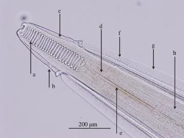
Figure 1.Anterior end of P.dromedarii.
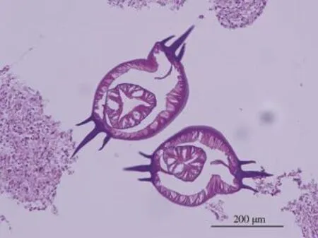
Figure 2.Cross section through the anterior ends of adult P.dromedarii at the level of the muscular oesophagus(centre)showing three pairs of lateral alae on each side.

Figure 3.Posterior end of a male P.dromedarii with spicules of unequal length(a,b).
2.Materials and methods
2.1.Isolation of infective larvae and experimental infection
In connection with a survey on possible intermediate hosts of P.dromedarii,large scarab beetles(Scarabaeus cristatus)were collected in the desert of the Dubai Emirate in spring 2014 and were examined for the presence of nematode larvae.For this,the body of each individual beetle was opened with scissors and was submerged for 20 min in 50 mL of 38°C warm artificial gastric juice on a magnet stirrer.Chitinous parts of the body were removed and the remaining liquid was poured in a Petri dish and examined for the presence of nematode larvae under a stereoscopic microscope.In order to determine their numbers,larvae wereconcentratedinthecentrebygentlyswayingthePetridish.In case of high numbers,all larvae were transferred into 15 mL centrifuge tubes and after 5 min of sedimentation time the supernatant was discharged.Depending on semi-quantitative estimationoflarvalnumbers,werefilledthetubeupto2,3,5or10mL with phosphate buffer solution.After vigorous mixing,an aliquot of 100μL larval suspension was extracted and placed on a glass slide.Larvae were counted and multiplied by the corresponding dilution factor(20,30,50 or 100).Isolated 3rd stage larvae were used to infect five adult dromedaries.Larval suspensions in dosages between 10 000 and 20 000 were orally inoculated.
2.2.Necropsy and reisolation of nematodes
The infection was terminated 14,42,70,84 and 280 days post infection(dpi)and dromedaries were necropsied.After removal from the carcass,the abomasum was opened and put into a bucket containing 5 L of 40°C warm normal saline and folds of the mucosa were vigorously scraped with a spoon to remove all hidden nematodes from abomasal folds.After 30 min of sedimentation time,the supernatant was discharged and the bucket was refilled.This procedure was repeated until the supernatant became clear.After vigorous stirring,a 20%aliquot was selected for counting the parasite burden.For this,the sediment was transferred into a Petri dish and was examined under the stereoscopic microscope.Isolated nematodes were fixed in hot(70°C)10%neutral formalin.Each 10 larvae,male and female worms were cleared in 30%lactic acid and were studied under the microscope(Olympus BX51)connected to a digital camera(Olympus DP71).Measurements of total length,length of pharynx,muscular and glandular oesophagus,distance of the cervical papillae,the nerve ring and the excretory pore to the anterior end and length of the spicules in males were taken by the aid of cellSens Dimension software.
3.Results
3.1.3rd larval stage
While 2nd stage larvae present in some of the beetles were destroyed during artificial digestion.3rd stage larvae remained viable and were quickly moving in the warm digestion fluid. They coiled spirally after the liquid was cooled(Figure 4).Alllarvae were in the size range of P.dromedarii and were characterized by the possession of cylindrical pharynx and a posterior end covered with minute spines[6].
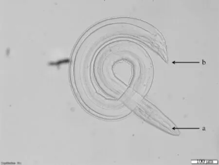
Figure 4.Third stage larva of P.dromedarii.
3.2.4th larval stage
Inthe camel(camel A)necropsied 14dpi(142 larvae),most of them in their 4th stage were recovered(Tables 1 and 2).These larvae were still surrounded by the sheet of the previous larval stage but showed already the typical for Physocephalus pharynx consisting of ring-like structures(Figure 5).The knoblike posterior end of the larval sheet was covered with minute spikes being characteristic for the 3rd larval stage(Figure 6).The distinctive inflation on the anterior end is not yet developed.Measuring 3.7mm,theaveragetotallengthhadmorethandoubledcompared to the original 3rd stage larva.Single 3rd stage larvae with a cylindrical pharynx were still present 2 weeks after infection.
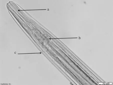
Figure 5.Fourth stage larva of P.dromedarii,anterior end.
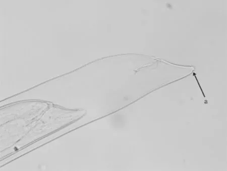
Figure 6.Fourth stage larva of P.dromedarii,anterior end.
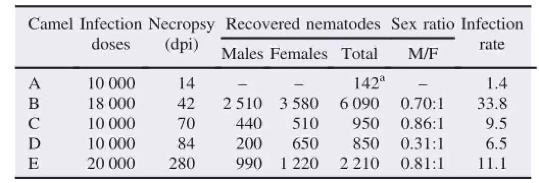
Table 1 Experimental infection of dromedaries with P.dromedarii.
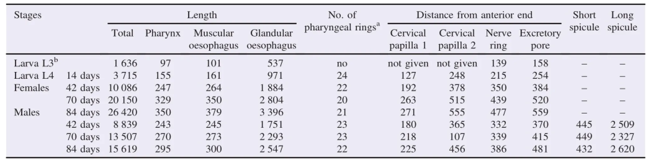
Table 2 Morphometrical data of larvae,male and female P.dromedarii of different age.
3.3.Juvenile nematodes(42 days old)
Juvenile 42 days old females were only slightly larger than males(10.1 versus 8.8 mm)(camel B)and showed also similar sizes in other parameters.At a closer look,a large number of thejuvenile adults were still in the sheet of the 4th stage larvae.In this case,males had the same shape as females and could only be distinguished by the presence of spicules.These spicules reached already the size of adult nematodes but were of inconspicuous colouration and difficult to recognize(Figure 7).Also,the caudal alae of the males were not fully developed.The uterus of females did not contain eggs.
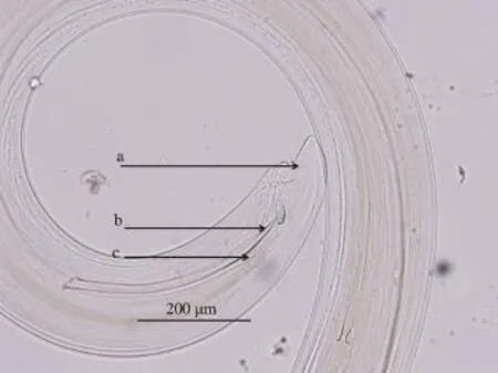
Figure 7.Juvenile male P.dromedarii 42 dpi in the sheet of the fourth stage larva(a).
3.4.Preadult nematodes(70 days old)
Considerable differences were seen between males and females in 70 days old preadult nematodes measuring 13.5 and 20.1 mm in length,respectively(camel C).The cuticular inflation at the anterior end in both sexes became noticeable but was less prominent in comparison to later stages.The uterus of the females started to fill up with eggs but the terminal part was still empty(Figure 8)indicating the prepatent status of this stage.Also,the typical swelling of the female adult worm in the midbody was absent.In preadult males that had nearly reached the final size,the posterior end became spirally corkscrew-like twisted.
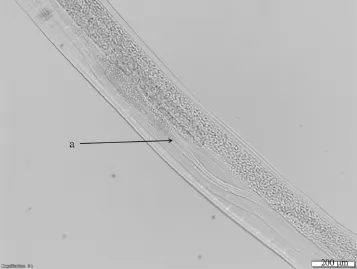
Figure 8.Preadult female P.dromedarii 70 dpi.
3.5.Adult nematodes
Camel D necropsied 84 dpi harboured 200 male and 650 female nematodes reaching a size given in the literature[5]and showing a prominent cuticular inflation at the anterior end resembling an arrow head.Asymmetrical deirids and lateral alae are distinctive.Female specimens with a total length of 24-28.2 mm showed a 2 mm long swelling in the midbody caused by uterine loops filled with eggs and the thickened distal part of the uterus(Figure 9).Their average size of 26.4 mm was considerably longer than in 10 week old females. In contrast,male nematodes at 12 weeks p.i.were only slightly longer,measuring 15.6 mm.No further growth was seen in male and female nematodes obtained from camel E necropsied 40 weeks p.i.Their average length amounted to 15.6 and 25.7 mm,respectively.
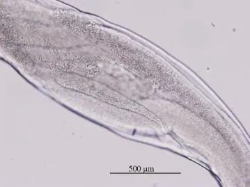
Figure 9.The swelling of the midbody of a 84 days old female P.dromedarii caused by loops of the anterior and posterior uterine horns.
4.Discussion
Nematodes of the genus Physocephalus were mentioned in papers on the helminth fauna of dromedaries in Algeria[1,7],Kuwait[8],Iraq[9],Iran[10,11],Turkmenistan[2-4,12,13]and Uzbekistan[14].Since Physocephalus eggs are small and cannot be determined with routine coproscopical procedures,diagnosis is based on findings of adult worms at necropsy.Contrary to Haemonchus contortus and Haemonchus longestipes,which are easily recognized by the red colour of their intestinal tube,pale pink specimens of P.dromedarii are less conspicuous and might be overlooked although this species has a size comparable to Haemonchus species.For this reason,it is suspected that P.dromedarii has a much wider geographical distribution and is more frequent in camel populations.
Dromedaries are the main host for P.dromedarii but equids also may become infected.Thus,“P.sexalatus”was found in donkeys in Uzbekistan[15]but the description of the nematodes matched rather with P.dromedarii.
Scarab beetles are the main intermediate hosts for Physocephalus spp.and terrestrial vertebrates can be involved in the life cycle as paratenic hosts.
For the development of Physocephalus spp.in final hosts only very few data are available.In guinea pigs that were used as experimental hosts in studies of the life cycle of Physocephalussexalatus dromedarii,only 0.2%of larvae survived up to 48 dpi and only few underwent further development but failed to become adult[16].
In rabbits and pigs,a first molt occurred 12-15 dpi.At this stage,the 4th stage larva measured 3.5-4.0 mm,the originally cylindrical pharynx became a structure of parallel arranged rings and the spines at the posterior end of the larva disappeared[16]. Our results confirmed these observations.A further molt was observed in rabbits and pigs between 23 and 25 dpi.When the nematodes reached a length of 6.8-8.2 mm,and 4 weeks later,on Day 42,lateral wings were present in both sexes and caudal alae and spicules were seen in males.Fully developed eggs containing the 1st larval stage were observed in females at 83 dpi[16].Our results obtained in camels suggest that the passage from 4th stage larva to juvenile adults in camels takes place later because 42 days old nematodes just reached half of their final size and a large number were still covered by the larval sheet of the 4th larval stage.Our data showed that females at 70 dpi are already fertilized but the terminal part of the uterus did not contain eggs.It can be concluded that the prepatent period of P.dromedarii lasts longer than 10 weeks but is completed in 12 weeks.
Some details on the development of P.sexalatus were reported in its final host,the pig[17].First changes of the larva were 2 weeks after infection when the originally cylindrically shaped pharynx becomes a structure consisting of 18 parallel arranged rings and the“stinkweed”kind of posterior end becomes a tail cap covered with minute spines.The whole development of P.sexalatus in the stomach of the pig is completed within 10-12 weeks but some specimens obtain sexual maturity already 6-7 weeks after infection.
Itwasassumedthat camelsbecome infectedviadrinkingwater that contains larval stages that had escaped from drowned beetles[12].Our experience showed that under desert conditions camels ingest scarab beetles or freshly dead paratenic hosts(reptiles,birds and rodents).The infection rate in our experimental infections ranged between 1.4%and 33.8%.The low number of recovered larvae 2 weeks after infection might be due to the small size of the stages.It is also possible that not all larvae had reached the abomasum.
The presence of P.dromedarii in the camel that was dissected 40 weeks after infection showed that this species has a long life span which is necessary because adult scarab beetles are available only during a limited time period,in spring and summer.
Conflict of interest statement
We declare that we have no conflict of interest.
[1]Seurat LG.[On the occurrence of Spiroptera sexalata Molin in the dromedary and the donkey].C R Seances Soc Biol Fil 1912;64: 174-6.French.
[2]Baskakov VP.[On the fauna of parasitic worms of Turkmenistan camels].Pap State Inst Exp Vet Med 1924;2:92-105.Russian.
[3]Badanin NV.[Helminths of the camel][dissertation].Moscow: Moscow Veterinary Academy;1939.Russian.
[4]Mushkambarova MG.[Scarab beetles in the lower reach of the rivers Murgab and Tedzen as intermediate hosts for helminthes][dissertation].Ashchabat:Turkmenistan Agricultural Institute;1967.Russian.
[5]Schuster RK,Wibbelt G,Kinne J.A re-description of Physocephalus dromedarii stat.nov.(Nematoda:Spirocercidae),an abomasalnematodeofdromedaries(Camelusdromedarius). J Helminthol 2013;88:499-505.
[6]Schuster RK,Wibbelt G,Sivakumar S,Reiczigel J.Light and scanning electron microscopical examination of the third stage larva of Physocephalus dromedarii(Nematoda:Spirocercidae)-an abomasal nematode of the one humped camel(Camelus dromedarius).Parasitol Res 2015;114:1913-20.
[7]Chauve M,Hamza-Cherif R,Marfoua K.[Parasites in the dromedary(Camelus dromedarius)in Algeria surveys in 4 wilayats(Adar Bechar-Laghouat-Ghardaia)].Maghreb Vet 1990;5:38-9. French.
[8]Abdul-Salam JM,Farah MA.Seasonal fluctuations of gastrointestinal helminths of camels in Kuwait.Vet Parasitol 1988;28:93-102.
[9]Altaif KI.Helminths in camels in Iraq.Trop Anim Health Prod 1974;6:55-7.
[10]Mirzayans A,Halim R.Parasitic infection of Camelus dromedarius from Iran.Bull Soc Exot Filiales 1980;73:442-5.
[11]Anvari-Tafti M,Sazmand A,Hekmatimoghaddam S,Moobedi I. Gastrointestinal helminthes of camels(Camelus dromedarius)in center of Iran.Trop Biomed 2013;30:56-61.
[12]Mushkambarova MG,Dobrynin MI.[Materials on physocephalosis of the one humped camel in Turkmenistan].Izv Akad Nauk Turkm SSR Ser Biol 1972;4:62-7.Russian.
[13]Mushkambarova MG,Dobrynin MI,Berdyev AB.[Helminthoses and their prophylaxis in camels in Turkmenistan].Ashgabat: Ylym;1989.Russian.
[14]Sultanov MA,Kabilov T,Atchanova C,Dadaev S.[Determination of the intermediate hosts of Physocephalus sexalatus dromedarii in the Kashkarinsk region].Dokl Akad Nauk Uzb SSR 1973;1:42-3. Russian.
[15]Zdanova MG.[Quantitative and qualitative analysis of the helminth fauna of donkeys in Uzbek SSR].Probl Parazitol 1969;4:18-20. Russian.
[16]Mushkambarova MG.The development of Physocephalus sexalatus in experimental definitive host.In:Academician KI,editor. Skrjabin and the development of helminthological science in Turkmenistan.Ashchabad:Ilim;1970.
[17]Hobmaier M.[The life history and pathological importance of Physocephalus sexalatus(Spiroptera sexalata,Molin)].M¨unch Tier¨arztl Wschr 1925;76:361-6,368-92,410-2,436-40.
2 Sep 2015
Rolf Karl Schuster,Central Veterinary Research Laboratory,PO Box 597,Dubai,United Arab Emirates.
Tel:+971 43375165
E-mail:rschuster@cvrl.ae
All experimental animals and treatment in this study were reviewed and approved by the Animal Ethic Committee of Central Veterinary Research Laboratory,and ministry of environment and water of the UAE,according to the Ministerial Decree No.384 of the year 2008 on the executive by-law of the Federal Law No.16 of the year 2007.
Peer review under responsibility of Hainan Medical University.The journal implements double-blind peer review practiced by specially invited international editorial board members.
2221-1691/Copyright©2016 Hainan Medical University.Production and hosting by Elsevier B.V.This is an open access article under the CC BY-NC-ND license(http:// creativecommons.org/licenses/by-nc-nd/4.0/).
 Asian Pacific Journal of Tropical Biomedicine2016年10期
- Asian Pacific Journal of Tropical Biomedicine的其它文章
- A review on promising phytochemical,nutritional and glycemic control studies on Moringa oleifera Lam.in tropical and sub-tropical regions
- A rare cause of acute abdomen-Spontaneous rectus sheath hematoma
- Evaluation of proline,chlorophyll,soluble sugar content and uptake of nutrients in the German chamomile(Matricaria chamomilla L.)under drought stress and organic fertilizer treatments
- ProductionofsecondarymetaboliteE2.2fromPhaleriamacrocarpaendophyticfungus
- Cytotoxic,genotoxic and apoptotic effects of naringenin-oxime relative to naringenin on normal and cancer cell lines
- Pandanusamaryllifoliusleafextractincreasesinsulinsensitivityinhigh-fatdiet-induced obese mice