
FTC-CD133纳米微粒抑制肝癌干细胞耐药性的研究
2016-02-23 05:17:37黄长文蒋星星殷香保黄跃英
中国全科医学 2016年2期
黄长文,蒋星星,殷香保,黄跃英,雷 康,熊 虎
FTC-CD133纳米微粒抑制肝癌干细胞耐药性的研究
黄长文,蒋星星,殷香保,黄跃英,雷 康,熊 虎
【摘要】目的制备包裹乳腺癌耐药蛋白(BCRP/ABCG2)多药转运体的特异抑制剂FTC且连接CD133抗体的纳米微粒,并评价其特性和治疗作用。方法运用复乳蒸发法制备以聚乳酸-聚乙醇酸(PLGA)为载体的包封FTC的载药纳米微粒,通过碳化二亚胺法在其表面连接CD133抗体,得到纳米微粒FTC-PLGA-NP-C;分析制备得到的纳米微粒的粒径分布、FTC包封率、载药量和纳米微粒的抗体连接效率;免疫磁珠法分选出细胞,MTT法检测细胞的增殖能力,Western blotting检测SP细胞中ABCG2的表达;荧光显微镜下观察FTC-PLGA-NP-C与细胞的结合效率;将细胞加入不同浓度的FTC-PLGA-NP-C纳米微粒,Western blotting检测其ABCG2的表达;以同体积PBS为对照,在细胞中加入FTC-PLGA-NP-C纳米微粒,同时加入阿霉素,HPLC检测孵育不同时间后细胞内阿霉素的浓度。裸鼠腋下荷瘤,成瘤后尾静脉注射FTC-PLGA-NP-C或者0.9%氯化钠溶液,48 h后再由尾静脉注射阿霉素,测量瘤体直径,并采用高效液相色谱法检测裸鼠肿瘤组织中阿霉素的浓度。结果FTC-PLGA-NP-C呈规则球形,粒径分布为60~120 nm,载药量为11.27%,包封率为37.18%,与细胞有较好的结合能力;Western blotting检测显示ABCG2在细胞中表达,且随着FTC-PLGA-NP-C纳米微粒剂量的增加,ABCG2的表达量逐渐降低,到4 mg/ml FTC-PLGA-NP-C时已几乎检测不到ABCG2的表达。从24 h开始,PBS中细胞内阿霉素浓度很快下降至1 μg/ml及以下;而FTC-PLGA-NP-C中细胞内阿霉素浓度一直维持在2~3 μg/ml。生理盐水组小鼠瘤体直径为(1.845±0.076)cm,高于FTC-PLGA-NP-C组的(1.238±0.072)cm(t=5.802,P<0.001);生理盐水组瘤内阿霉素浓度为(2.005±0.154)μg/ml,低于FTC-PLGA-NP-C组的(3.853±0.261)μg/ml(t=6.088,P<0.001)。结论制备的载药纳米微粒FTC-PLGA-NP-C可较好地靶向肝癌干细胞,并抑制其耐药蛋白ABCG2的表达,降低肿瘤细胞对常规治疗药物的耐药性,提高化疗药物的治疗效果。
肿瘤是一种严重危害人类健康的疾病。化学药物治疗是目前常用的治疗手段。研究发现,肿瘤中存在含量极少但具有无限增殖潜能的肿瘤干细胞,这些细胞具有高致瘤性,是肿瘤发生和转移的根源[1]。有学者通过体外实验发现,某些化疗药物可以杀死肿瘤细胞,但对肿瘤干细胞却无效[2-3],这就决定了肿瘤复发的必然性。因此,只有探索杀灭肿瘤干细胞的治疗方式,才能最终彻底治愈肿瘤。

ABCG2(ATP-binding cassette superfamily G member 2)是ABC转运蛋白(ATP-binding cassette)家族中的成员,主要存在于干细胞、某些肿瘤细胞和上皮细胞的顶膜,是具有广泛底物作用的特异性外排泵,在SP细胞中呈高表达,还可使进入细胞中的药物被排出胞外,故该功能被认为参与了肿瘤细胞的多药耐药性[5-6]。
本研究拟应用包裹乳腺癌耐药蛋白(BCRP/ABCG2)多药转运体的特异抑制剂FTC(fumitremorgin C)且连接CD133抗体的纳米微粒,通过其特异性作用于肝癌SP细胞,使纳米微粒中FTC在SP细胞中高效、持续发挥作用,有效逆转SP细胞的耐药性,进而使普通化疗药物(如阿霉素)更容易进入SP细胞并大量蓄积。这种直接以肿瘤干细胞为靶向,并且针对肿瘤干细胞耐药性的治疗思路或许能寻找到彻底治愈肿瘤的突破口。
1材料与方法
1.1试剂与动物阿霉素(Adriamycin,ADM),购自意大利Farmitalia公司;FTC,购自上海博升生物科技有限公司;聚乳酸-羟基乙酸共聚物(PLGA),购自济南岱罡生物科技有限公司;碳化二亚胺(EDC),购自美国Pierce公司;Micro BCA蛋白质定量试剂,购自美国Pierce公司;离心机,Eppendorf 5810R式,德国Eppendorf公司;流式细胞仪,BD FACSCalibur,美国BD公司;Zeta分析仪,英国Malvern公司;酶标仪,美国Bio-Rad公司;荧光显微镜,日本Olympus公司;Chemidoc XRS凝胶成像仪,美国Alpha公司;人肝癌细胞株MHCC97-H,本实验室培养保存;裸鼠,购自北京华阜康生物科技有限公司。
1.2包裹FTC且连接CD133抗体纳米微粒的制备将适量FTC溶液在超声条件下加入PLGA二氯甲烷溶液中,在所得溶液中加入少量Tween 80与Span 80混合乳化剂(Tween 80∶Span 80=5∶1),超声5 min使之成为均一稳定的初乳液。将所得初乳液在超声条件下滴加到50 ml O-羧甲基壳聚糖水溶液中,超声10 min形成复乳液。然后将此复乳液在室温下电磁搅拌4 h以上,使有机溶剂完全挥发,形成FTC-PLGA-NP混悬液。将此混悬液以2 000 r/min离心5 min,离心半径17.8 cm,取沉淀用蒸馏水离心洗涤3次,除去未包埋药物,将所得沉淀冷冻干燥即得FTC-PLGA-NP。将CD133抗体通过EDC法(每1 mg FTC-PLGA-NP中加入2 mg CD133抗体)连接在FTC-PLGA-NP上,制备得到纳米微粒FTC-PLGA-NP-C。
1.3表征及FTC载药量、包封率和抗体连接效率
1.3.1微粒表征的检测将样品稀释后取约1 ml加至样品池中,然后将样品池放入仪器中进行检测。
1.3.2FTC载药量和包封率检测通过MicroBCA蛋白测定试剂盒测定FTC浓度,严格按照说明书进行操作。载药量(%)=纳米粒中药物重量/纳米粒重量×100%;包封率(%)=纳米粒中药物重量/给药量×100%。
1.3.3CD133连接效率的检测将样品稀释不同倍数,通过MicroBCA蛋白测定试剂盒测定CD133的连接效率,严格按照说明书操作。
1.5MTT法检测SP细胞的增殖能力将分离得到的SP细胞富集并重悬,调整细胞浓度为5×10⁴/ml。取96孔板,每孔加细胞悬液200 μl,然后放入37 ℃,5% CO2恒温箱孵育24 h~1周。加入MTT 15 μl/孔,敲打均匀后放入培养箱中孵育4 h。取出培养板,以2 500 r/min离心25 min,完全弃上清液,加入200 μl DMSO,吹匀后放入酶标仪,在630 nm波长下读取吸光度A50值(OD值)。
1.6Western blotting检测SP细胞中ABCG2的表达细胞处理完成后提取细胞总蛋白,BCA法测定蛋白浓度。采用SDS-PAGE电泳法电泳分离,转移至PVDF膜上;结合特异性抗体及相应二抗孵育;将PVDF膜放入Chemidoc XRS凝胶成像仪,滴加适量显影液后获取样品蛋白图像。
1.7FTC-PLGA-NP-C与SP细胞结合取分离后的SP细胞接种于6孔板中,加入FTC-PLGA-NP-C,使其终浓度为5 g/L。培养24 h后将培养基换成PBS,加入FITC-CD133抗体,避光孵育1 h后在荧光显微镜下观察纳米微粒与细胞的结合效率。
1.9FTC-PLGA-NP-C对瘤体及其耐药性的影响裸鼠腋下荷瘤。成瘤后尾静脉注射5 mg/kg的FTC-PLGA-NP-C或者0.9%氯化钠溶液,48 h后,再由尾静脉注射阿霉素,测量瘤体直径;然后将肿瘤研碎制成单细胞悬液,取相同数量的细胞,破壁后用1 ml PBS悬起,离心后取上清液,高效液相色谱法检测裸鼠肿瘤组织中阿霉素的浓度。

2结果
2.1FTC-PLGA-NP-C检测
2.1.1FTC-PLGA-NP-C的粒径分布制备得到FTC-PLGA-NP-C,其粒径分布为60~120 nm,平均粒径为107 nm(见图1)。
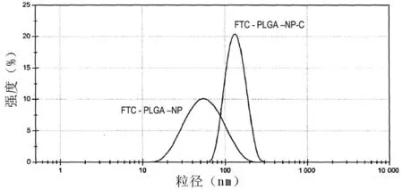
图1 FTC-PLGA-NP-C的粒径分布
2.1.2载药量和包封率FTC载药量为11.27%,包封率为37.18%。
2.1.3抗体连接效率通过MicroBCA蛋白测定试剂盒测定CD133的连接效率,结果显示该载药纳米微粒表现出对靶抗原良好的免疫结合活性,其效价与游离抗CD133抗体基本相当(见图2)。
2.1.4FTC-PLGA-NP-C与SP细胞的结合能力通过荧光显微镜观察纳米微粒与SP细胞的结合能力,结果显示,FTC-PLGA-NP-C与SP细胞有较好的结合能力(见图3)。
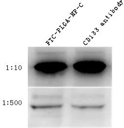
图2 FTC-PLGA-NP-C中CD133表达
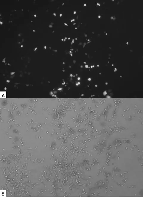
注:A为在绿色荧光下观察,B为在白光下观察
图3FTC-PLGA-NP-C与SP细胞的结合效率
Figure 3Binding efficiency of FTC-PLGA-NP-C and SP cells

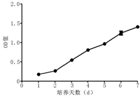
图5 MTT法检测细胞的增殖
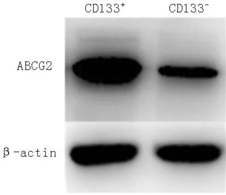
图6 ABCG2在细胞中的表达
2.3.1最佳FTC-PLGA-NP-C纳米微粒剂量的确定随着纳米微粒剂量的增加,ABCG2的表达量逐渐降低,到4 mg/ml FTC-PLGA-NP-C时已几乎检测不到ABCG2的表达(见图7)。

图7 不同浓度FTC-PLGA-NP-C对ABCG2表达的影响
Figure 7Effect of different concentrations of FTC-PLGA-NP-C on the expression of ABCG2

图8 培养不同时间后 SP细胞内阿霉素浓度

2.4FTC-PLGA-NP-C对瘤体及其耐药性的影响生理盐水组小鼠瘤体直径为(1.845±0.076)cm,FTC-PLGA-NP-C组小鼠瘤体直径为(1.238±0.072)cm,两组比较,差异有统计学意义(t=5.802,P<0.001)。生理盐水组瘤内阿霉素浓度为(2.005±0.154)μg/ml,FTC-PLGA-NP-C组瘤内阿霉素浓度为(3.853±0.261)μg/ml,两组比较,差异有统计学意义(t=6.088,P<0.001)。
3讨论
肿瘤是人类健康的一大杀手,肿瘤的治疗方法和效果也是广泛关注的热点。化学药物治疗在经历了长期的发展和完善后,仍然是目前肿瘤治疗的主要手段。但长期使用化学药物治疗存在两大难题:一是大部分抗肿瘤药物靶向性差,在杀死肿瘤的同时对正常的组织器官也存在严重的损伤;二是长期大量使用化学药物治疗后,肿瘤细胞会产生耐药性,即对药物的杀伤作用不敏感或是将进入细胞的药物排出胞外,需加大给药量才能起到抗肿瘤作用。
实验和临床均证实,肿瘤对化疗药物有耐药性主要表现为肿瘤干细胞耐药。肿瘤干细胞虽然数量少,但有无限增殖的潜能,同时对化学药物具有耐药性,所以决定了肿瘤复发和转移的必然性[9-10]。因此,针对肿瘤干细胞的治疗方式才是有效的肿瘤治疗方案,才能最终彻底治愈肿瘤。

ABCG2是ABC转运蛋白家族中的一个成员,多药抗性的产生常常伴随ABC家族中一个或几个成员的过表达,特别是ABCG2蛋白的过表达。该转运蛋白是P-糖蛋白,由多药抗性蛋白1(multidrug resistance protein 1,MDR1)和多药抗性相关蛋白(multidrug resistance-associated protein,MAP)的基因编码。研究发现,ABCG2是SP细胞主要的ABC转运蛋白,SP细胞通过ABCG2将荧光活性染料Hoechst33342排出细胞外,正是据于此,SP细胞被鉴定、分离。ABCG2在SP中呈高表达可使进入细胞中的药物被排出胞外,故该功能被认为参与了肿瘤细胞的多药耐药性[21-22]。
综上所述,本研究合成的FTC-PLGA-NP-C,由于纳米微粒良好的靶向作用和ABCG2抑制效果,可明显削弱肝癌干细胞的耐药性,提高胞内化疗药物的浓度、增强药物的治疗效果,是一种安全有效的新型肝癌靶向治疗制剂,具备良好的临床应用前景。
本文链接:

作者贡献:黄长文进行试验设计,殷香保进行试验设计与实施,黄跃英资料收集整理,蒋星星撰写论文,黄长文成文并对文章负责;黄跃英、雷康、熊虎进行实验实施、评估、资料收集;黄长文进行质量控制及审校。
本文无利益冲突。
参考文献
[1]Korkaya H,Wicha MS.Selective targeting of cancer stem cells:a new concept in cancer therapeutics[J].BioDrugs,2007,21(5):299-310.
[2]Reya T,Morrison SJ,Clarke MF,et al.Stem cells,cancer,and cancer stem cells[J].Nature,2001,414(6859):105-111.
[3]Zhou S,Li F,Xiao J,et al.Isolation and identification of cancer stem cells from human osteosarcom by serum-free three-dimensional culture combined with anticancer drugs[J].J Huazhong Univ Sci Technolog Med Sci,2010,30(1):81-84.

[5]Xie C,Pei XT.ABCG2/Bcrp1 transporter as phenotype markets and regulators of side population stem cells[J].Progress in Physiological Sciences,2003,34(1):57-59.(in Chinese)
谢超,裴雪涛.ABCG2/Bcrp1转运蛋白:侧群干细胞的表型标记与功能调控蛋白[J].生理科学进展,2003,34(1):57-59.
[6]Xie J,Jin B,Li DW,et al.ABCG2 regulated by MAPK pathways is associated with cancer progression in laryngeal squamous cell carcinoma[J].Am J Cancer Res,2014,4(6):698-709.
[7]Zhao X,Ran YL,Yu L,et al.Isolation,cultivation and identification of cancer stem-like cells from human liver cancer tissues[J].Chinese Journal of Cancer Biotherapy,2009,16(5):436-441.(in Chinese)
赵璇,冉宇靓,遇珑,等.人肝癌组织中肿瘤干细胞样细胞的分离培养及鉴定[J].中国肿瘤生物治疗杂志,2009,16(5):436-441.
[8]Nigavekar SS,Sung LY,Llanes M,et al.3H dendrimer nanoparticle organ/tumor distribution[J].Pharm Res,2004,21(3):476-483.
[9]Chiba T,Kita K,Zheng YW,et al.Side population purified from hepatocellular carcinoma cells harbors cancer stem cell-like properties[J].Hepatology,2006,44(1):240-251.
[10]Phesse TJ,Clarke AR.Normal stem cells in cancer prone epithelial tissues[J].Br J Cancer,2009,100(2):221-227.
[11]Lai DM,Liu T,Huang Y,et al.Identification and characterization of ovarian cancer stem-like cells from primary tumor[J].Zhonghua Fu Chan Ke Za Zhi,2009,44(12):936-940.
[12]Kuzmanov A,Hayrabedyan S,Karaivanov M,et al.Basal cell subpopulation as putative human prostate carcinoma stem cells[J].Folia Histochem Cytobiol,2007,45(2):75-80.
[13]O′Brien CA,Pollett A,Gallinger S,et al.A human colon cancer cell capable of initiating tumour growth in immunodeficient mice[J].Nature,2007,445(7123):106-110.
[14]Ma S,Chan KW,Hu L,et al.Identification and characterization of tumorigenic liver cancer stem/progenitor cells[J].Gastroenterology,2007,132(7):2542-2556.
[15]Yin S,Li J,Hu C,et al.CD133positive hepatocellular carcinoma cells possess high capacity for tumorigenicity[J].Int J Cancer,2007,120(7):1444-1450.
[16]Zhang H,Li SY.Research progression of CD133as a marker of cancer stem cells[J].Chinese J Cancer,2010,29(3):243-247.
[17]陈书德,宋文杰,张福琴,等.干细胞表面标志物CD133在肝癌组织中的表达及其临床意义[J].第四军医大学学报,2009,30(5):458-461.
[18]Bari S,Seah KK,Poon Z,et al.Expansion and homing of umbilical cord blood hematopoietic stem and progenitor cells for clinical transplantation[J].Biol Blood Marrow Transplant,2015,21(6):1008-1019.
[19]Bahnassy AA,Zekri AR,El-Bastawisy A,et al.Circulating tumor and cancer stem cells in hepatitis C virus-associated liver disease[J].World J Gastroenterol,2014,20(48):18240-18248.
[20]Varna M,Gapihan G,Feugeas JP,et al.Stem cells increase in numbers in perinecrotic areas in human renal cancer[J].Clin Cancer Res,2015,21(4):916-924.
[21]Basu S,Hertsenberg AJ,Funderburgh ML,et al.Human limbal biopsy-derived stromal stem cells prevent corneal scarring[J].Sci Transl Med,2014,6(266):266ra172.
[22]Weiss J,Theile D,Dvorak Z,et al.Interaction potential of the multitargeted receptor tyrosine kinase inhibitor dovitinib with drug transporters and drug metabolising enzymes assessed in vitro[J].Pharmaceutics,2014,6(4):632-650.
(本文编辑:贾萌萌)
·论著·
【关键词】纳米复合物;肿瘤干细胞;FTC;CD133;ABCG2
黄长文,蒋星星,殷香保,等.FTC-CD133纳米微粒抑制肝癌干细胞耐药性的研究[J].中国全科医学,2016,19(2):184-189.[www.chinagp.net]
Huang CW,Jiang XX,Yin XB,et al.Research of FTC-CD133nanoparticles inhibiting the drug resistance of liver cancer stem cell[J].Chinese General Practice,2016,19(2):184-189.
Research of FTC-CD133Nanoparticles Inhibiting the Drug Resistance of Liver Cancer Stem CellHUANGChang-wen,JIANGXing-xing,YINXiang-bao,etal.DepartmentofHepatobiliarySurgery,theSecondAffiliatedHospitalofNanchangUniversity,Nanchang330006,China
【Abstract】ObjectiveTo prepare the nanoparticles packing FTC,a specific inhibitor of multidrug transporter BCRP/ABCG2,and connecting CD133antibody,and to evaluate its features and therapeutic effect.MethodsNanoparticles packing FTC and with PLGA as carrier were prepared using multiple emulsion-solvent evaporation method,and carbodiimide reaction was undertaken to connect CD133antibody on the surface of the nanoparticles,forming the nanoparticles of FTC-PLGA-NP-C;the particle size distribution,FTC encapsulation efficiency,drug loading capacity and the antibody connection efficiency of nanoparticles were analyzed; SP cells were sorted by magnetic activated cell sorting(MACS),MTT method was used to measure the multiplication capacity of SP cells,and western blotting method was employed to detect the expression of ABCG2 in SP cells;the combination efficiency of FTC-PLGA-NP-C and SP was observed under fluorescence microscope; SP cells were added into nanoparticles of FTC-PLGA-NP-C of different concentrations,and western blotting was used to detect the expression of ABCG2;with PBS of the same volume taken as control,FTC-PLGA-NP-C nanoparticles were added into SP cells with adriamycin added at the same time,and the concentration of adriamycin in the cells was determined by HPLC at different time points of incubation.Tumor was transplanted to the armpit of nude mice,and tail intravenous injection of FTC-PLGA-NP-C or 0.9% sodium chloride solution was undertaken after the formation of tumor;48 hours later,tail intravenous injection of adriamycin was conducted,tumor diameter was measured,and high efficiency liquid chromatography was employed to measure the concentration of adriamycin in the tumor tissue of mude mice.ResultsFTC-PLGA-NP-C was in sphere shape,with a size distribution of 60-120 nm,a drug loading capacity of 11.27% and an encapsulation efficiency of 37.18%,and had a better combining capacity with SP cells;western blotting showed that ABCG2 expression existed in SP cells and decreased with the increase of the dosage of FTC-PLGA-NP-C nanoparticles with no ABCG2 expression detected when the dosage of FTC-PLGA-NP-C was 4 mg/ml.At 24 hours of incubation,the concentration of adriamycin began to decrease quickly to below 1 μg/ml in the SP cells in PBS,while the concentration of adriamycin in the SP cells in FTC-PLGA-NP-C remained at 2-3 μg/ml.The diameter of the mice tumor of normal saline group was(1.845±0.076)cm,higher than(1.238±0.072)cm of FTC-PLGA-NP-C group(t=5.802,P<0.001);the concentration of adriamycin in the tumor of normal saline group was(2.005±0.154)μg/ml,lower than (1.238±0.072)cm of FTC-PLGA-NP-C group(t=6.088,P<0.001).ConclusionPrepared drug-loading nanoparticle FTC-PLGA-NP-C could effectively target liver cancer stem cells,inhibit the expression of drug resistance protein ABCG2,reduce the drug resistance of tumor cells to conventional drugs and improve the therapeutical effect of chemotherapeutic drug.
【Key words】Nanocomposites;Neoplastic stem cells;FTC;CD133;ABCG2
收稿日期:(2015-10-13;修回日期:2015-12-12)
【中图分类号】R 730.21
【文献标识码】A
doi:10.3969/j.issn.1007-9572.2016.02.013
通信作者:黄长文,330006 江西省南昌市,南昌大学第二附属医院肝胆外科;E-mail:hcw02ys@163.com
基金项目:作者单位:330006 江西省南昌市,南昌大学第二附属医院肝胆外科(黄长文,殷香保,黄跃英,雷康,熊虎);新余市人民医院外科(蒋星星)
