黏蛋白2在结肠癌中的表达及其相关临床意义的研究
左晓旭朱袭嘉李盛国罗喜顺甘泽林王海鹏
黏蛋白2在结肠癌中的表达及其相关临床意义的研究
左晓旭①朱袭嘉①李盛国①罗喜顺①甘泽林①王海鹏①
目的:研究黏蛋白(MUC2)在结肠正常组织、结肠良性腺瘤组织和结肠癌组织中的表达状况,以及MUC2的表达情况与结肠癌患者临床资料的关联。方法:选择2011年3月-2015年3月本院手术切除的结肠正常组织15例,结肠良性腺瘤组织41例及结肠癌组织121例,应用免疫组织化学技术检测MUC2在不同组织中的表达。结果:(1)MUC2在结肠正常组织、结肠良性腺瘤组织和结肠癌组织中的阳性表达率分别为100%(15/15)、73.17%(30/41)和46.28%(56/121),不同组织中MUC2阳性表达率比较,差异有统计学意义(P<0.01)。(2)MUC2高表达的结肠癌患者组织分型、淋巴结转移及肿瘤分期均优于MUC2低表达患者,比较差异均有统计学意义(P<0.05)。结论:结肠癌中MUC2的表达状况与组织分型、淋巴结转移和肿瘤分期有关。MUC2的表达下调可能参与了结肠癌的发生、发展、浸润和转移等过程。
黏蛋白2; 结肠腺瘤; 结肠癌
结肠癌(colorectal cancer,CRC)是目前世界上排名第三的常见恶性肿瘤,也是肿瘤引起相关死亡的常见原因之一[1-2]。在发展中国家,结肠癌是男性排名第二、女性排名第三的肿瘤相关性疾病引起死亡的原因[3]。因此,结肠癌的早期预防、早期诊断及积极治疗等对于降低结肠癌的发病率及死亡率显得尤为重要。黏蛋白(mucins,MUC)是由胃肠道、呼吸道和卵巢等上皮组织分泌的大分子糖蛋白,有润滑和保护的作用,目前已发现有21个家族成员[4]。MUC2最早是由美国学者于1989年在人类小肠组织的cDNA表达文库中克隆到的一种黏蛋白核心肽基因,定位于人类染色体的11p 15.3~15.5[5]。MUC2是一种由MUC2基因编码的分泌型黏蛋白,MUC2基因是由可变数量的重复序列(VNTR)构成,VNTR的氨基酸排序为:PTTTPISTTTTVTPTPTPTGTQT[6]。近年来,研究发现MUC2的表达异常与结肠癌、胃癌及胰腺导管腺癌等癌症相关。本研究旨在讨论MUC2在结肠正常组织、结肠良性腺瘤组织和结肠癌中的表达情况及各自的关联,并对MUC2与结肠癌患者的临床相关资料进行研究,现具体报道如下。
1 资料与方法
1.1 标本来源 标本来自于本院2011年3月-2015年3月手术切除的原发性结肠癌组织121例,癌旁正常组织15例,结肠良性腺瘤组织41例,所有病例均有完整的随访资料。其中,121例结肠癌患者中,男72例,女49例;发病年龄20~60岁。根据WTO肿瘤组织学分类,低分化21例,中、高分化100例。肿瘤分期中,Ⅰ、Ⅱ期63例,Ⅲ、Ⅳ期58例。
1.2 方法
1.2.1 主 要 试 剂 (1)MUC2(NCL-MUC-2:mouse monoclonal antibody/ Novocastra);(2) 抗 体 稀 释 液(antibody diluent with background reducing components/ DAKO);(3)ABC免 疫 组 化 试 剂 盒(ultra tech hrp streptavidin-itoin universal detection system/ Immunotech);(4)DAB显色试剂盒(DAB核染色液/ MERCK)。
1.2.2 免疫组织化学染色(ABC法) 操作步骤严格按照说明书进行:(1)载玻片处理:将清洗干净的载玻片放置于70%乙醇-1%盐酸中充分浸泡,蒸馏水冲洗,烤箱烘干。用10%多聚赖氨酸涂片,风干过夜。(2)石蜡切片常规脱蜡至水,3%H2O2室温孵育5 min,蒸馏水冲洗,浸泡5 min。微波抗原修复,PH 6.0枸橼酸盐缓冲液95 ℃、15 min,室温孵育10 min;10%羊血清封闭,室温孵育10 min。(3)弃去血清,未冲洗,每张切片滴加两滴(100 μL)第一抗体MUC2(1∶100稀释),4 ℃过夜。PBS液冲洗,5 min×3次,滴加生物素标记的二抗液体,37 ℃温箱孵育30 min;再次用PBS液冲洗,5 min×3次。滴加链霉素抗生素-过氧化物酶连接物,室温孵育30 min;再一次PBS液冲洗,5 min×3次。(4)DAB显色,常温1~5 min,自来水彻底冲洗、复染、脱水、透明、封片、观察。
1.2.3 免疫组织化学染色结果判定 MUC2定位于细胞浆,根据阳性细胞所占比例分为:“-”为阳性细胞小于5%(低表达);“+”为阳性细胞小于10%;“++”为阳性细胞介于10%~20%,“+++”为阳性细胞大于20%。
1.3 统计学处理 应用SPSS 20.0统计软件对数据进行处理,计数资料以率(%)表示,比较采用 χ2检验,以P<0.05表示差异有统计学意义。
2 结果
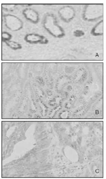
2.1 MUC2在结肠正常组织、结肠良性腺瘤组织及结肠癌组织中的表达 MUC2在结肠正常组织中的阳性表达(高表达)率为100%(15/15)(图1A),在结肠良性腺瘤组织中的阳性表达率为73.17%(30/41)(图1B),在结肠癌组织中阳性表达率为46.28%(56/121)(图1C),不同组织中MUC2阳性表达率比较,差异有统计学意义(P<0.01)。

图1 MUC2在不同结肠组织中的表达模式(HE染色,SPX400)
2.2 MUC2的表达与结肠癌相关临床参数的关联 MUC2高表达的结肠癌患者组织分型、淋巴结转移及肿瘤分期均优于MUC2低表达患者,比较差异均有统计学意义(P<0.05);MUC2高表达与低表达患者肿瘤大小、淋巴管侵袭、血管侵袭及性别构成方面比较差异均无统计学意义(P>0.05),见表1。

表1 结肠癌中MUC2的表达与临床病理因子的关联 例
3 讨论
随着研究的不断深入,MUC家族中的一些成员与相应疾病的关联日益得到广泛重视。研究表明,在胃腺瘤和胃癌中,MUC1在细胞质中的阳性表达是缺失的[7-8]。MUC2在正常胃组织中是不表达的,但在肠型胃癌中其表达是明显增加的[9]。报道称MUC2是Barrett食管的特异性标志之一[10]。同时,研究发现在胃癌中,MUC3的阳性表达与肿瘤大小、转移和预后有一定的关联[11]。MUC4在正常胰腺导管上皮不表达,但在胰腺癌细胞株中过量表达[12]。此外,MUC5AC在正常结直肠组织中是不表达的,但在结肠绒毛状腺瘤中被检测到阳性表达[13]。
本研究中,MUC2在结肠正常组织、结肠良性腺瘤和结肠癌中的阳性表达率分别为100%(15/15)、73.17%(30/41) 和 46.28%(56/121), 本 次 研究呈现的递减趋势与相关文献[14]是相符的。同时,本次试验收集整理了结肠癌患者的相关临床资料,发现MUC2高表达的结肠癌患者具有较好的组织分型、较少的淋巴结转移和较低的肿瘤分期。关于MUC2参与肿瘤发生和发展的具体机制并未明确,但有研究报道认为MUC2与结肠癌细胞的远处转移及细胞外基质和血管内皮细胞的结合有重要作用,即MUC2可介导肿瘤细胞的迁移[15]。同时研究表明MUC2启动子的甲基化在杯状细胞中低于柱状细胞[16]。同时,组蛋白修饰和micRNA的沉默都是MUC2表达下调的原因[17]。
总之,本研究表明,MUC2在结肠正常组织、结肠良性腺瘤组织和结肠癌组织中的表达趋势是递减的。同时,MUC2高表达的结肠癌患者有较好的临床病理资料。由此,笔者认为MUC2可能将会成为结肠癌临床诊断、治疗及判断预后的潜在标志物。
[1] Klimczak A,Kempinska-Miroslawska B,Mik M,et al.Incidence of colorectal cancer in Poland in 1999-2008[J].Arch Med Sci,2011,7 (4):673-678.
[2] Ferlay J,Steliarova-Foucher E,Lortet-Tieulent J,et al.Cancer incidence and mortality patterns in Europe:estimates for 40 countries in 2012[J].Eur J Cancer,2013,49(6):1374-1403.
[3] Garcia M,Jemal A,Ward E M,et al.Global cancer facts and figures[M].Atlanta:American Cancer Society,2007:1-6.
[4] Shet T,Valsangar S,Dhende S.Secretory carcinoma of breast:pattern of MUC 2/MUC 4/MUC 6 expression[J].Breast J,2013,19 (2):222-224.
[5]陈淑敏,唐忠辉CDX2和MUC2在胃癌癌变多阶段组织中的表达及意义的研究近况[J].医学综述,2011,17(14):2111-2113.
[6] Inoue M,Takahashi S,Yamashina I,et al.High density O-glycosylation of the MUC2 tandem repeat unit by N-acetylgalactosaminyltransfera se-3 in colonic adenocarcinoma extracts[J].Cancer Res,2001,61 (3):950-956.
[7]李娟,吴丽华,石岩,等.MUC1,MUC2和MUC3在胃增生性息肉中的表达模式[J].世界华人消化杂志,2011,19(35):3591-3596.
[8] Benjamin J B,Jayanthi V,Devaraj H.MUC1 expression and its association with other aetiological factors and localization to mitochondria in preneoplastic and neoplastic gastric tissues[J].Clin Chim Acta,2010,411(23-24):2067-2072.
[9] Conze T,Carvalho A S,Landegren U,et al.MUC2 mucin is a major carrier of the cancer-associated sialyl-Tn antigen in intestinal metaplasia and gastric carcinomas[J].Glycobiology,2010,20(2):199-206.
[10] Gulmann C,Shaqaq O A,Grace A,et al.Gytokeratin 7/20 and MUC 1,2,5AC and 6 expression patterns in Barrett’s esophagus and intestinal metaplsia of the stomach:intestinal metaplsia of the cardia is related to Barrett’s esophagus[J].Appl Immunohistochem Mol Morphol,2004,12(2):142-147.
[11] Wang R Q,Fang D C.Alterations of MUC1 and MUC3 expression in gastric carcinoma:relevance to patient clinicopathological features[J]. J Clin Pathol,2003,56(5):378-384.
[12]倪竟全,高军,李兆申,等.粘蛋白MUC1、MUC2、MUC4和MUC5AC在胰腺导管腺癌中的表达和意义[J].中华胰腺病杂志,2008,8(2):122-124.
[13] Longman R J,Douthwaite J,Sylvester P A,et al.Lack of mucin MUC5AC field change expression associated with tubulovillous and villous colorectal adenomas[J].J Clin Pathol,2000,53(2):100-104.
[14]鲁明良,何瑾,叶卫东.MUC1联合MUC2在结直肠腺瘤及结直肠癌中联合表达与临床病理的相关性研究[J].结直肠肛门外科,2014,20(3):158-163.
[15]肖春卫,易永芳.MUC1、MUC2及MUC5AC在大肠黏液腺癌组织中的表达及意义[J].实用癌症杂志,2007,22(1):44-49.
[16] Gratchev A,Siedow A,Bumke-Vogt C,et al.Regulation of the intestinal mucin MUC2 gene expression in vivo:evidence for the role of promoter methylation[J].Cancer Lett,2001,168(1):71-80.
[17] Kim Y S,Samuel B.Intestinal goblet cells and mucins in health and disease recent insights and progress[J].Curr Gastroenterol Rep,2010,12(5),319-330.
Research of the Expression and Its Associated Clinical Significance of MUC2 in Colorectal Carcinomas/
ZUO Xiao-xu,ZHU Xi-jia,LI Sheng-guo,et al.//Medical Innovation of China,2015,12(29):012-014
Objective:To investigate the expression of MUC2 in normal colorectal tissue,colorectal adenomas and colorectal carcinomas,and to explore the relation between the expression of MUC2 and its associated clinical data.Method:15 cases of normal colorectal tissues,41 cases of colorectal adenomas tissues and 121 cases of colorectal carcinomas tissues in our hospital were selected from March 2011 to March 2015.The expression of MUC2 in different tissues were detected by immunohistochemistry.Result:(1)The positive expression rates of MUC2 in normal colorectal tissues,colorectal adenomas tissues and colorectal carcinomas tissues were 100%(15/15),73.17%(30/41) and 46.28%(56/121) respectively.The differences of the positive expression rates in different tissues were statistically significant(P<0.01).(2)The histological differentiation,lymph node metastasis and tumor stage in patients with high expression rates of MUC2 were better than those in patients with low expression rates of MUC2,the differences were statistically significant(P<0.05).Conclusion:The expression of MUC2 in colorectal carcinomas has relationships with histological differentiation,lymph node metastasis and tumor stage.The down-regulation of MUC2 expression may play a role in the carcinogenesis,progression,infiltration,metastasis and other processes of colorectal carcinomas.
MUC2; Colorectal adenoma; Colorectal carcinoma
10.3969/j.issn.1674-4985.2015.29.004
2015-07-01)(本文编辑:王利)
①桂林医学院第二附属医院 广西 桂林 541000
王海鹏
First-author’s address:The Second Affiliated Hospital of Guilin Medical University,Guilin 541000,China

