
Cough-induced intercostal lung herniation
2015-07-12 07:37:14EvaGenbruggeJayantKichariMedischSpectrumTwenteDepartmentofRadiologyHaaksbergerstraat557513EREnschedetheNetherlands
Journal of Acute Disease 2015年2期
Eva Genbrugge, Jayant R. KichariMedisch Spectrum Twente, Department of Radiology, Haaksbergerstraat 55, 7513 ER Enschede, the Netherlands
Cough-induced intercostal lung herniation
Eva Genbrugge, Jayant R. Kichari
Medisch Spectrum Twente, Department of Radiology, Haaksbergerstraat 55, 7513 ER Enschede, the Netherlands
ARTICLE INFO ABSTRACT
Article history:
Received 25 November 2014
Received in revised form 15 December 2014 Accepted 25 January 2015
Available online 28 January 2015
Keywords:
Lung
Herniation
Spontaneous
Intercostal lung herniation is an uncommon entity, defined as protrusion of lung tissue between the ribs. The disease is mostly diagnosed either congenitally or acquired following post-surgery or after chest wall trauma. A spontaneous lung herniation is rarely described.
Tel: +31534873200
Fax: +31534873232
E-mail: jkichari@hotmail.com
1. Introduction
Spontaneous intercostal lung herniation is a rare entity mostly seen in male patients, with smoking and obesity as predisposing factors, following sudden increase in intrathoracic pressure. Surgical repair is recommended. We present a case of a 73-year-old man with an intercostal lung herniation following a period of coughing.
2. Case report
A 73-year-old man presented with acute left sided pleuritic chest pain since 1 day following a period of coughing. There was no associated fever, haemoptysis or dyspnoea. There was no history of trauma. His medical history included hypertension, myocardial infarction, diabetes, obesity and smoking. Physical examination revealed no abnormalities apart from painful palpation of the left chest.
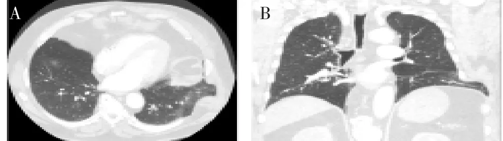
Figure 1. Contrast-enhanced computed tomography of the chest showed findings consistent with an intercostal lung herniation.
A : Axial CECT of the chest shows lung tissue and pleura protruding between the ribs outside the thoracic cavity; B: Coronal images clearly depict an intercostal muscle defect with widening of the intercostal space of rib 7 and 8 left.
A chest radiograph showed normal findings. Subsequent contrast-enhanced computed tomography of the chest to rule out a possible pulmonary embolism showed findings consistent with an intercostal lung herniation (Figure 1A and 1B). No pulmonary embolism was found. Our patient underwent left posterolateral thoracotomy showing a full rupture of the intercostal muscles between rib 7 and 8 over a length of 40 cm. The defect was repaired with a synthetic mesh. The patient recovered quickly. No recurrence of the lung herniation was reported almost 2 years after surgery.
3. Discussion
Intercostal lung herniation is an uncommon entity, defined as protrusion of lung tissue between the ribs outside the thoracic cage. Mostly diagnosed either congenitally or acquired following surgery or chest wall trauma. Less than 25 cases of spontaneous lung herniation have been reported in literature[1,2]. A spontaneous lung herniation may occur with a sudden increase in intrathoracic pressure following coughing with chronic bronchitis, playing wind instruments, sneezing or heavy lifting[1, 2]. Spontaneous lung herniation tends to occur more in men, while smoking and obesity are predisposing factors[2]. Literature regarding the management of intercostal lung herniation is limited[1,3]. Early surgical repair is recommended preventing incarceration of lung tissue or extension of the defect[2,3]. There is no consensus regarding the use of synthetic or biological repair materials.
In conclusion, spontaneous lung herniation is a rare entity for which surgical repair is recommended.
Conflict of interest statement
We declare that we have no conflict of interest.
References
[1] Sulaiman A, Cottin V, De Souza Neto EP, Orsini A, Cordier JF,
Gamondes JP, et al. Cough-induced intercostal lung herniation requiring surgery: report of a case. Surg Today 2006; 36: 978-980.
[2] Brock MV, Heitmiller RF. Spontaneous anterior thoracic lung hernias. J Thorac Cardiovasc Surg 2000; 119: 1046-1047.
[3] Weissberg D, Refaely Y. Hernia of the lung. Ann Thorac Surg 2002; 74: 1963-1966.
doi:Document heading
*Corresponding author:Jayant R. Kichari, Medisch Spectrum Twente, Department of Radiology, Haaksbergerstraat 55, 7513 ER Enschede, the Netherlands.
 Journal of Acute Disease2015年2期
- Journal of Acute Disease的其它文章
- A report of acute atrial fibrillation induced by misapplication of epinephrine
- Clinical manifestation as acute coronary syndrome without electrocardiographically ischemia: a clue for aortic dissection
- Report of a child with acute herpes zoster ophthalmicus induced partial third nerve palsy
- Report of a pregnant lady with bilateral elbow dislocation caused by acute fall injury
- An unusual presentation of acute electrocution
- A retrospective study of acute pertussis in Hasan Sadikin Hospital-Indonesia