一种新型通用型牙本质粘结剂对牙本质粘结强度的体外评价
一种新型通用型牙本质粘结剂对牙本质粘结强度的体外评价
米长江1,2,喻洁1,朱虹倩1,朱万春2,米方林2,刘兴容1
(1.泸州医学院附属口腔医院,泸州医学院口颌面修复重建和再生实验室,四川泸州646000; 2.川北医学院附属医院口腔科,四川南充637000)
【摘要】目的:评价一种新型通用型牙本质粘结剂(All Bond Universal,ABU)对牙本质的粘结强度,为临床选择合适的粘结剂提供参考依据。方法:选取人体12颗无龋坏磨牙,磨除牙釉质暴露牙本质面,分别用酸蚀-冲洗法和一步自酸蚀法使用All Bond Universal粘结剂,在其上堆砌树脂,并与酸蚀-冲洗粘结剂Prime&Bond NT(PBN)和一步法自酸蚀粘结剂G Bond (GB)进行对照。试样于37℃去离子水储存24 h后,每颗牙齿垂直于粘结面制备出0.81 mm2的树脂/牙本质试件,进行牙本质粘结微拉伸强度(μTBS)测试。结果:通用型牙本质粘结剂All Bond Universal在两种粘结方法下,其粘结力均高于对照组;就All Bond Universal本身而言,酸蚀-冲洗技术可以提高其粘结强度。结论:这种新型牙本质粘结剂具有较高的粘结强度,并且酸蚀-冲洗技术能提高其粘结力。
【关键词】新型通用型粘结剂,微拉伸强度,牙本质
自1955年Bunocore[1]成功的将酸蚀技术应用于牙釉质粘结以来,牙釉质的粘结技术已逐渐成熟,但是牙本质粘结因其自身结构的特殊性,使牙本质粘结仍未取得与牙釉质粘结一样令人满意的效果。目前牙本质粘结系统已发展到第七代,这一代牙本质粘结剂集酸蚀、偶联、粘合等诸多功能为一体,最大限度地简化临床操作,被称为“all-in-one”粘结剂系统[2]。在此基础上发展而来的新型通用牙本质粘结剂[3-4](universal adhesive)不但可以使用酸蚀-冲洗技术也可以使用自酸蚀技术,可以粘结树脂等直接修复材料,也可以粘结氧化锆,金属等间接修复材料,真正做到了“一瓶通用”,引起研究者及口腔临床医生的关注,但由于对其粘结性能了解不很透彻,故临床应用并不十分广泛。
本研究拟采用微拉伸强度测试(microtensile bond strength,μTBS),测量这种新型牙本质粘结剂对牙本质的粘结强度,并与临床广泛应用的酸蚀-冲洗粘结剂Prime&Bond NT和一步法自酸蚀粘结剂G Bond进行比较,为临床上选择和使用牙本质粘结剂提供参考依据。
1 材料和方法
1.1实验材料
实验标本的收集与保存:选取12颗新鲜拔除的无龋下颌第三磨牙,保存于4℃1%氯胺溶液中,1个月内使用。
实验仪器和设备: All Bond Universal(Bisco公司,美国),Prime&Bond NT(Dentsply公司,美国),G Bond(GC公司,日本),GC幻彩复合树脂(GC公司,日本),氰基丙烯酸粘合剂(Zapit,Bisco公司,美国),体视显微镜(SMZ100,尼康公司,日本),光固化机(QHL75,Dentsply公司,美国),Isomet慢速锯(Buehler,美国),微拉伸测试仪(Mierotensiletester,Bisco公司,美国)。
各组的粘结剂主要成分和使用方法,见表1。

表1 粘结剂主要成分及使用方法
1.2实验方法
1.2.1实验分组将牙齿随机分为4组,每组3个,分别使用Ⅰ组: All Bond Universal酸蚀-冲洗法,Ⅱ组: All Bond Universal自酸蚀法,Ⅲ组: Prime&Bond NT(酸蚀-冲洗法),Ⅳ组: G bond(自酸蚀法)进行粘结。
1.2.1粘结前样本预备每组样本均用Iosmet慢速锯于牙尖下4 mm处垂直于牙体长轴去除牙合面的釉质,暴露下方的牙本质,用600目碳化硅水砂纸打磨牙本质表面1 min,形成用于牙本质粘结的玷污层,并在体视显微镜下观察无残留釉质。超声清洗,按分组使用粘结剂后,其上均使用GC幻彩树脂修复达5~6 mm厚。所有粘结完成的牙齿储存于37℃的去离子水中24 h,再用Iosmet慢速锯流水条件下从釉牙骨质界下约2 mm处磨除牙根,然后沿(牙合)龈方向将实验牙切成0.9 mm厚的薄片,再将每片切成0.9 mm×0.9 mm的小柱(其中只有G Bond组出现3个试件的样本断裂)。所得样本先人工剔除因牙本质部分较短不利于粘结到测试模型的样本后,再利用体视显微镜将有微裂纹的样本剔除。在筛选后的试件中,利用SPSS20.0软件每组随机抽取30个,用游标卡尺准确测量条状试样粘接面的长度和宽度,计算出实际的粘接面积(mm2)。
1.2.3微抗拉伸粘结强度测试用氰基丙烯酸粘合剂将实验试件两端粘结固定在特制的试件夹持固定装置上,在微拉伸测试仪上测量试件拉伸断裂时的最大载荷(N),拉伸速度为1 mm/min,根据粘接面积和载荷峰值计算微拉伸粘接强度。公式为:微拉伸粘接强度(MPa)=载荷峰值(N)/粘接面积(mm2)。
1.3统计学分析
2 结果
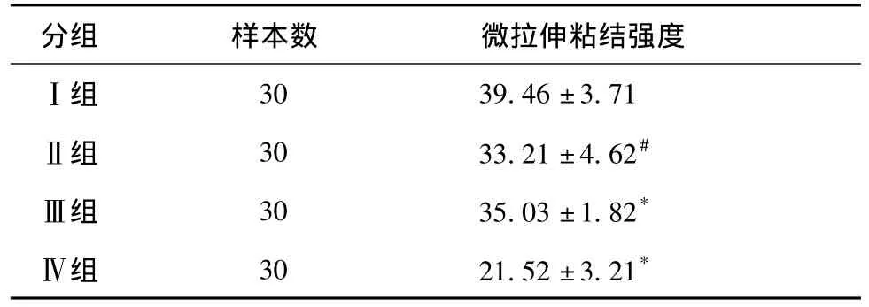
各组粘结系统微拉伸强度,见表2。All Bond Universal在酸蚀-冲洗和自酸蚀两种方式下的粘结强度均大于同样使用方法的相应的对照组(Prime&Bond NT和G bond),差异有统计学意义(P<0.05),就All Bond Universal本身而言,酸蚀-冲洗组的粘结力更大。
表2 不同种类牙本质粘结系统的微拉伸强度(±s,MPa)

表2 不同种类牙本质粘结系统的微拉伸强度(±s,MPa)
* P<0.05,同种使用方法,与对照组比较; #P<0.05,同一实验对象,不同使用方法。
分组 样本数 微拉伸粘结强度Ⅰ组30 39.46±3.71Ⅱ组 30 33.21±4.62#Ⅲ组 30 35.03±1.82*Ⅳ组 30 21.52±3.21*
3 讨论
微拉伸测试法(microtensile method)是由Sano等[5]于1994年提出,认为:当粘结面积在1.6~1.8 mm2以下时,绝大多数的粘结破坏发生在粘结层,其结果能更好的反映粘结剂的强度。之后,许多学者的测试都印证了该结论,认为该方法所产生的牙本质、复合树脂内聚力破坏较少,较传统方法能更准确地反映所测试粘结系统的强度[6]。本研究将粘结界面面积定在0.9 mm×0.9 mm,较小的面积使应力分布更加均匀,从而降低了被粘结体内聚破坏的可能性。同时增加了从同一个牙上获取多个样本的可能,大大降低了由于样本来自不同的牙而造成的误差。
通用型牙本质粘结剂是目前市场上新推出的牙本质粘结剂,对其研究较少,国外有研究显示不同品牌的通用型粘结剂粘结强度相差较大,更多与其组成成分有关[3,7-8]。
在本实验中,无论酸蚀-冲洗法还是自酸蚀法,与Ⅲ、Ⅳ组相比,All Bond Universal在粘结方法一致时都显示了较强的粘结力。这可能与其所含的功能单体MDP(10-甲基丙烯酰氧葵基二羟基磷酸酯)有关。磷酸酯类功能单体MDP最早被可乐丽公司(Osaka,Japan)合成,其功能性基团可以在水中分解为两个磷酸二氢盐集团质子[9],与羟基磷灰石反应生成溶解度很低的钙盐[10],从而与羟基磷灰石发生化学吸附作用[11-12]。MDP是目前最被认可的功能单体之一,被广泛应用在多种自酸蝕粘接剂中,包括Clearfil SE Bond,Clearfil S3 Bond等[13-14],近期,Van Meerbeek等[15]通过X射线衍射(XRD)和透射电镜(TEM)研究发现,羟基磷灰石粉末与MDP反应后,可以形成一个厚约4 mn的纳米交互层结构,并推测高粘接强度与该纳米交互层结构有关,这一发现也被Yoshida等[16]证实。所以,含有MDP单体的ABU在粘结力上要优于同样酸蚀方式的PBN和GB。
另外,许多实验室和临床研究[11,16-17]都证明了沿纳米层沉积的稳定的MDP-钙盐沉可以提高粘结力的稳定性,但是含MDP单体的ABU的远期粘结效能还需要进一步的研究。
就ABU本身而言,用磷酸预酸蚀的“酸蚀-冲洗”技术可以获得更大的粘结力。这一结果与Muħ-oz等[7]一致。这是由于在使用自酸蚀粘结剂之前,用磷酸预酸蚀牙本质表面,有利于去除牙本质表面玷污层及牙本质小管口的玷污栓,使管周和管间牙本质明显脱矿,可以使含有功能性单体的粘结剂更好的渗透到牙本质中,从而产生更长的树脂突[8,18]以及更厚的混合层[19]。但是,也有研究显示相反的结果[8],即虽然酸蚀-冲洗法可以产生更长的树脂突,但是其粘结强度要比自酸蚀法小,这可能是与实验所用粘结剂及其成分不同有关。
虽然,本研究结果显示自酸蚀技术的粘结强度较酸蚀-冲洗技术稍低,但实验证明其粘结强度可满足临床要求。而且,自酸技术也有其自身优势,比如操作简便快捷;减少了常规酸蚀冲洗后可能造成的粘接面的唾液污染;部分溶解玷污层,玷污层残成分与渗入的树脂一起参与形成混合层,纳米渗漏及术后敏感的发生率比全酸蚀粘结系统小等。因此,在临床运用中,要根据实际情况选择不同类型的牙本质粘结剂及选择恰当的粘结方法。
参考文献
[1]Buonocore MG.A simple method of increasing the adhesion of acrylic filling materials to enamel surfaces[J].J Dent Res,1955,34(6):849-853.
[2]Atash R,Van den Abbeele A.Bond strengths of eight contemporary adhesives to enamel and to dentine: an in vitro study on bovine primary teeth[J].Int J Paediatr Den,2005,15(4):264-273.
[3]Hanabusa M,Mine A,Kuboki T,et al.Bonding effectiveness of a new‘multimode’adhesive to enamel and dentine[J].J Dent,2012,40(6):475-484.
[4]Perdigão J,Sezinando A,Monteiro PC.Laboratory bonding ability of a multi-purpose dentin adhesive[J].Am J Dent,2012,25(3): 153-158.
[5]Sano H,Shono T,Sonoda H,et al.Relationship between surface area for adhesion and tensile bond strength—evaluation of a microtensile bond test[J].Dent Mater,1994,10(4):236-240.
[6]Tanumiharja M,Burrow MF,Tyas MJ.Microtensile bond strengths of seven dentin adhesive systems[J].Dent Mater,2000,16(3): 180-187.
[7]Muñoz MA,Luque I,Hass V,et al.Immediate bonding properties of universal adhesives to dentine[J].J Dent,2013,41(5):404-411.
[8]Wagner A,Wendler M,Petschelt A,et al.Bonding performance of universal adhesives in different etching modes[J].J Dent,2014, 42(7):800-807.
[9]Hayakawa T,Kikutake K,Nemoto K.Influence of self-etching primer treatment on the adhesion of resin composite to polished dentin and enamel[J].Dent Mater,1998,14(2):99-105.
[10]Inoue S,Koshiro K,Yoshida Y,et al.Hydrolytic stability of selfetch adhesives bonded to dentin[J].J Dent Res,2005,84(12): 1160-1164.
[11]Waidyasekera K,Nikaido T,Weerasinghe DS,et al.J.Reinforcement of dentin in self-etch adhesive technology: a new concept[J].J Dent,2009,37(8):604-609.
[12]Peumans M,De Munck J,Van Landuyt KL,et al.Eight-year clinical evaluation of a 2-step self-etch adhesive with and without selective enamel etching[J].Dent Mater,2010,26(12):1176-1184.
[13]Yu X,Liang B,Jin X,et al.Comparative in vivo study on the desensitizing efficacy of dentin desensitizers one-bottle self-etching adhesives[J].Oper Dent,2010,35(3):279-286.
[14]Jiang Q,Pan H,Liang B,et al.Effect of saliva contamination and decontamination on bovine enamel bond strength of four self-etching adhesives[J].Oper Dent,2010,35(2):194-202.
[15]Van Meerbeek B,Peumans M,Poitevin A,et al.Relationship between bond-strength tests and clinical outcomes[J].Dent Mater 2010,26(2): e100-121.
[16]Yoshida Y,Yoshihara K,Nagaoka N,et al.Self-assembled nanolayering at the adhesive interface[J].J Dent Res,2012,91(4): 376-381.
[17]Feitosa VP,Leme AA,Sauro S,et al.Hydrolytic degradation of the resin-dentine interface induced by the simulated pulpal pressure,direct and indirect water ageing[J].J Dent,2012,40 (12): 1134-1143.
[18]Langer A,Ilie N.Dentin infiltration ability of different classes of adhesive systems[J].Clin Oral Investig,2013,17 (1): 205-216.
[19]Margvelashvili M,Goracci C,Beloica M,et al.In vitro evaluation of bonding effectiveness to dentin of all-in-one adhesives[J].J Dent,2010,38(2):106-112.
(学术编辑:刘英)
网络出版时间: 2015-3-5 12∶47网络出版地址: http://www.cnki.net/kcms/detail/51.1254.R.20150305.1247.013.html
Evaluation of bond strength of a new universal adhesive to dentin in vitro
MI Chang-jiang1,2,YU Jie1,ZHU Hong-qian1,ZHU Wan-chun2,MI Fang-lin2,LIU Xing-rong1
(1.Affiliated Stomatology Hospital of Luzhou Medical College,Oral and Maxillofacial Reconstruction and Regeneration Laboratory,Luzhou Medical College,Luzhou 646000; 2.Department of Stomotology,Affiliated Hospital of North Sichuan Medical College,Nanchong 637000,Sichuan,China)
【Abstract】Objective: The aim of this study was to evaluate the microtensile bond strength (μTBS)of a new universal adhesive (All Bond Universal,ABU)applied in two different etching modes (i.e.self-etch and etch-and-rinse).It provides a referent data for choosing a proper adhesive of clinical application.Methods: The superficial occlusal dentins of 12 sound human molars were removed and the exposed surfaces were treated with universal adhesives: All-Bond Universal in self-etch and etch-and-rinse mode.One etch-andrinse adhesive (Prime&Bond NT,PBU )and one one-step self-etch adhesive (G bond,GB)were applied in additional teeth as control.After composite build up,the specimens were stored for 24h in distilled water at 37℃.Composite/dentine beams were prepared (0.81mm2)and microtensile test was performed.Results: The dentin bond strength of the All Bond Universal adhesive(both etch-andrinse and self-etch)is superior to its control groups.And etch-and-rinse application mode has higher μTBS than its self-etch application mode.Conclusion: The bond strength of All Bond Universal adhesive to dentin is higher than Prime&Bond NT and G bond.And application of an etching step prior to All Bond Universal improves its bond strength.
【Key words】New universal adhesive; Microtensile bond strength; Dentine
通讯作者:刘兴容,E-mail: liuxingrong163@163.com
作者简介:米长江(1981-),男,四川广安人,硕士研究生,主要从事牙体牙髓病基础与临床研究。
基金项目:四川省教育厅基金(11ZB188)
收稿日期:2014-08-26
doi:10.3969/j.issn.1005-3697.2015.01.13
【文章编号】1005-3697(2015)01-0056-04
【中图分类号】R783.1
【文献标志码】A

