
Investigation of optically pumped continuous terahertz laser in biological imaging
2014-05-12 07:56ZhangMinQuanRunaiSuHongandHuXuejuan
深圳大学学报(理工版) 2014年2期
Zhang Min,Quan Run'ai,Su Hong,and Hu Xuejuan
Shenzhen Key Laboratory of Laser Engineering,College of Electronics Science and Technology,Shenzhen University,Shenzhen 518060,P.R.China
Investigation of optically pumped continuous terahertz laser in biological imaging
Zhang Min†,Quan Run'ai,Su Hong,and Hu Xuejuan
Shenzhen Key Laboratory of Laser Engineering,College of Electronics Science and Technology,Shenzhen University,Shenzhen 518060,P.R.China
Terahertz imaging is of great potential in biological and biomedical researches for bio-sample structure analysis and pathological-tissues diagnosis.For optically pumped terahertz laser imaging,it can offer high output power,large dynamic range and high signal-to-noise ratio.In this paper,continuous wave terahertz laser(SIFIR 50)was employed for terahertz bio-sample imaging.The internal information of the sample was obtained and it confirmed the application of continuous wave terahertz imaging system.In order to make it more convenient for the naked eye to watch,false-color image processing was applied to the original terahertz images.
optoelectronics and laser engineering;continuous wave terahertz;terahertz;bio-sample;scanning imaging;image processing;false-color image
Terahertz(THz)radiation lies between infrared light and microwave region of the electromagnetic spectroscopy.Due to its good penetration in non-polar materials and low photonic energy[1],terahertz imaging has potential for biomedical and biological areas.During the past few years,there have been extensive developments in THz bio-imaging[2-4].Hu et al[5]first reported THz imaging of leaf based on THz time domain spectroscopy in 1995,and water concentration of the dry and fresh leaf can be distinguished.Thereafter,a lot of biological tissues were used as imaging samples such as tumor[6],corneal[7],oral cancer[8],and so on.These projects either focused on coherent imaging of amplitude and phase information,their configurations were relatively complex,or they employed quantum cascade laser as illumination,requiring extremely low temperature to run the laser.While for optically pumped terahertz laser(OPTL),it can offer high output power,large dynamic range and high signal-to-noise ratio of THz imaging process.Meanwhile fast imaging speed is provided in OPTL continuous-wave imaging.
Owing to the water-concentration distribution and different tissues fabric inside bio-samples,a terahertz image including intensity information will be obtained.On the other hand,terahertz wave can penetrate the animal fat with low attenuation.We utilized two lenses to focalize the THz beam,and measured the focal spot size by means of blade.An ant was set as sample.After image processing,a clear THz image of the sample was achieved.
1 Experimental setup
The continuous wave(CW)terahertz laser used in this experiment is SIFIR 50 THz laser system which based on a tunable CO2laser.The CO2laser pumped low-pressure methylic gas(CH3OH,CH2F2,etc.)and generated a narrow band terahertz radiation.In this configuration,2.52 THz laser was used as the illumination,and the average output power was above 50 mW.The detector was made by our research group,containing two pyroelectric units.It has a relatively good respondence,especially for weak signal(output power less than 10 mW).
It is demonstrated that parabolic mirror has a better effect on focalizing the laser beam.For the sake of smaller focal spot,we exploited a picarin lens(f=150 mm)and a parabolic mirror(f=50 mm)to focus the incident THz radiation.The experimental setup is shown in Fig 1.Considering the effective area of the detector,terahertz radiation was focused by another picarin lens(f=50 mm).At last,the sample's image was displayed on the computer.

Fig.1 Terahertz scanning imaging setup图1 扫描太赫兹成像的实验光路
The living sample was firstly soaked in alcohol,and then fitted on translation stage when it was stable.The sample stage was controlled by the computer.It could move along x and z axis,respectively.Meanwhile,as the signal received by the detector was very weak,a signal amplifier was needed.
Spot size is a very important factor for terahertz imaging resolution.In order to evaluate the focal spot,knife-edge method[9]was adopted to measure the terahertz focused beam.10%and 90%of the maximum terahertz radiation was marked as x1and x2respectively,and then the focus spot size was x1-x2.By means of knife-edge method in our experiment,the spot diameter in the position of the sample was 0.169 mm(x)and 0.283 mm(y),which determined the spatial resolution of terahertz imaging.And the scanning step of the experimental setup was 0.1 mm.Chopper frequency also affects the imaging quality:the smaller chopper frequency is,the better is the imaging quality.However,when the chopper frequency is too small,the chopper can not work stably.In our experiment,it was selected as 4 Hz.
Considering that the naked eye is more sensitive to chromatic images,we conducted false color processing to the gray images taken by our terahertz scanning imaging configuration.False color processing is such a procedure[10]:for each pixel,the gray level has done red-transformation,green-transformation and blue transformation individually.At last,the three transformations will be synthesized to one false-color image.
2 Imaging results and processing
According to the diffraction limit formula:

where θ is the angular resolution,λ is the wavelength,D is a lens diameter.When θ is small,sinθ approximately equal tanθ,approximately equal Δ /f,of which Δ is the minimum resolution size,f is the focal length.
Derive the spatial resolution limit of the imaging equation:

As the frequency of light source is 2.52 THz,λ =118.83 μm,the lens diameter D=50 mm,the lens focal length f=50 mm,the calculated limit of the spatial resolution of approximately equals 145 μm.
Fig 2(a)shows the optical image of the ant sample.After the measurement,the ant length is 7.32 mm,head length is 1.84 mm/width is 1.24 mm,tail length is 2.42 mm/width is 1.78 mm,the spacing between the roots behind the three legs is 0.76 mm,and the diameter of the legs is 0.28 mm.From Fig 2(b),the image of ant leg is clear,indicating that the spatial resolution is up to 280 μm,less than the spatial resolution limit of 145 μm.The theoretic analysis is in the agreement with experimental data.As the B-ultrasonic wave can only be used to measure objects over 0.3 mm,and X-rays penetrate most biological tissue,Fig 2(b)also proved terahertz imaging in biological advantages.
The ant sample is stuck in a thin plastic,which is nearly transparent to terahertz radiation.Its scanning terahertz image is shown in Fig 2(b),which contained 140×130 pixels.As a result of the low output power of the terahertz laser(only more than 10 mW),the image contrast was a little weak:ant's internal information was hardly distinguished.With the increase of output power,imaging contrast will get better.On the other hand,because the output power was not stable during the imaging procedure,the background appeared relatively non-uniform.Fig 2(c)shows the image after false-color transformation.It apparently appeared the intensity differences of the ant legs,which was caused by the size of the leg itself.
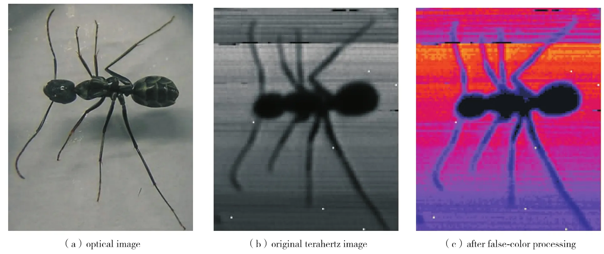
Fig.2 Images of the ant sample图2 整个蚂蚁样品的照片及太赫兹图像
Fig 3(a)shows the original terahertz scanning image of the ant's head.Terahertz power was about 32 mW.Scanning step was set as 0.1 mm with 45 × 50 pixels.Due to the water content or other tissues which absorbed little terahertz radiation,there was a difference in the head in the terahertz image.It is demonstrated that when the terahertz radiation increases,the imaging contrast gets better too.Fig 3(b)shows the terahertz image of the ant head after false-color processing.We can see that in Fig 3(b),the head's characteristic is more obvious.

Fig.3 Images of the ant's head图3 蚂蚁头部的太赫兹图像
The terahertz image in the middle of the ant is shown in Fig 4(a).The output power of the terahertz laser was 50 mW.Scanning step of the system was 0.1 mm and the pixel number was 45×50.The head's characteristic can dimly be seen,meanwhile,we find that the tail part of the ant sample has a relatively bright area.After image processing,the false-color image of the middle part is shown in Fig 4(b).The internal contrast of terahertz image was enhanced.

Fig.4 Images of the ant's middle part图4 蚂蚁中间部分的太赫兹图像
Depending on the clue implied in Fig 4,there may be a district which absorbs less terahertz radiation compared with the other parts of the ant's tail.Fig 5(a)shows the scanning terahertz image of the ant's tail part.Terahertz power was about 40 mW,and the pixel number was 45 ×50 with scanning step of 0.1 mm.At the end of the ant's tail,there was a point where terahertz radiation transmitted with a very low loss.It can be speculated that maybe this point is a water container for the ant;here the ant was dry,so terahertz can penetrated it with low loss.Fig 5(b)is the false-color image of the original terahertz image.
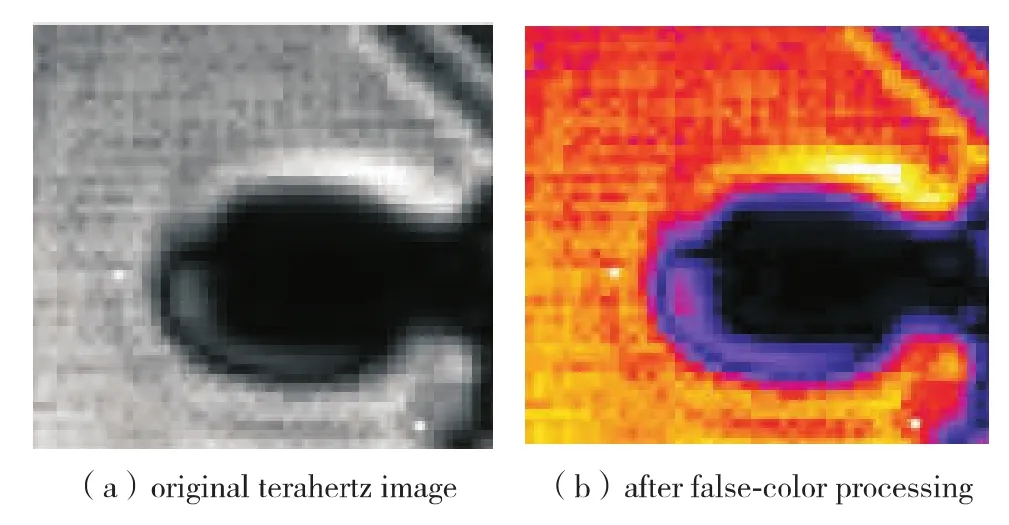
Fig.5 Images of the ant's tail图5 蚂蚁尾部的太赫兹图像
Conclusions
Terahertz scanning imaging configuration is of great potential in biological and biomedical researches for bio-sample structure analysis and pathological-tissues diagnosis.In this paper,the imaging results of the ant sample with the terahertz scanning imaging setup confirm the bio-application of this setup.From the experimental results we demonstrate that the output power of the terahertz laser plays an important role in imaging progress.Whereas,when output power is high enough,the effect caused by it will be low.Because naked eyes are more sensitive to chromatic images,false color processing has been done to the terahertz images.In our experiments,the signal to noise ratio was not so good,which might be related with the surrounding environment and the detector itself.
In our future work,we will focus on improving the signal to noise ratio of the imaging system from the two aspects:decreasing surrounding temperature and humidity to realize low noise,on the other hand,improving the detector's dynamic range.Meanwhile,for terahertz imaging,some imaging process will be operated to improve the image quality.
[1] Zhou Zekui,Zhang Tongjun,Zhang Guangxin.Science and technology of terahertz wave[J].Process Automation Instrumentation,2006,27(3):1-6.(in Chinese)
周泽魁,张同军,张光新.太赫兹波科学与技术[J].自动化仪表,2006,27(3):1-6.
[2]Jeong Young Uk,Kazakevitch Grigori M,Cha Hyuk Jin,et al.Demonstration of a wide-band compact free electron laser to the THz imaging of bio samples[J].Nuclear Instruments and Methods in Physics Research A,2007,575(1/2):58-62.
[3]Woodward R M,Cole B E,Wallace V P,et al.Terahertz pulse imaging in reflection geometry of human skin cancer and skin tissue [J].Physics in Medicine and Biology,2002,47(21):3853-3863.
[4]Knobloch P,Schildknecht C,Kleine-Ostmann T,et al.Medical THz imaging:an investigation of histo-pathological samples[J].Physics in Medicine and Biology,2002,47(21):3875-3884.
[5]Hu B B,Nuss M.Imaging with terahertz waves[J].Optics Letters,1995,20(16):1716-1717.
[6]Miura Y,Kamataki A,Uzuki M,et al.Terahertz-wave spectroscopy for precise histopathological imaging of tumor and non-tumor lesions in paraffin sections[J].The Tohoku Journal of Experimental Medicine,2011,223(4):291-296.
[7]Bennett D B,Taylor Z D,Tewari P,et al.Terahertz sensing in corneal tissues[J].Journal of Biomedical Optics,2011,16(5):057003-1-057003-8.
[8]Sim Y C,Park J Y,Ahn K M,et al.Terahertz imaging of excised oral cancer at frozen temperature[J].Biomedical Optics Express,2013,4(8):1413-1421.
[9]Fan Xinmin,Zheng Yi,Wang Guanjun,et al.Two calculation methods for measuring the beam waist of Gaussian laser beam using a 90/10 knife-edge method [J].Applied Laser,2008,28(2):139-141.(in Chinese)
樊心民,郑 义,王冠军,等.90/10刀口法测量高斯激光光束束腰的两种计算方法 [J].应用激光,2008,28(2):139-141.
[10] Gonzalez R C,Woods R E.Digital image processing[M].2nd ed.Beijing:Upper Saddle River(USA):Prentice Hall,2007:243-247.
光泵连续太赫兹波在生物成像中的应用研究
张 敏,权润爱,苏 红,胡学娟
深圳市激光工程重点实验室,深圳大学电子科学与技术学院,深圳 518060
采用连续太赫兹激光器 (SIFIR 50)作为光源对生物样品进行太赫兹成像,得到具有较高分辨率的太赫兹图像.由于生物体内部组织结构的不同,同时得到了内部明暗对比显著的图像,鉴于人眼对彩色图像更敏感的特点,对成像结果进行伪彩色图像处理,得到了较好的结果.
光电子与激光技术;连续太赫兹波;太赫兹;生物样品;扫描成像;图像处理;伪彩色
广东省科技计划资助项目 (2006B3733002);深圳市科技基础研究计划资助项目 (JC201105170626)
张 敏 (1978-),女 (汉族),湖北省宜昌市人,深圳大学高级实验师、博士.E-mail:zhangmin@szu.edu.cn
/参考文献:
TN 219
A
10.3724/SP.J.1249.2014.02160
2013-09-01;Revised:2013-12-20;
2014-01-18
Foundation:Science and Technology Planning Project of Guangdong Province(2006B3733002);Shenzhen Science and Technology Research Foundation for Basic Project(JC201105170626)
†
Senior lab master Zhang Min.E-mail:zhangmin@szu.edu.cn
:Zhang Min,Quan Run'ai,Su Hong,et al.Investigation of optically pumped continuous terahertz laser in biological imaging [J].Journal of Shenzhen University Science and Engineering,2014,31(2):160-163.
引 文:张 敏,权润爱,苏 红,等.光泵连续太赫兹波在生物成像中的应用研究[J].深圳大学学报理工版,2014,31(2):160-163.(英文版)
【中文责编:方 圆;英文责编:海 潮】
猜你喜欢
表面工程与再制造(2022年1期)2022-05-25
深圳大学学报(理工版)(2021年6期)2021-11-29
深圳大学学报(理工版)(2020年6期)2020-11-14
雷达学报(2018年1期)2018-04-04
雷达学报(2018年1期)2018-04-04
雷达学报(2018年1期)2018-04-04
深圳大学学报(理工版)(2017年5期)2017-09-23
生物工程学报(2016年1期)2016-07-04
学周刊·下旬刊(2016年3期)2016-01-31
学周刊·下旬刊(2014年12期)2015-06-02