
Influence of edaravone on growth arrest and DNA damage-inducible protein 34 expression following focal cerebral ischemia-reperfusion in rats
2014-03-22 12:22WeiWangXiaoMeiWuBoJiangChunYuWangHaiNanZhangXiangMinShen
Wei Wang, Xiao-Mei Wu, Bo Jiang, Chun-Yu Wang, Hai-Nan Zhang, Xiang-Min Shen
Department of Neurology, the Second Xiangya Hospital, Central South University, Changsha 410011, China
Influence of edaravone on growth arrest and DNA damage-inducible protein 34 expression following focal cerebral ischemia-reperfusion in rats
Wei Wang, Xiao-Mei Wu, Bo Jiang, Chun-Yu Wang, Hai-Nan Zhang, Xiang-Min Shen*
Department of Neurology, the Second Xiangya Hospital, Central South University, Changsha 410011, China
PEER REVIEW
Peer reviewer
Professor Viroj Wiwanitkit, Visiting professor, Hainan Medical University, China; visiting professor, Faculty of Medicine, University of Nis, Serbia; adjunct professor, Joseph Ayobabalola University, Nigeria; professor, senior expert, Surin Rajabhat University, Thailand; honorary professor, Patil Medical University, India.
Tel: 6624132436
E-mail: wviroj@yahoo.com
Comments
This work explores the effect of the new agent, edaravone on GADD34 expression following focal cerebral ischemia-reperfusion in rats. The animal model is new and shows new finding that can be useful for further referencing in antioxidant research.
Details on Page 717
Objective:To investigate the influence of edaravone on the expression of growth arrest and DNA damage-inducible protein 34 (GADD34).
Edaravone, Cerebral ischemia-reperfusion, Growth arrest and DNA damage-inducible protein 34
1. Introduction
Growth arrest and DNA damage-inducible protein 34 (GADD34) is a kind of cell cycle protein that can be upregulated under conditions of DNA damage, cell cycle arrest and endoplasmic reticulum (ER) dysfunction,etc[1]. Some animal models with cerebral ischemia indicated that GADD34 expression was up-regulated at certain time windows after cerebral ischemia, but there was no consistent conclusion on the dynamic changes of GADD34 expression due to the different animal breeds and models used in the researches[2,3]. Edaravone can scavenge free radicals and is verified to have neuroprotection function in animal experiments. However, its influence on GADD34 expression has been rarely reported in recent years. In this study, edaravone was used as an interventional agent to detect the GADD34 expression changes in peri-infarct regions on ischemic parietal cortex, hoping to provide experimental basis for the prevention and treatment of cerebral infarction.
2. Materials and methods
2.1. Materials
Edaravone was supplied by Jiangsu Simcere Company, GADD34 antibody was purchased from American Santa Cruz Company, and streptavidin-biotin complex (SABC) immunohistochemical kits and diaminobenzidine stain were brought from Wuhan Boster Biological Technology, Ltd.
2.2. Animal models and grouping
A total of 108 healthy male Sprague-Dawley (SD) rats (230-280 g) aged 10-12 weeks were fed by full-price nutritional fodders at 18-25 °C room temperature, 50%-60% relative humidity and 12 h diurnal cycle of illumination. All rats could eat and drink freely. Then they were randomly divided into sham operation group, model group and edaravone group by random number table (36 cases for each group). Suture-occluded method reported by Paschenet al.was adopted to prepare left focal middle cerebral artery (MCA) occlusion models[4]. The occlusion was maintained for 120 min and then unplugged to form reperfusion. After model rats revived, neurological deficit score was conducted based on the level V of Paschen’s standard scoring method[4]. Rats with 1-3 scores in the initial neurological deficit score were included, whereas the dead rats, or whose with subarachnoid hemorrhage and without contralateral hemiplegia signs were considered as failed ones. Successful rat models in the same period were selected to supplement the object numbers. And then, the two groups were divided into six subgroups according to the reperfusion times such as reperfusion 1, 3, 6, 12, 24 and 72 h groups (12 cases for each group). At each perfusion corresponding time, the rats were sacrificed.
2.3. Methods
2.3.1. Edaravone group
A volume of 10 mg edaravone injection (Trade name: Edaravone; Batch number: H20031342; Specification: 10 mg/5 mL) was diluted by 5 mL normal saline to prepare 1 mg/mL solution. About 3 mg/kg edaravone injection was injected into the caudal veins immediately after reperfusion.
2.3.2. Model group
A total of 3 mL/kg normal saline was injected into the caudal veins immediately after reperfusion.
2.3.3. Sham operation group
This group was divided into six subgroups according to the above reperfusion corresponding times (six cases for each). In this group, suture occlusions were inserted 8-10 mm in depth so as to maintain the smooth of anterior and posterior MCA. The rest operations were similar to those in other groups.
2.4. Preparation of paraffin sections
At reperfusion corresponding time-point, 10% chloral hydrate was used to narcotize the rats, and then 250 mL 0.9% normal saline was infused from apex cordis. When the solution discharged from the right atrial appendage was clear, 300 mL 4% paraformaldehyde was infused. All rat brains were decollated and the part from antinion to occipital lobe was divided into five equal sections marked as A, B, C, D and E. C section was embedded by paraffin to prepare the consistent slices (4 µm in thickness).
2.5. Immunohistochemical staining
SABC was used. The potency of GADD34 polyclonal antibodies was verified to be 1:100 by the pre-experiment, and the detailed operations were as follows. The slices were routinely deparaffinaged into water, soaked in 3% H2O2at room temperature for 5-10 min, and washed by double distilled water by 2 min×3 times. The antigen was repaired by microwave heating, washed by 0.1 mol/L phosphate buffer solution (PBS) by 2 min×2 times after natural refrigeration; added with normal goat serum blocking buffer, incubated at room temperature for 20 min and the serum was discarded; then added with the primary antibodies of rabbit antimice GADD34 polyclonal antibodies (1:100), incubated at 4 °C overnight, washed by 0.1 mol/L PBS by 2 min×3 times; added with biotinylated goat anti-rabbit IgG antibodies (carried with kits with potency 1:100), incubated at 37 °C for 20 min and washed by 0.1 mol/L PBS by 2 min×3 times; added with SABC, incubated at 37 °C for 30 min and washed by 0.1 mol/ L PBS by 5 min×4 times; added with diaminobenzidine to develop color; and then dehydrated, transparentized and sealed. The above steps were performed in sequence. Five fields were randomly collected in the peri-infarct regions of ischemic cortex of each rat under microscope (×400), and the mean value was regarded as the estimated value. Highdefinition pathological image analysis system (HPIAS-1000) was applied to analyze the images of slices and determine the grey value of positive protein expression.
2.6. Statistical data analysis
SPSS 11.5 software was applied for all data analysis. All data were expressed as mean±SD. One-way ANOVA was used for the comparisons of multi-sample means while student’st-test was for comparisons among groups with α=0.05.P<0.05 was considered to be statistically significant.
3. Results
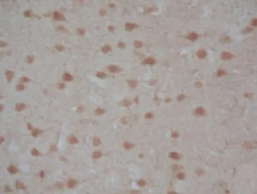
In sham operation group, there were positive GADD34 immunoreactive cell expressions in the left side of the parietal cortex, but ANOVA test showed no significant differences in GADD34 immunoreactive cell expressions at each time point (P>0.05). In model group, 1 h after reperfusion, GADD34 expression increased in periinfarct regions on ischemic parietal cortex, whose grey values decreased along with the prolonged reperfusion times, indicating that the counts of positive GADD34 cells increased gradually and reached its peak at 24 h, and even 72 h after reperfusion, the positive cells were still visible. However, the GADD34 expression, marked by dynamic changes, were evidently higher in edaravone group 6, 12 and 24 h after reperfusion than in model group (P<0.05), but the differences at other time-points were not statistically significant (P>0.05), as shown in Table 1 and Figures 1 and 2.
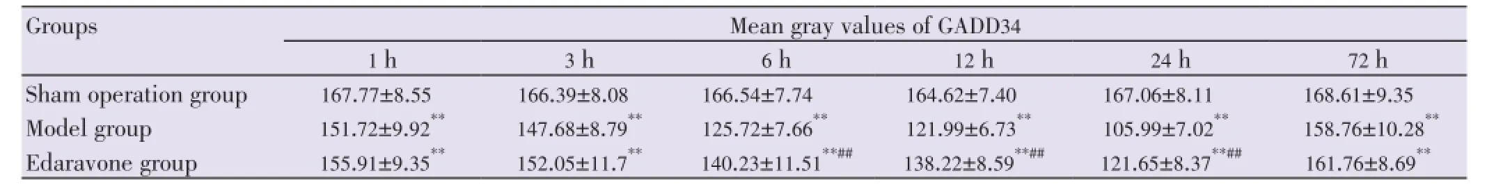
Table 1 Comparison of mean gray values of GADD34 expression on left parietal cortex (mean±SD).

Figure 1. GADD34 positive cell expression in peri-infarct regions of left parietal cortex 24 h after reperfusion in model group.
Fgure 2. GADD34 positive cell expression in peri-infarct regions of left parietal cortex 24 h after reperfusion in edaravone group.
4. Discussion
GADD34 is a kind of cell cycle protein that can be upregulated under conditions of DNA damage, cell cycle arrest and ER dysfunction,etc[4]. A recent study on GADD34 genes in fibroblasts in rats proved that this protein could dephosphorylate eIF2α and repair endoplasmic reticulum stress (ERS) associated protein synthesis inhibition[5,6]. Therefore, it can be said that GADD34 gene expression is limited by ERS, becoming a critical factor for the successful recovery of protein synthesis function.
According to the known functions of GADD34, it can be concluded that the increased protein compound expressions of GADD34 serve the purposes of alleviating ischemia induced protein synthesis inhibition, repairing damaged DNA and influencing programmed cell death (apoptosis)[7-10]. However, the intriguing point is that the pre-treatment of ischemia could obviously reduce the cerebral ischemia-reperfusion induced protein synthesis inhibition[11]. Garciaet al.reported that GADD34 was one of the proteins induced by the pre-stimulation of ischemia (5 min sub-fatal stimulation) and could be strongly translated during reperfusion after fatal ischemia, for which the pre-treatment of ischemia could remarkably protect the brains so as to response to the fatal ischemic stimulation for 30 min[11].
The existing animal experiments proved that cerebral ischemia could induce ERS and improve unfolded protein response. However, the dynamic changes were not consistent due to the different animal breeds and models used in researches. Huet al.made male SD rat models with focal cerebral ischemia by electrocoagulation to observe the GADD34 expression changes at different time points after ischemia[12], and the results revealed that 4 h after operation, the ischemic brains were detected with GADD34 immune positive cells (mainly neurons). The cells were the most intensive in periinfarct regions, and there was significant difference when compared with those in contralateral cerebral cortex. And 24 h after operation, the immune positive cell expressions reached the peak, and the double staining of GADD34 and terminal deoxynucleotidyl transferase mediated nick end labeling immunofluorescence showed deficient co-localization of neurons in the ischemic cortex. In this study, it was found that GADD34 expression, which was up-regulated at certain time after cerebral ischemia-reperfusion, could be detected 1 h after reperfusion and reached to the peak 24 h after reperfusion, which further proved that reperfusion could induce ERS and improve unfolded protein response target gene expression.
Edaravone (3-methyl-1-phenyl-2-pyrazon-5-ketone), as a new colorless transparent free radical scavenger, can effectively scavenge hydroxyl free radicals by inhibiting activities of hydroxyl free radicals, lipid peroxidation and ferric ion induced peroxidation injury. Additionally, it can also inhibit the activities of xanthine oxidase and hypoxanthine oxidase, improve the activity of superoxide dismutase, strengthen the total anti-oxidation function of cells, inhibit the peroxidation of cell membranes as well as stimulate the formation of prostaglandin, reduce the level of inflammatory factor leukotriene so as to significantly promote the cell stability, protect neurocytes, decrease cell apoptosis and increase cell survival rates. Previous study showed that edaravone had neuroprotective effect in that it could down-regulate the ERS gene expressions after cerebral ischemia-reperfusion, such as PKR-like endoplasmic reticulum kinase and C/EBP homology protein, and reduce the apoptosis of nerve cells in peri-infarct regions[13-15]. Other clinical trials also demonstrated that the mechanism of neuroprotective effect of edaravone was that it could alleviate the damage of vascular endothelial cell, inhibit the delayed neuron necrosis and apparently reduce the cerebral infarction volume injured by the obstruction-reperfusion of unilateral MCA[16-19] .
This study found that edaravone could reduce GADD34 expression in SD rat models with cerebral ischemiareperfusion, while GADD34 was verified to be effective in protecting nerves and beneficial to cell survivals. Therefore, it is controversial that whether the cerebral ischemia-reperfusion may be aggravated if GADD34 expression is inhibited byedaravone. The cell outcome mainly depends on the strength and duration of ERS because ERS has both increased expression of apoptosis-promoting molecule C/EBP homology protein and up-regulated expressions of pro-survival molecules GADD34 and GRP78, which means that the relationship between GADD34 expression and ERS is the power comparison of survival and death. So it was predicated that, through scavenging free radicals, edaravone could evidently relieve oxidative stress in ischemic penumbra and protect ER against lipid peroxidation so as to inhibit the ERS and ERS gene expression in upper stream and play its neuroprotective effect.
Conflict of interest statement
We declare that we have no conflict of interest.
Acknowledgements
The authors are thankful with our deepest core of heart to Jiangsu Simcere Company and to those who contribute much to the study.
Comments
Background
This is a basic laboratory study on influence of edaravone on GADD34 expression following focal cerebral ischemiareperfusion in rats. It is a good experimental study in animal model.
Research frontiers
The work explores a new aspect on effect of the new chemical edaravone on GADD34 expression following focal cerebral ischemia-reperfusion in animal model. It is a new finding in antioxidant research.
Related reports
There is no previous report using this model for exploration on the effect of edaravone.
Innovations and breakthroughs
This is an actual innovation in antioxidant research. It is also a new report on the new agent, edaravone. The study on GADD34 expression following focal cerebral ischemia-reperfusion in rats is a new model study.
Applications
It can be further applied in the field of antioxidant research. The work can be further referenced in the field of pharmacology as well.
Peer review
This work explores the effect of the new agent, edaravone on GADD34 expression following focal cerebral ischemiareperfusion in rats. The animal model is new and shows new finding that can be useful for further referencing in antioxidant research.
[1] McCabe C, White F, Brown SM, Macrae IM. GADD34 gene restores virulence in viral vector used in experimental stroke study. J Cereb Blood Flow Metab 2008; 28(4): 747-751.
[2] Imai H, Harland J, McCulloch J, Graham DI, Brown SM, Macrae IM. Specific expression of the cell cycle regulation proteins, GADD34 and PCNA, in the peri-infarct zone after focal cerebral ischaemia in the rat. Eur J Neurosci 2002; 15(12): 1929-1936.
[3] McCaig D, Imai H, Gallagher L, Graham DI, Harland J, Moira Brown S, et al. Evolution of GADD34 expression after focal cerebral ischemia. Brain Res 2005; 1034(1-2): 51-61.
[4] Paschen W, Hayashi T, Saito A, Chan PH. GADD34 protein levels increase after transient ischemia in the cortex but not in the CA1 subfield: implications for post-ischemic recovery of protein synthesis in ischemia-resistant cells. J Neurochem 2004; 90(3): 694-701.
[5] Farook JM, Shields J, Tawfik A, Markand S, Sen T, Smith SB, et al. GADD34 induces cell death through inactivation of Akt following traumatic brain injury. Cell Death Dis 2013; 4: e754.
[6] Connell BJ, Saleh MC, Kucukkaya I, Abd-El-Aziz AS, Khan BV, Saleh TM. UPEI-300, a conjugate of lipoic acid and edaravone, mediates neuroprotection inischemia/reperfusion. Neurosci Lett 2014; 561: 151-155.
[7] Kojima E, Tacheuchi A, Haneda M, Yagi A, Hasegawa T, Yamaki K, et al. The function of GADD34 is a recovery from a shutoff of protein synthesis induced by ER stress: elucidation by GADD34-deficient mice. FASEB J 2003; 17(11): 1573-1575.
[8] Novoa I, Zeng H, Harding HP, Ron D. Feedback inhibition of the unfolded protein response by GADD34-mediated dephosphorylation of eIF2alpha. J Cell Biol 2001; 153(5): 1011-1022.
[9] Hasegawa T, Isobe K. Evidence for the interaction between Translin and GADD34 in mammalian cells. Biochim Biophys Acta 1999; 1428(2-3): 161-168.
[10] Hollander MC, Poola-Kella S, Fornace AJ Jr. Gadd34 functional domains involved in growth suppression and apoptosis. Oncogene 2003; 22(25): 3827-3832.
[11] Garcia L, Burda J, Hrehorovská M, Burda R, Martín ME, Salinas M, et al. Ischaemic preconditioning in the rat brain: effect on the activity of several initiation factors, Akt and extracellular signal-regulated protein kinase phosphorylation, and GRP78 and GADD34 expression. J Neurochem 2004; 88(1): 136-147.
[12] Hu LW, Yen JH, Shen YT, Wu KY, Wu MJ. Luteolin modulates 6-hydroxydopamine-induced transcriptional changes of stress response pathways in PC12 cells. PLoS One 2014; 9(5): e97880.
[13] Tian F, Yamashita T, Deguchi K, Omote Y, Kawai H, Ohta Y, et al. In vivo optical imaging correlates with improvement of cerebral ischemia treated by intravenous bone marrow stromal cells (BMSCs) and edaravone. Neurol Res 2013; 35(10): 1051-1058.
[14] Kikuchi K, Kawahara KI, Uchikado H, Miyagi N, Kuramoto T, Miyagi T, et al. Potential of edaravone for neuroprotection in neurologic diseases that do not involve cerebral infarction. Exp Ther Med 2011; 2(5): 771-775.
[15] Shen XM, Tan LM, Liu YH, Zhang HN, Wang CY, Yang QD, et al. Neuroprotective role of edaravone and the effects of endoplasmic reticulum stress in an adult rat model of focal cerebral ischemia/ reperfusion. Neural Regen Res 2010; 5(3): 197-204.
[16] Wu S, Sena E, Egan K, Macleod M, Mead G. Edaravone improves functional and structural outcomes in animal models of focal cerebral ischemia: a systematic review. Int J Stroke 2014; 9(1): 101-106.
[17] Okamura K, Tsubokawa T, Johshita H, Miyazaki H, Shiokawa Y. Edaravone, a free radical scavenger, attenuates cerebral infarction and hemorrhagic infarctionin rats with hyperglycemia. Neurol Res 2014; 36(1): 65-69.
[18] Mizuno D, Kawahara M. The molecular mechanisms of zinc neurotoxicity and the pathogenesis of vascular type senile dementia. Int J Mol Sci 2013; 14(11): 22067-22081.
[19] Ahmad A, Khan MM, Javed H, Raza SS, Ishrat T, Khan MB, et al. Edaravone ameliorates oxidative stress associated cholinergic dysfunction and limits apoptotic response following focal cerebral ischemia in rat. Mol Cell Biochem 2012; 367(1-2): 215-225.
*Corresponding author: Xiang-Min Shen, Department of Neurology, the Second Xiangya Hospital, Central South University, Changsha 410011, China.
Tel: +86 13873170903
E-mail: sxm010001@126.com
Foundation Project: Supported by Clinical Special Funds of Chinese University Medical Journals, China (Grant No: 11321937).
Article history:
Received 8 Jul 2014
Received in revised form 12 Jul, 2nd evised form 18 Jul. 3rd revised form 25 Jul 2014
Accepted 15 Aug 2014
Available online 24 Aug 2014
10.12980/APJTB.4.201414B291
Methods:A total of 108 healthy male Sprague-Dawley rats were randomly divided into sham operation group, model group and edaravone group (36 cases for each group). Transient focal cerebral ischemia was induced by middle cerebral artery occlusion for 2 h followed by reperfusion in Sprague-Dawley rats. Then, GADD34 expression was measured with immunohistochemistry at different time-points after reperfusion in the peri-infarct regions of all rats.
Results:The GADD34 expression was detected in the peri-infarct regions of rats 1 h after reperfusion, which reached its peak 24 h after reperfusion. And edaravone could significantly down-regulate the GADD34 expression.
Conclusions:Edaravon could down-regulate GADD34 expression, which suggests that edaravone may exert an important function in inhibiting endoplasmic reticulum stress reaction by scavenging free radicals in the upper stream.
 Asian Pacific Journal of Tropical Biomedicine2014年9期
- Asian Pacific Journal of Tropical Biomedicine的其它文章
- Hematological and serum biochemical aspects associated with a camel (Camelus dromedarius) naturally infected by Trypanosoma evansi with severe parasitemia in Semnan, Iran
- Formulation and evaluation of novel stomach specific floating microspheres bearing famotidine for treatment of gastric ulcer and their radiographic study
- Rate of carcass and offal condemnation in animals slaughtered at Yazd Slaughterhouse, central Iran
- Phytochemical screening and antioxidant activity of ethanol extract of Tithonia diversifolia (Hemsl) A. Gray dry flowers
- Acute brucellosis as unusual cause of immune thrombocytopenia: a case report and review of the literature
- A case report of cutaneous larva migrans in a Mexican population of high marginalization