
Hematological and serum biochemical aspects associated with a camel (Camelus dromedarius) naturally infected by Trypanosoma evansi with severe parasitemia in Semnan, Iran
2014-03-22 12:22:38MahmoodAhmadihamedaniKhosroGhazvinianMohammadMehdiDarvishi
Mahmood Ahmadi-hamedani, Khosro Ghazvinian, Mohammad Mehdi Darvishi
1Department of Pathobiology, Faculty of Veterinary Medicine, Semnan University, Semnan, Iran
2Department of Animal Sciences and Food Industries, Faculty of Veterinary Medicine, Semnan University, Semnan, Iran
3Department of Veterinary Parasitology, Faculty of Veterinary Medicine, Semnan University, Semnan, Iran
Hematological and serum biochemical aspects associated with a camel (Camelus dromedarius) naturally infected by Trypanosoma evansi with severe parasitemia in Semnan, Iran
Mahmood Ahmadi-hamedani1*, Khosro Ghazvinian2, Mohammad Mehdi Darvishi3
1Department of Pathobiology, Faculty of Veterinary Medicine, Semnan University, Semnan, Iran
2Department of Animal Sciences and Food Industries, Faculty of Veterinary Medicine, Semnan University, Semnan, Iran
3Department of Veterinary Parasitology, Faculty of Veterinary Medicine, Semnan University, Semnan, Iran
ARTICLE INFO
Article history:
Received 13 Jan 2014
Received in revised form 15 Apr 2014
Accepted 6 Jun 2014
Available online 3 Jul 2014
Biochemistry
Camel
Hematology
Semnan
Trypanosoma evansi
Objective:To determine the presence of Trypanosoma evansi (T. evansi) and the effect of trypanosomosis on hemato-biochemical profile of dromedary camels in Semnan, Iran, which has not been reported yet.
1. Introduction
Trypanosomosis in camels is a protozoal disease caused byTrypanosoma evansi(T. evansi) which is transmitted by hematophagous flies includingTabanusandStomoxys. Although trypanosomosis of camels occurs in both acute and chronic forms, it commonly occurs in the chronic form. Chronically infected camels show an intermittent fever, pale mucous membranes, corneal opacity,emaciation, thigh muscle atrophy, abortion and the loss of production[1]. Camel trypanosomiasis, also known as surra, is a disease causing morbidity up to 30.0% and mortality of around 3.0%[2]. Geographically,T. evansihas a wide distribution and affects many domestic animals, including camels, equines, donkeys, cattle, cats, dogs, buffaloes, small ruminants, carnivores and pigs. Surra disease in camels occurs in Asia, Africa, Central and South America and causes important economic losses[3-5]. According to available information, the prevalence of these diseases in camels in Iran is reported to be 10.0%[6]. Trypanosomosis diagnosis is made by identifying protozoa by direct microscopic smear preparations of blood. Trypanosomosis is responsible for significant changes in hematology and biochemistry parameters of infected racing camels[1].
However, few studies have been done on hematological and biochemical alterations induced by natural trypanosomosis, but the influence of heavy infection withTrypanosomaon these parameters has not been investigated. Therefore, the purpose of this report was to evaluate the effects of heavy infection withT. evansion hematological and biochemical profiles in a dromedary camel.
2. Materials and methods
For the determination of the presence ofT. evansiinfection in camels, 21 camels (12 males and 9 females) from 4 different herds located in Semnan county (36°0’ N, 54°0’ E) were randomly selected. The age of all camels used in this study ranged from 3 to 18 years old. Blood samples were collected by venipuncture into plain and EDTA-K2-containing vacutainer tubes from each camel.
The blood samples collected in ethylene diamine tetraacetic acid containing tubes were used to prepare thin blood smears for microscopic examination and to evaluate hematology profiles.
Thin blood smears prepared from each camel were stained by Giemsa and hematoxylin and eosin based on standard procedures and examined microscopically for the presence of trypanosome in circulation. The measured hematology parameters were red blood cell (RBC) counts, white blood cell (WBC) counts, packed cell volume (PCV), hemoglobin (Hb) concentrations, mean corpuscular volumes (MCVs), mean corpuscular hemoglobins (MCHs) and mean corpuscular hemoglobin concentrations (MCHCs). These parameters were estimated manually according to the method described by Tornquist[7].
To evaluate serum biochemical profiles, the blood samples in plain tubes were allowed to clot, and the serum after centrifugation at 3 000 r/min for 10 min was stored in single test tube at -20 °C until processing. The serum biochemical parameters measured included: total proteins, albumin, globulins, albumin to globulin ratios (A/G) and γ-glutamyl transferase (GGT) levels. Biochemical analyses were carried out using commercial kits (Pars Azmun, Tehran, Iran) according to manufacturer’s instructions. The colorimetric reactions were measured using a spectrophotometer (Biochrom WPA Biowave II). Serum protein fractions (α1-, α2-, β-, and γ-globulin) were separated using electrophoresis on cellulose acetate plate according to manufacturer’s instructions (Helena Biosciences, UK).
3. Results
Microscopical examination of the stained blood films determined the presence ofT. evansiin one of the samples. However, it should be noted that this sample showed a very high parasitemia (more than 5 trypomastigote were visible per microscopic field at a magnification of 1 000×) (Figure 1). This heavy parasitemia was associated with an 18-yearold female camel that showed symptoms of corneal opacity, intense emaciation and pale mucous membranes.

Figure 1. Microscopical examination of the stained blood smear from a naturally camel infected with T. evansi (arrow) (1 000×).a: Hematoxylin and eosin-stained blood smear; b: Giemsa-stained blood smear.
Hematological parameters between the camel infected withT. evansiand uninfected camels are compared in Table 1. The parameters of PCV, Hb concentration, RBC counts, MCV and MCHC values were decreased, while WBC counts were increased in theT. evansi-positive sample (Table 1).

Table 1 Hematological parameters of T. evansi-negative and T. evansi-positive groups.
Serum biochemical parameters between the camel infected withT. evansiand uninfected camels are compared and showed in Table 2. Parameters of albumin, A/G, α2- and β-globulin were decreased, while total protein, globulins, α1-, γ-globulin and GGT were increased in theT. evansi-positive sample (Table 2).
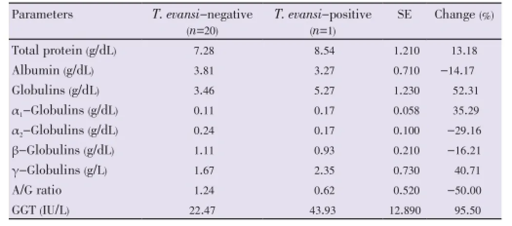
Table 2 Biochemical parameters of T. evansi-negative and T. evansi-positive groups.
4. Discussion
One of the most important diseases among camels that causes serious economic losses in America, Africa and Asia, including Iran, is surra[8]. This study is the first report of trypanosomosis in camels in Semnan, Iran.
In the present study, decreased hematocrite level, Hb concentration, total erythrocyte counts, MCV and MCHC values and increased WBC counts inT. evansiinfected camel compared to uninfected camels were observed. Anemia and leukocytosis are common features of trypanosomosis in camels[9]. Anemia appears to be predominantly caused by hemolytic associated with decrease life span of erythrocytes and extensive erythrophagocytosis[10]. Mechanisms involvedin the development of anemia include: hemolysis, free fatty acids, immunologic mechanisms, hemodilution, coagulation disorders, depression of erythrogenesis and release of trypanosomal sialidase[10-13]. The most important oxidative enzyme during trypanosomosis is sialidase. Sialic acid in erythrocyte surface membrane is hydrolysed by sialidase[14].
Leukocytosis is the result of increased activity of the mononuclear phagocytic system during trypanosomiasis. These results are similar to the findings of Chaudhary and Iqbal, and Padmaja[1,15].
To determine the functional status of various organs investigated serum biochemical profile is a good indicator. Total protein, albumin, globulins, A/G ratio, α1, α2, β and γ-globulin and GGT enzyme were studied in one smear positive sample and 20 smear negative samples. Hyperproteinemia and hyperglobulinemia in theT. evansiinfected camel compared with uninfected camels may be related to the presence of trypanosomes in the blood stream, which stimulates the immune system to secrete immunoglobulins[16]. In response to the animal trypanosomosis, especially the chronic form of infection, IgM levels often increase[16,17]. Therefore, increased immunoglobulin levels can lead to increased concentrations of serum total protein in our study.
Severe degenerative changes in the liver, which was confirmed cytologically by the presence of hepatic necrosis, may be hypoalbuminemia[1,18]. With hyperglobulinemia in trypanosomosis, hypoalbuminemia may be a compensatory mechanism to maintain osmolarity[3].
The decrease in the A/G ratio is related to the presence of hypoalbuminemia and hyperglobulinemia[1,18]. The increase in γ-globulin can be caused by increased immunoglobulin concentration, as was previously reported by Khosraviet al.,and Taylor and Authié[4,16]. Serum GGT activity is largely derived from the hepatobiliary system. Increasing serum GGT activity observed in the present study may be a result of the fatty degeneration of the liver cells and subsequent liver necrosis[19].
Results of the present study revealed that: first, trypanosomosis is present in dromedary camels of Semnan County, Iran (infection rate is 4.76%) and this is the first study of this protozoa infection in the region of Iran. Secondly, hematological and biochemical parameters are markedly affected by camel trypanosomosis.
Conflict of interest statement
We declare that we have no conflict of interest.
Acknowledgements
The authors thank Masoum M . and Karkeh-abadi M. for their assistance in collecting samples. This work was supported by research fund of Semnan University, Semnan, Iran (Grant No. 266/92/3040).
[1] Chaudhary ZI, Iqbal J. Incidence, biochemical and haematological alterations induced by natural trypanosomosis in racing dromedary camels. Acta Trop 2000; 77: 209-213.
[2] Allam L, Ogwu D, Agbede RI, Sackey AK. Hematological and serum biochemical changes in gilts experimentally infected with Trypanosoma brucei. Vet Arhiv 2011; 81: 597-609.
[3] Da Silva AS, Wolkmer P, Costa MM, Tonin AA, Eilers TL, Gressler LT, et al. Biochemical changes in cats infected with Trypanosoma evansi. Vet Parasitol 2010; 171: 48-52.
[4] Khosravi A, Hakimi Parizi M, Bamorovat M, Borhani Zarandi M, Mohammadi MA. Prevalence of Trypanosoma evansi in camels using molecular and parasitological methods in the southeast of Iran, 2011. J Parasit Dis 2013; doi: 10.1007/s12639-013-0355-9.
[5] Sow A, Sidibé I, Kalandi M, Bathily A, Ndiaye NP, Ouédraogo M, et al. Biochemical changes induced by natural infection of trypanosomosis in Burkinabese local donkey breeds. Comp Clin Pathol 2014; 23: 103-109.
[6] Zarif-Fard MR, Hashemi-Fesharki R. Study on tissue and blood protozoa of camels in Southern Iran. J Camel Pract Res 2000; 7: 193-194.
[7] Tornquist SJ. Hematology of Camelids. In: Weiss DJ, Wordrop KJ, editors. Schalm’s veterinary haematology. USA: Wiley-Blackwell Publishing Ltd.; 2010, p. 910-917.
[8] Enwezor FN, Sackey AK. Camel trypanosomosis-a review. Vet Arhiv 2005; 75: 439-452.
[9] Gutierrez C, Corbera JA, Juste MC, Doreste F, Morales I. Clinical, hematological, and biochemical findings in an outbreak of abortion and neonatal mortality associated with Trypanosoma evansi infection in dromedary camels. Ann N Y Acad Sci 2006; 1081: 325-327.
[10] Habila N, Inuwa MH, Aimola IA, Udeh MU, Haruna E. Pathogenic mechanisms of Trypanosoma evansi infections. Res Vet Sci 2012; 93: 13-17.
[11] Adamu S, Ibrahim ND, Nok AJ, Esievo KA. Sialyl transferase activity probably counteracts that of sialidase as one of the possible mechanisms of natural recovery of stabilization of erythrocyte mass in trypanosome-infected animals-a perspective. Afr J Biotechnol 2008; 7: 4992-5001.
[12] Megahed GA, Abd Ellah MR, Abdel-Rady A. Comparative biochemical studies on natural Trypanosoma evansi infection in she-camels. Comp Clin Pathol 2012; 21: 1121-1124.
[13] Omer HO, Mousa HM, Al-Wabel N. Study on the antioxidant status of rats experimentally infected with Trypanosoma evansi. Vet Parasitol 2007; 145: 142-145.
[14] Sallau AB, Ibrahim MA, Salihu A, Yusuf IA. Bloodstream form of Trypanosoma evansi contains galactosidase. Middle-East J Sci Res 2008; 3: 49-52.
[15] Padmaja K. Haemato-biochemical studies of camels infested with trypanosomiasis. Vet World 2012; 5: 356-358.
[16] Taylor K, Authié EM. Pathogenesis of animal trypanosomiasis. In: Maudlin I, Holmes PH, Miles MA, editors. The trypanosomiases. Wallingford: CABI Publishing; 2004.
[17] Baral TN, De Baetselier P, Brombacher F, Magez S. Control of Trypanosoma evansi infection is IgM mediated and does not require a type I inflammatory response. J Infect Dis 2007; 195: 1513-1520.
[18] Ahmed S, Butt AA, Muhammad G, Athar M, Khan MZ. Haematobiochemical studies on the haemoparasitized camels. Int J Agric Biol 2004; 6: 331-334.
[19] Abd El-Baky AA, Salem SI. Clinicopathological and cytological studies on naturally infected camels and experimentally infected rats with Trypanosoma evansi. World Appl Sci J 2011; 14: 42-50.
10.12980/APJTB.4.2014APJTB-2014-0053
*Corresponding author: Dr. M. Ahmadi-hamedani, Assistant Prof., Veterinary Clinical Pathology, Department of Pathobiology, Faculty of Veterinary Medicine, Semnan University, Semnan, Iran.
Tel: +98 232 3664892
Fax: +98 232 3664894
E-mail: m.ahmadi@profs.semnan.ac.ir
Foundation Project: Supported by research fund of Semnan University, Semnan, Iran (Grant No. 266/92/3040).
Methods:To perform this project, blood samples were collected by venipuncture into plain and EDTA-K2-containing vacutainer tubes from 21 dromedary camels (12 males and 9 females) aged 3-18 years, from 4 different regions of Semnan.
Results:Microscopic examination of stained thin blood smears revealed the presence of T. evansi in one of the samples. However, it should be noted that this sample showed a very high parasitemia (more than 5 trypomastigote were visible per microscopic field with MGG, 1 000×). This heavy parasitemia was associated with an 18-year-old female camel that showed symptoms of corneal opacity, intense emaciation and pale mucous membranes. Comparison of hematologyical and serum biochemical profiles between the camel infected by T. evansi and uninfected camels indicated anemia, leukocytosis, hyperproteinemia, hypoalbuminemia, hyperglobulinemia, reduction A/G ratio, increased α1, β and globulins and decreased of α2globulins and increased the concentration of gamma-glutamyl transferase enzyme.
Conclusions:Results of the present study revealed that trypanosomosis was present in dromedary camels of Semnan, Iran (infection rate is 4.76%) and hemato-biochemical parameters were markedly affected by camel trypanosomosis.
 Asian Pacific Journal of Tropical Biomedicine2014年9期
- Asian Pacific Journal of Tropical Biomedicine的其它文章
- A case report of cutaneous larva migrans in a Mexican population of high marginalization
- Acute brucellosis as unusual cause of immune thrombocytopenia: a case report and review of the literature
- Chemical composition and larvicidal activity of essential oil of Origanum majorana (Lamiaceae) cultivated in Morocco against Culex pipiens (Diptera: Culicidae)
- Phytochemical screening and antioxidant activity of ethanol extract of Tithonia diversifolia (Hemsl) A. Gray dry flowers
- Rate of carcass and offal condemnation in animals slaughtered at Yazd Slaughterhouse, central Iran
- Formulation and evaluation of novel stomach specific floating microspheres bearing famotidine for treatment of gastric ulcer and their radiographic study