花姜酮对胰腺癌PANC1细胞株增殖和凋亡的影响
2010-11-23 08:13:40陈晓燕王卫星丁佑铭殷涛阿布力克木阿布力孜
中华胰腺病杂志 2010年6期
陈晓燕 王卫星 丁佑铭 殷涛 阿布力克木·阿布力孜
·论著·
花姜酮对胰腺癌PANC1细胞株增殖和凋亡的影响
陈晓燕 王卫星 丁佑铭 殷涛 阿布力克木·阿布力孜
目的探讨花姜酮对胰腺癌PANC1细胞增殖和凋亡的影响,探讨其作用机制。方法应用3.75、7.5、15、30、60 μg/ml的花姜酮处理PANC1细胞,以未处理细胞作为对照。CCK-8法检测细胞增殖抑制率,Hoechst33342染色观察细胞形态,流式细胞术检测细胞凋亡率,Western blotting法检测细胞磷酸化STAT1(p-STAT1)、Bax和Bcl-2蛋白的表达。结果花姜酮呈时间-剂量依赖性抑制PANC1细胞的生长,15 μg/ml花姜酮作用48 h后,细胞增殖抑制率达(72.8±2.72)%,并可观察到典型的细胞凋亡形态学改变,细胞凋亡率达14.2%;同时,PANC1细胞p-STAT1和Bax蛋白表达明显增加(0.654±0.048对0.074±0.011,0.577±0.044对0.218±0.027,P<0.05),Bcl-2蛋白表达明显下降(0.162±0.029 对 0.459±0.034,P<0.05)。结论花姜酮可能通过上调STAT1活性,升高Bax/Bcl-2比率,从而诱导PANC1细胞的凋亡和抑制细胞增殖。
胰腺肿瘤; 转录激活因子1; 花姜酮; 细胞凋亡
花姜酮是一种从球姜的根茎中分离出的倍半萜类化合物,已被证明对结肠癌、皮肤癌均具有好的化学预防作用[1]。联合应用花姜酮和顺铂也可延缓宫颈上皮瘤样病变向宫颈癌发展的进程[2]。新近研究发现,花姜酮参与调控胰腺癌细胞内某些信号转导通路。信号转导与转录激活因子1(signal transducer and activator of transcription 1,STAT1)信号通路是近年来发现的比较重要的信号通路,具有调控细胞生长、分化等多种生理功能。本研究观察花姜酮对PANC1细胞的增殖、凋亡以及STAT1活性的影响,探讨其可能的机制。
材料与方法
一、细胞培养
胰腺癌细胞系PANC1由中国科学院上海细胞生物研究所细胞库提供。常规培养传代。取对数生长期的PANC1细胞进行实验。每个浓度设5个复孔,实验重复3次。
二、CCK-8法检测细胞增殖抑制率
CCK-8细胞计数试剂盒购自日本同仁化学公司。以每孔5×10⁷个细胞接种于96孔板,培养24 h。换含15%胎牛血清的DMEM培养48 h。分别加入0、3.75、7.5、15、30、60 μg/ml的花姜酮继续培养12、24、48 h,以无细胞孔作为空白对照。按说明书操作。抑制率=(空白对照A450值-实验组A450值)/(空白对照A450值-对照组A450值)×100%。
三、Hoechst33342染色观察细胞核形态的变化
细胞接种于24孔细胞培养板培养24 h,加入0、7.5、15 μg/ml花姜酮继续培养48 h。吸弃半量培养液,加入Hoechst33342,终浓度为10 μg/ml,室温避光作用10 min,弃上清培养液。于荧光倒置显微镜下观察、摄片。
四、流式细胞术检测细胞凋亡率
细胞接种于6孔板培养24 h,加入0、7.5、15 μg/ml花姜酮继续培养48 h。收集各孔细胞,分别制成单细胞悬液,加入5 μl Annexin V-FITC和10 μl PI,室温避光5 min,然后上流式细胞仪检测。
五、Western blotting检测细胞p-STAT1、 Bax和Bcl-2蛋白表达水平
分别收集0、7.5、15 μg/ml花姜酮处理48 h的细胞,提取蛋白。取100 μg常规行Western blotting,最后ECL化学发光,X线胶片曝光,凝胶成像系统扫描,以目的条带与actin条带灰度值的比值代表蛋白的表达水平。
六、统计学处理

结 果
一、花姜酮对PANC1细胞增殖的抑制作用
花姜酮呈浓度和时间依赖性抑制PANC1细胞的增殖 (表1)。
表1 花姜酮对PANC1细胞增殖的影响
二、花姜酮对PANC1细胞凋亡的影响
PANC1细胞核经Hoechst33258染色呈圆形,淡蓝色,内有较深的蓝色颗粒。花姜酮处理后,细胞数减少,出现较多核固缩或呈分叶状的凋亡细胞。随着药物浓度的升高,凋亡细胞的数也增加(图1)。对照组PANC1细胞凋亡率1.1%,7.5 μg/ml和15 μg/ml花姜酮处理组细胞凋亡率分别为3.9%和14.2%,三组间差异显著(P<0.01,图1)。

图10(a,d)、7.5(b,e)、15(c,f) μg/ml花姜酮处理48 h后PANC1细胞的形态改变(Hoechst33258染色 ×200)及细胞凋亡率(流式细胞仪)
三、花姜酮对PANC1细胞p-STAT1、 Bax和Bcl-2蛋白表达的影响
花姜酮处理后细胞p-STAT1和Bax蛋白表达水平明显升高;而Bcl-2蛋白表达水平明显降低(P<0.05,表2、图2)。
表2 各组p-STAT1、Bax、Bcl-2蛋白的表达
注:与对照组比较,aP<0.05;与7.5 μg/ml组比较,bP<0.05
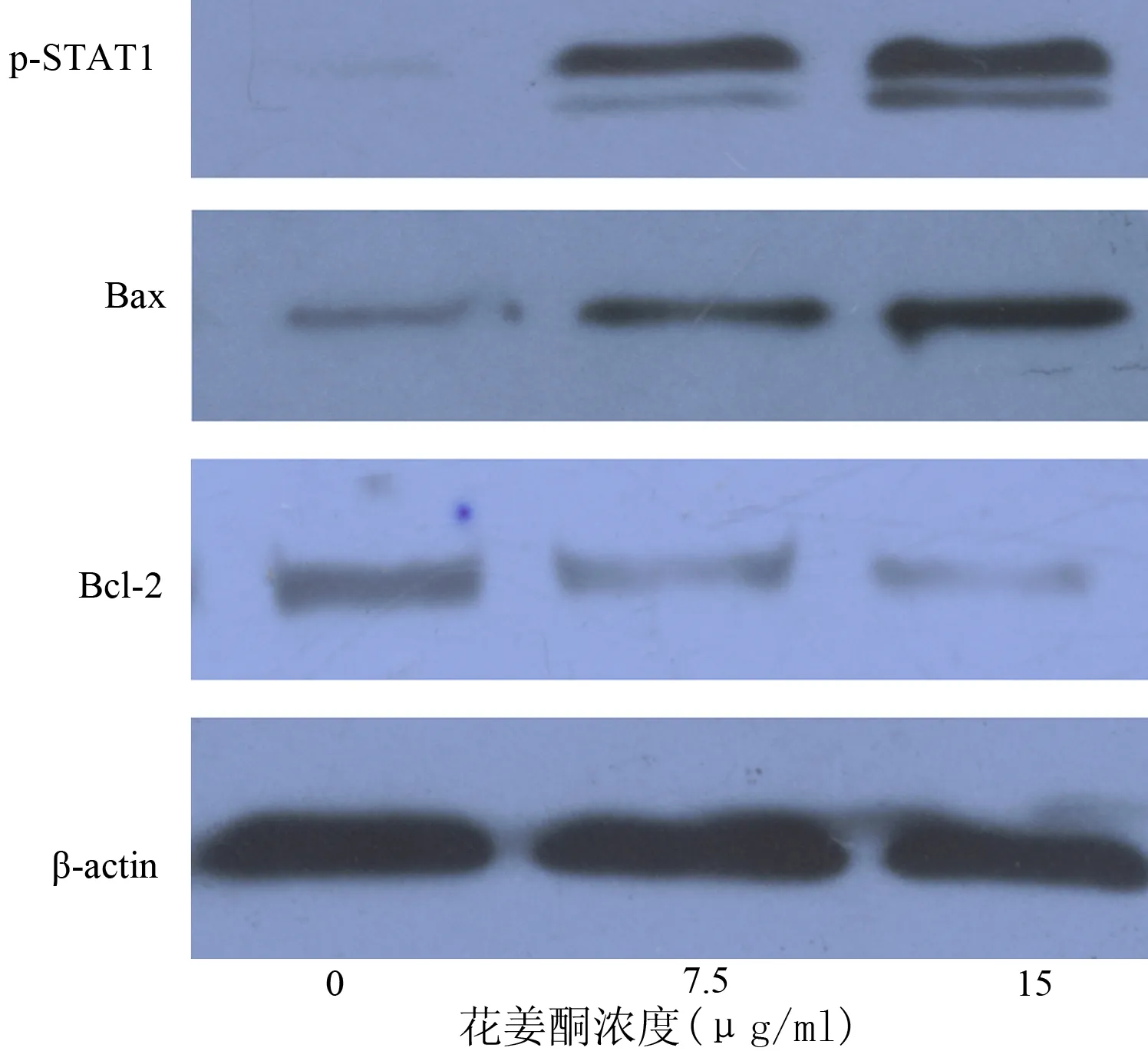
图2不同浓度花姜酮处理48 h后PANC1细胞p-STAT1、Bax和Bcl-2蛋白的表达(Western blotting)
讨 论
花姜酮具有抗炎、抗肿瘤等多种生物活性,本研究结果也证明花姜酮能够明显抑制胰腺癌细胞的生长。有研究证实,花姜酮能通过抑制NF-κB及其相关基因的表达,从而起到预防和治疗癌症的作用[3]。Sakinah等[4]发现,花姜酮能够通过调节Bax/Bcl-2比率来诱导肝癌细胞凋亡[5]。
STAT是一组能与DNA结合的蛋白质,介导多种细胞因子和生长因子向细胞核内传导,影响靶基因的转录,调节人体免疫反应、炎症反应和细胞的生长、分化等[5]。大量研究证明,通过增强或抑制STAT的活性,可影响多种癌细胞系的增殖和凋亡[6-8]。其成员STAT1在多种肿瘤组织中呈现低表达趋势[9-10]。通过药物刺激上调STAT1水平能起到促进胰腺癌细胞凋亡和增强癌细胞化疗敏感性的作用[11-12]。本研究结果显示,随着花姜酮浓度的升高,发生凋亡的细胞明显增多,同时磷酸化STAT1和Bax蛋白表达显著增强, Bcl-2表达降低。因此我们推测,花姜酮通过激活STAT1信号转导通路进而影响凋亡相关蛋白Bax和Bcl-2的表达,促进胰腺癌细胞凋亡。
[1] Murakami A,Tanaka T,Lee JY,et al.Zerumbone, a sesquiterpene in subtropical ginger,suppresses skin tumor initiation and promotion stages in ICR mice.Int J Cancer,2004,110:481-490.
[2] Abdul AB,Abdelwahab SI,Bin Jalinas J,et al.Combination of zerumbone and cisplatin to treat cervical intraepithelial neoplasia in female BALB/c mice.Int J Gyn Cancer,2009,19:1004-1010.
[3] Takada Y,Murakami A,Aggarwal BB.Zerumbone abolishes NF-kappaB and IkappaBalpha kinase activation leading to suppression of antiapoptotic and metastatic gene expression,upregulation of apoptosis,and downregulation of invasion.Oncogene,2005,24:6957-6969.
[4] Sakinah SA,Handayani ST,Hawariah LP.Zerumbone induced apoptosis in liver cancer cells via modulation of Bax/Bcl-2 ratio.Cancer Cell Int,2007,7:4.
[5] Darnell JE,Kerr IM,Stark GR.JAK-STAT pathways and transcriptional activation in response to IFNs and other extracelluar signaling proteins.Science,1994,264:1415-1421.
[6] Thoennissen NH,Iwanski GB,Doan NB,et al.Cucurbitacin B induces apoptosis by inhibition of the JAK/STAT pathway and potentiates antiproliferative effects of gemcitabine on pancreatic cancer cells.Cancer Res,2009,69:5876-5884.
[7] Ulivi P,Arienti C,Amadori D,et al. Role of RAF/MEK/ERK pathway,p-STAT-3 and Mcl-1 in sorafenib activity in human pancreatic cancer cell lines.J Cell Physiol,2009,200:214-221.
[8] Sahu RP,Srivastava SK.The role of STAT-3 in the induction of apoptosis in pancreatic cancer cells by benzyl isothiocyanate.J Natl Inst,2009,101:176-193.
[9] Xi S,Dyer KF,Kimak M,et al.Decreased STAT1 expression by promoter methylation in squamous cell carcinogenesis.J Natl Cancer Inst,2006,98:181-189.
[10] Yu H,Jove R.The STATs of cancer-new molecular targets come of age.Nat Rev Cancer,2004,4:97-105.
[11] Wang SO,Koromilas AE.Stat1 is an inhibitor of Ras-MAPK signaling and Rho small GTPase expression with implications in the transcriptional signature of Ras transformed cells.Cell Cycle,2009,8:2070-2079.
[12] Muerkoster SS,Werbing V,Koch D,et al.Role of myofibroblasts in innate chemoresistance of pancreatic carcinoma-Epigenetic downregulation of caspases.Int J Cancer,2008,123:1751-1760.
2010-03-17)
(本文编辑:屠振兴)
EffectsofzerumboneontheproliferationandapoptosisofhumanpancreaticcancercelllinePANC1
CHENXiao-yan,WANGWei-xing,DINGYou-ming,YINTao,ABLIZAblikim.
DepartmentofHepatobiliaryandLaparoscopicSurgery,RenminHospital,WuhanUniversity,Wuhan430060,China
WANGWei-xing,Email:sate.llite@163.com
ObjectiveTo investigate the effect of zerumbone on the proliferation and apoptosis of human pancreatic cancer cell line PANC1 and its possible mechanism.MethodsZerumbone of various concentrations (3.75, 7.5, 15, 30, 60 μg/ml) were used to treat PANC1, and cells without treatment were used as control. CCK-8 assay was used to detect the inhibitory rate of cell proliferation. Cell apoptosis analysis was determined by using Hoechst 33342 staining and flow cytometry. Western blotting was performed to evaluate the phosphorylation Stat1 (p-STAT1), and Bax and Bcl2 protein expression.ResultsZerumbone caused a time- and dose-dependent reduction of cell viability in PANC1 cells. After 48h treatment of Zerumbone of 15 μg/ml, cells inhibitory rate was increased to (72.8±2.72)%, and classic apoptosis morphology was observed, with apoptosis rate was 14.2%. At the same time, p-STAT1, and Bax protein expression was significantly increased (0.654±0.048vs0.074±0.011, 0.577±0.044vs0.218±0.027,P<0.05); Bcl-2 protein expression was significantly decreased (0.162±0.029vs0.459±0.034,P<0.05).ConclusionsZerumbone may inhibit the proliferation of PANC1 cells and inducing cell apoptosis, which may be related to the up-regulation of STAT1′s activity and Bcl-2/Bax ratio.
Pancreatic neoplasms; Activator transcription factor 1; Zerumbone; Apoptosis
10.3760/cma.j.issn.1674-1935.2010.06.014
430060 武汉,武汉大学人民医院肝胆外科
王卫星,Email: sate.llite@163.com
猜你喜欢
黑龙江科学(2023年8期)2023-06-04 08:40:58
保健医苑(2022年6期)2022-07-08 01:25:22
中国循证心血管医学杂志(2021年10期)2021-11-05 06:56:00
台州学院学报(2018年6期)2018-02-26 06:14:38
中成药(2017年8期)2017-11-22 03:19:00
天津医药(2016年9期)2016-10-20 03:19:39
中国现代医学杂志(2015年26期)2015-12-23 11:04:22
上海工运(2015年11期)2015-08-21 07:27:00
医学研究杂志(2015年7期)2015-06-22 11:01:42
西安交通大学学报(医学版)(2015年2期)2015-02-28 17:59:21