不同分度大肠黑变病伴发息肉的内镜特点分析
张雪 秦佳敏 文黎明


基金項目:四川省科技计划项目(2023YFG0262);2021年四川省医学会科研课题(S21074)
作者单位:646000 泸州,西南医科大学临床医学院(张雪,文黎明);621000 绵阳,四川绵阳四〇四医院(张雪,秦佳敏,文黎明)
通信作者:文黎明,E-mail: wenlm404@sohu.com
【摘要】目的 探讨不同分度大肠黑变病(MC)伴发息肉的内镜特点。方法 回顾性分析597例行结肠镜检查并诊断为MC患者的临床资料及内镜资料。分析不同分度MC与年龄、性别、伴发肠道疾病的相关性。并将不同分度MC合并息肉患者的性别、年龄、息肉大小、息肉病理类型等进行统计学分析。结果 Ⅰ度MC比Ⅱ、Ⅲ度MC更易检出合并息肉。不同分度MC合并息肉大小比较有统计学意义,其中<0.5 cm息肉在Ⅲ度MC合并息肉组检出率(37.5%)低于Ⅰ度MC合并息肉组(67.11%)组和Ⅱ度MC合并息肉组(60.18%);0.6~1.0 cm息肉在Ⅲ度MC合并息肉组检出率(57.50%)高于Ⅰ度MC合并息肉组(23.49%)组和Ⅱ度MC合并息肉组(25.66%)。增生性息肉、管状腺瘤在不同分度MC合并息肉患者的分布比较差异有统计学意义(P均< 0.05);炎性息肉、潴留性息肉、锯齿状腺瘤、管状-锯齿状腺瘤在不同分度MC合并息肉患者的分布比较差异无统计学意义(P均> 0.05)。结论 Ⅰ度MC更易检出息肉,Ⅲ度MC更易检出0.6~1.0 cm息肉,Ⅰ度MC更易检出增生性息肉,而Ⅱ、Ⅲ度MC更易检出管状腺瘤。
【关键词】大肠黑变病;不同分度;结直肠息肉;管状腺瘤
Endoscopic characteristics of different grades of melanosis coli complicated with polyps Zhang Xue△, Qin Jiamin, Wen Liming.△Clinical School of Medicine, Southwest Medical University, Luzhou 646000, China
Corresponding author: Wen Liming, E-mail: wenlm404@sohu.com
【Abstract】Objective To investigate the endoscopic characteristics of different grades of melanosis coli (MC) complicated with polyps. Methods Clinical and endoscopic data of 597 patients diagnosed with MC who underwent colonoscopy were retrospectively analyzed. The correlation of different grades of MC with age, gender and intestinal diseases was analyzed. The gender, age, polyp size and pathological type of polyps in patients with different grades of MC were analyzed statistically. Results Polyps were more commonly detected in gradeⅠMC than in gradeⅡand gradeⅢMC. The size of polyps significantly differed among different grades of MC. The detection rate of < 0.5 cm polyps in gradeⅢMC was 37.50%, significantly lower than 67.11% in gradeⅠand 60.18% in gradeⅡMC. The detection rate of 0.6-1.0 cm polyps in gradeⅢMC was 57.50%, significantly higher than 23.49% in grade Ⅰand 25.66% in gradeⅡMC. The detection rates of hyperplastic polyps and tubular adenoma significantly differed among different grades of MC complicated with polyp (all P < 0.05). No statistical significance was observed in the distribution of inflammatory polyp, retention polyp, serrated adenoma and tubular-serrated adenoma among different grades of MC complicated with polyp (all P > 0.05). Conclusions Polyps are more commonly detected in gradeⅠMC. Polyps of 0.6-1.0 cm in size are more likely to be detected in grade ⅢMC. Hyperplastic polyps are more commonly detected in gradeⅠMC, whereas tubular adenoma in gradeⅡandⅢMC.
【Key words】Melanosis coli ; Different grades; Colorectal polyp; Tubular adenoma
大肠黑变病(MC) 是一种可逆的、大肠黏膜固有层中巨噬细胞内色素聚集的一种以黏膜色素沉着为特征的非炎症性疾病,病因和发病机制尚不清楚[1-2]。目前公认的病因主要为长期便秘及蒽醌类泻剂的使用,但缺乏明确的证据支持[3]。随着人们健康意识越来越强、医学技术的进步,MC的检出率逐渐升高。此外也有不少MC与大肠息肉及结直肠癌相关性的报道,然而关于MC与结直肠癌的关系存在争论,但多数研究支持MC患者的结直肠腺瘤发生率较非大肠黑变病患者更高,故应警惕大肠黑变病合并结直肠息肉进一步发生癌变。因此加深对该病的认识,对早期发现结直肠癌的意义重大。
对象与方法
一、研究对象
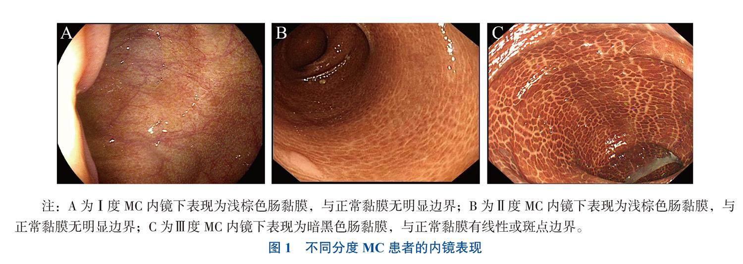
收集2019年1月至2023年8月于四川绵阳四〇四医院消化内科内镜中心行肠镜检查并诊断为MC的患者597例,根据MC镜下分度标准[4-5]将其分为3组,分别为Ⅰ度组(350例)、Ⅱ度组(197例)、Ⅲ度组(50例),见图1。
二、方 法
收集其性别、年龄、合并肠道疾病等资料并进行分析。另外根据是否合并肠息肉分为Ⅰ度MC合并息肉组(149例)、Ⅱ度MC合并息肉组(113例)、Ⅲ度MC合并息肉组(40例),收集其临床信息、息肉大小、息肉分布、病理资料等。纳入标准:①肠道准备充分、结肠镜检查完整者;②内镜资料、病理活组织检查资料、临床资料收集完善者。排除标准:①既往结直肠癌史;②既往内镜下息肉切除术史;③炎症性肠病、肠结核、白塞病、家族性腺瘤性息肉病或其他慢性结肠炎的患者。本研究经医院伦理委员会审查通过(批件号:2024001),所有入组人员签署知情同意书。
三、统计学处理
采用SPSS 25.0进行统计学分析。采用K-S正态性检验或直方图检验计量资料的正态性,服从正态分布的计量资料以表示,采用单因素方差分析比较组间差异,并采用LSD方法进行事后两两比较;非正态分布的资料以M(P25,P75)表示,通过K-W检验比较组间差异;计数资料以n(%)表示,通过χ 2检验或Fisher确切概率法比较组间差异。P < 0.05为差异有统计学意义。
结果
一、不同分度MC与性别、年龄的比较
行结肠镜检查并诊断为MC共计597例,其中Ⅰ度MC组350例,男125例(35.7%)、女225例(64.3%),年龄67 (55,74)岁;Ⅱ度MC组197例,男77例(39.1%)、女120例(60.9%),年龄66(55,75)岁;Ⅲ度MC组50例,男26例(52.0%)、女24例(48.0%),年龄60(52,72)岁。3组性别比较差异无统计学意义(P = 0.081);Ⅲ度MC组年龄与Ⅰ度MC组年龄比较差异有统计学意义(P = 0.002)。
二、不同分度MC合并肠道疾病的比较
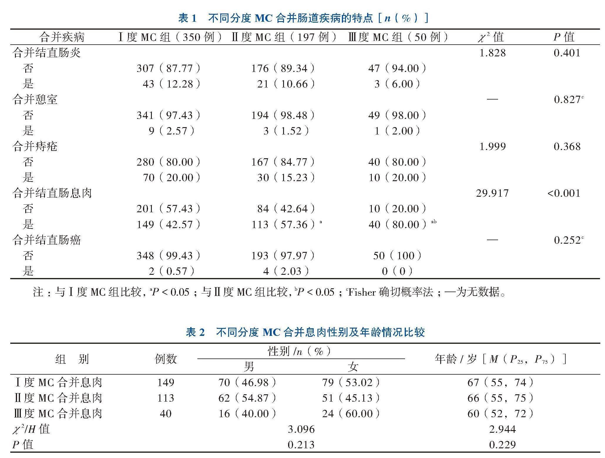
MC合并的肠道疾病主要包括结直肠炎、憩室、痔疮、结直肠息肉、结直肠癌,不同分度MC合并的肠道疾病进行比较,其中只有不同分度MC合并结直肠息肉两两比较差异有统计学意义(P < 0.001)。见表1。
三、不同分度MC合并息肉性别、年龄的比较
不同分度MC合并息肉患者年龄及性别比较差异均无统计学意义(P均> 0.05)。见表2。
四、不同分度MC合并息肉镜下分布情况
不同分度MC合并息肉数量比较差异无统计学意义(P = 0.771),不同分度MC合并息肉其息肉大小比较差异有统计学意义(P = 0.019),进一步两两比较结果显示,Ⅲ度MC合并息肉与Ⅰ度MC合并息肉其息肉大小比较差异有统计学意义。见表3。
五、不同分度MC合并息肉的息肉病理类型比较
增生性息肉、管状腺瘤在不同分度MC合并息肉患者的比较差异均有统计学意义(P均< 0.001);炎性息肉、潴留性息肉、锯齿状腺瘤、管状-锯齿状腺瘤在不同分度MC合并息肉患者的比较差异均无统计学意义(P均> 0.05)。见表4。
讨论
MC被认为是一种可逆性、良性、非炎症性疾病,目前关于MC的发病机制存在争议,但公认与便秘、蒽醌类泻药及凋亡机制有关[6-8]。随着MC的检出率逐渐升高,出现不少关于MC与大腸息肉及结直肠癌相关性的报道,虽存在争论,但大多数研究表明MC患者的结直肠腺瘤发生率高于非MC患者。
结直肠息肉具有一定的癌变潜能,腺瘤性息肉是常见的病理类型,可通过“腺瘤-癌”发展为结肠癌,目前已成为公认的结肠癌的癌前病变[9]。结直肠癌是我国最常见的恶性肿瘤之一,因此对癌前病变的早诊断、早治疗是预防结直肠癌发生的重要手段[10]。Liu等[4]研究发现MC患者的低度腺瘤发生率和非腺瘤息肉发生率高于对照组。
一项研究表明,MC患者更有可能同时患有增生性息肉和低级别腺瘤[11]。Blackett等[12]研究也表明MC与腺瘤检出率增加有关。研究表明,其色素沉着是黏膜损失的一种表现,而非无害[4]。MC与结直肠腺瘤增加有关,但与息肉组织学进展无关[13]。但也有学者认为结直肠腺瘤不含黑色素样色素沉着,腺瘤与大肠黑变病的关联可能与在深色结肠黏膜内微小息肉表现为白点而易于检出有关[14]。徐晓光等[15] 对MC患者与经高清智能电子染色内镜检查的非MC患者进行比较,两者结肠息肉的检出率无明显差别,MC与结肠息肉的发生并无相关性,与黏膜背景变化有关。但本研究结果表明,管状腺瘤在Ⅱ度、Ⅲ度MC合并息肉组的检出比例较高,在Ⅰ度MC合并息肉组则相反,然而增生性息肉的检出情况则相反,这一结果可能与最佳增强效应有关,非腺瘤性息肉如炎症性息肉是有色素的,而腺瘤在MC患者的黏膜上没有色素,这可能导致严重MC患者的腺瘤检出率较高。
笔者通过对不同分度MC的临床特征进行比较发现,Ⅲ度MC易发生在年龄偏大的患者中。一项大样本多中心研究也表示随着年龄的增长,无论性别,检出率逐年递增,≥60岁的患者检出率快速上升[16]。而田甜等[17]的回顾分析发现女性患MC居多,发病年龄及息肉检出率女性均低于男性,其另一研究表明年龄≥65岁的腹型肥胖男性更容易在MC的基础上出现结肠息肉[18]。柴海娜等[19]的研究发现MC结肠息肉检出以多发为主,<0.5 cm的息肉最多见。本研究发现<0.5 cm息肉在Ⅲ度MC合并息肉组检出率低于Ⅰ度、Ⅱ度MC合并息肉组,然而0.6~1.0 cm息肉中則呈现相反的结果。这与先前报道的严重MC的检出率更高结果相符[20]。
综上所述,MC不同分度与年龄相关,Ⅲ度MC合并息肉有更高管状腺瘤的检出率,对于Ⅲ度MC合并息肉的患者,建议早期息肉切除,以预防发生结直肠癌,但具体机制待未来进行组织学和分子研究来阐明。
参 考 文 献
[1] Zhou Y, Yang T, Wu T, et al. Mucinous colorectal adenocar-cinoma in a patient with familial adenomatous polyposis and melanosis coli[J]. Asian J Surg, 2023, 46(12): 5773-5775.
[2] Lombardi N, Crescioli G, Maggini V, et al. Anthraquinone laxatives use and colorectal cancer: a systematic review and meta-analysis of observational studies[J]. Phytother Res, 2022, 36(3): 1093-1102.
[3] Zhang R, Huang C, Wu F, et al. Review on melanosis coli and anthraquinone-containing traditional Chinese herbs that cause melanosis coli[J]. Front Pharmacol, 2023, 14: 1160480.
[4] Liu Z H, Foo D C C, Law W L, et al. Melanosis coli: harmless pigmentation? A case-control retrospective study of 657 cases [J]. PLoS One, 2017, 12(10): e0186668.
[5] 杨玄, 付雯, 崔月萌, 等. 结肠黑变病研究进展[J]. 中国临床研究, 2022, 35(8): 1147-1150.
Yang X, Fu W, Cui Y M, et al. Research progress of melanosis coli[J]. Chin J Clin Res, 2022, 35(8): 1147-1150.
[6] Yang N, Ruan M, Jin S. Melanosis coli: a comprehensive review[J]. Gastroenterol Hepatol, 2020, 43(5): 266-272.
[7] Hua X, Chen J, Wu L. Identification of candidate biomarkers associated with apoptosis in melanosis coli: GNG5, LPAR3, MAPK8, and PSMC6[J]. Biosci Rep, 2019, 39(1): BSR20181369.
[8] Cheng Y, Zhang H, Qu L, et al. Identification of Rhein as the metabolite responsible for toxicity of rhubarb anthraquinones[J]. Food Chem, 2020, 331: 127363.
[9] Keum N, Giovannucci E. Global burden of colorectal cancer: emerging trends, risk factors and prevention strategies[J]. Nat Rev Gastroenterol Hepatol, 2019, 16(12): 713-732.
[10] 赵利娜, 王恺疌, 罗琦, 等. 251例结肠息肉切除术后再发危险因素分析[J]. 新医学, 2022, 53(7): 491-495.
Zhao L N, Wang K J, Luo Q, et al. Analysis of risk factors of postoperative colorectal polyps recurrence in 251 patients[J]. J New Med, 2022, 53(7): 491-495.
[11] Kassim S A, Abbas M, Tang W, et al. Retrospective study on melanosis coli as risk factor of colorectal neoplasm: a 3-year colonoscopic finding in Zhuhai Hospital, China[J]. Int J Colorectal Dis, 2020, 35(2): 213-222.
[12] Blackett J W, Rosenberg R, Mahadev S, et al. Adenoma detection is increased in the setting of melanosis coli[J]. J Clin Gastroenterol, 2018, 52(4): 313-318.
[13] Abu Baker F, Mari A, Feldman D, et al. Melanosis coli: a helpful contrast effect or a harmful pigmentation[J]. Clin Med Insights Gastroenterol, 2018, 11: 1179552218817321.
[14] Nusko G, Schneider B, Ernst H, et al. Melanosis coli--a harmless pigmentation or a precancerous condition[J]. Z Gastroenterol, 1997, 35(5): 313-318.
[15] 徐曉光, 管恩翠, 薛方喜, 等. 结肠息肉在结肠黑变病与经i-scan内镜检出率的比较[J]. 浙江临床医学, 2017, 19(7): 1329-1330.
Xu X G, Guan E C, Xue F X, et al. Comparison of the detection rate of colonic polyps in melanosis coli and by i-scan endoscopy[J]. Zhejiang Clin Med J, 2017, 19(7): 1329-1330.
[16] Wang S, Wang Z, Peng L, et al. Gender, age, and concomitant diseases of melanosis coli in China: a multicenter study of 6, 090
cases[J]. PeerJ, 2018, 6: e4483.
[17] 田甜, 胡文炜, 王瑞玲, 等. 结肠黑变病合并结肠息肉与性别的相关性研究[J]. 长春中医药大学学报, 2020, 36(4): 763-765.
Tian T, Hu W W, Wang R L, et al. Study on the correlation between gender and melanosis coli complicated with colonic polyps [J]. J Changchun Univ Chin Med, 2020, 36(4): 763-765.
[18] 田甜, 胡文炜, 刘明浩, 等. 结肠黑变病发生结肠息肉的相关因素及与代谢综合征的相关性研究[J]. 解放军医药杂志, 2019, 31(7): 59-63.
Tian T, Hu W W, Liu M H, et al. A study on related factors and their correlation with metabolic syndrome in melanosis coli patients with colon polyps[J]. Med Pharm J Chin Peoples Liberation Army, 2019, 31(7): 59-63.
[19] 柴海娜, 刘军, 吴莹莹, 等. 结肠黑变病的临床特点分析[J]. 中国内镜杂志, 2021, 27(10): 24-29.
Chai H N, Liu J, Wu Y Y, et al. Analysis of clinical characteristics of melanosis coli[J]. China J Endosc, 2021, 27(10): 24-29.
[20] Katsumata R, Manabe N, Monobe Y, et al. Severe grade of melanosis coli is associated with a higher detection rate of colorectal adenoma[J]. J Clin Biochem Nutr, 2022, 71(2): 165-171.
(收稿日期:2023-12-13)
(责任编辑:杨江瑜)

