Icariin plus curcumol enhances autophagy through the mTOR pathway and promotes cathepsin B-mediated pyroptosis of prostate cancer cells
Xu-Yun Wang ,Wen-Jing Xu ,Bo-Nan Li ,Tian-Song Sun ,Wen Sheng
1Department of Andrology, Beijing Hospital of Traditional Chinese Medicine, Capital Medical University, Beijing, 100010, China
2Department of Dermatology, The First Affiliated Hospital of Hunan University of Chinese Medicine, Changsha, 410021, China
3School of Integrated Chinese and Western Medicine, Hunan University of Chinese Medicine, Changsha, 410208, China
4Andrology Laboratory, Hunan University of Chinese Medicine, Changsha, 410208, China
5School of Rehabilitation Medicine and Health Care, Hunan University of Medicine, Huaihua, 418000, China
6School of Traditional Chinese Medicine, Hunan University of Medicine, Huaihua, 418000, China
ABSTRACT Objective:To examine the effect of icariin plus curcumol on prostate cancer cells PC3 and elucidate the underlying mechanisms.Methods:We employed the Cell Counting Kit 8 assay and colony formation assay to assess cell viability and proliferation.Autophagy expression was analyzed using monodansylcadaverine staining.Immunofluorescence and Western blot analyses were used to evaluate protein expressions related to autophagy,pyroptosis,and the mTOR pathway.Cellular damage was examined using the lactate dehydrogenase assay.Moreover,cathepsin B and NLRP3 were detected by co-immunoprecipitation.Results:Icariin plus curcumol led to a decrease in PC3 cell proliferation and an enhancement of autophagy.The levels of LC3-Ⅱ/LC3-Ⅰ and beclin-1 were increased,while the levels of p62 and mTOR were decreased after treatment with icariin plus curcumol.These changes were reversed upon overexpression of mTOR.Furthermore,3-methyladenine resulted in a decrease in inflammatory cytokines,pyroptosis-related protein levels,and lactate dehydrogenase concentration,compared to the icariin plus curcumol group.Inhibiting cathepsin B reversed the regulatory effects of icariin plus curcumol.Conclusions:Icariin plus curcumol demonstrates great potential as a therapeutic agent for castration-resistant prostate cancer by enhancing autophagy via the mTOR pathway and promoting pyroptosis mediated by cathepsin B.These findings provide valuable insights into the molecular mechanisms underlying the therapeutic potential of icariin and curcumol for prostate cancer treatment.
KEYWORDS: Icariin;Curcumol;Autophagy;mTOR;Cathepsin B;Pyroptosis;Prostate cancer
1.Introduction
Prostate cancer is a common malignant disease that affects men worldwide,ranking as the third leading cause of cancer-related death in men[1].Androgen deprivation therapy is the primary treatment for patients diagnosed with prostate cancer for the first time[2].Initially,androgen deprivation therapy was effective in over 90%of patients,but the disease eventually developed into castrationresistant prostate cancer (CRPC)[3].Only a few medications have been approved specifically for treating CRPC.Therefore,researchers focus on developing novel drugs to address this condition.
Icariin (ICA),the main active ingredient found in the traditional herbEpimedium,possesses anti-apoptotic,antioxidant,and antineuroinflammatory properties[4].Curcumol,a sesquiterpene extracted from curcumin,exhibits remarkable neuroprotective,antiinflammatory,and anti-tumor properties[5].Moreover,curcumol can be used in conjunction with various synthetic drugs as an antibiotic or anti-cancer agent[6].Though both ICA[7] and curcumol[8]have been found to have considerable anti-cancer effects,few studies have explored the therapeutic benefits of using the two compounds in combination to treat CRPC.The mammalian target of rapamycin (mTOR) is an essential factor in progression and drug resistance[9].Previous research has revealed that ICA and curcumol can synergistically regulate the miR-7/mTOR/SREBP1 pathway,resulting in autophagy in prostate cancer cells[10].Additionally,curcumol has been shown to inhibit the malignant progression of prostate cancer by regulating the PDK1/AKT/mTOR pathway[6].Therefore,our research aimed to investigate the effect of ICA plus curcumol on CRPC and its underlying mechanisms.
Autophagy is the cellular self-digestion process that facilitates the breakdown of misfolded proteins and dysfunctional organelles[11],playing a significant role in the survival mechanism of CRPC.Inhibition of autophagy severely reduces the survival of prostate cancer cells[12].Pyroptosis is a form of cell death that is mediated by Caspase-1[13],and it is closely associated with prostate cancer[14].Cathepsin B (CTSB),a crucial cysteine protease in preventing bone metastasis in prostate cancer[15],is released by the increase of autophagy and the breakdown of autophagy polymers.It activates the NLRP3 inflammasome and promotes pyroptosis[16].In the present study,we aimed to explore whether treating CRPC with ICA plus curcumol affects the regulation of pyroptosis by CTSBviaautophagy regulation.
2.Materials and methods
2.1.Cell culture and grouping
PC3 cells (SNL-102,SUNNCELL,China) were purified with 10%fetal bovine serum (10099141,Thermo Fisher Scientific,USA)and 1% penicillin-streptomycin (SV30010,Beyotime,China) in Dulbecco’s Modified Eagle Medium (DMEM) (D5796,Merck,Germany) and incubated at 37 ℃ in a humidified incubator with 5%CO2(DH-160I,SANTN,China).The logarithmically grown cell plates were cultured in 6-well plates.
Next,the cells were divided into 5 groups: Dimethyl sulfoxide(DMSO),ICA,ethanol,curcumol,and ICA+curcumol groups.The DMSO group served as the control for the ICA group.The ethanol group served as the control for the curcumol group.The ICA group and the curcumol group were treated with 30 μM ICA and 50 μg/mL curcumol,respectively,for 48 h.The ICA+curcumol group was treated with 30 μM ICA and 50 μg/mL curcumol for 48 h.
In addition,to investigate the impact of the mTOR pathway,we divided the cells into two additional groups: ICA+Curcumol+oe-NC and ICA+Curcumol+oe-mTOR groups.In the ICA+Curcumol+oe-NC group,the cells were treated with 30 μM ICA and 50 μg/mL curcumol for 48 h and then transfected with oe-NC.In the ICA+Curcumol+oe-mTOR group,the cells were treated with 30 μM ICA and 50 μg/mL curcumol for 48 h and then transfected with oemTOR.
To further study the effect of ICA plus curcumol on PC3 cell autophagy,we divided the cells into 5 groups: Control,ICA,curcumol,ICA+curcumol,and ICA+curcumol+3-methyladenine(3-MA).In the control group,PC3 cells were cultured normally.The ICA,curcumol,and ICA+curcumol groups were treated as mentioned previously.In the ICA+curcumol+3-MA group,the cells were pretreated with 10 mM 3-MA for 3 h and then received a combination of 30 μM ICA and 50 μg/mL curcumol for 48 h.
To determine whether CTSB regulated pyroptosis induced by ICA plus curcumol through enhanced autophagy in PC3 cells,we divided the cells into 2 groups: ICA+curcumol+si-NC and ICA+curcumol+si-CTSB.In the ICA+curcumol+si-NC and ICA+curcumol+si-CTSB groups,the cells were transfected with sinegative control (NC) and si-CTSB for 6 h,respectively,and then received a combination of 30 μM ICA and 50 μg/mL curcumol for 48 h.
2.2.Cell Counting Kit 8 (CCK8) assay
Different groups of PC3 cells were digested and counted.The cells were then cultured in 96-well plates (0030730119,Eppendorf,Germany) at 1×10⁴cells/well density.Each well contained 100 μL of culture medium,and three replicates were used for each group.After the cells had adhered to the plate and started to grow,100 μL of fresh complete DMEM medium containing 10% CCK8 solution was added to each well following the instructions provided with the CCK8 kit (NU679,DOJINDO,Japan).The plate was then incubated at 37 ℃ with 5% CO2for 4 h.The optical density (OD) value at 450 nm was measured using the microplate reader (MB-530,HEALCES,China).The OD value was regarded as proportional to cell viability[17].Cell viability=(OD of experimental wells -OD of blank wells)/(OD of control wells -OD of blank wells)×100%.
2.3.Colony formation assay
The cells in each group were inoculated into 6-well plates with 200 cells per well and 1 mL of culture medium.The cells were then cultured at 37 ℃ with 5% CO2and saturated humidity for 2 weeks.The culture was terminated when visible clones appeared in the Petri dish.Later,1 mL of 4% paraformaldehyde (AWI0070a,Abiowell,China) was added to fix the cells for 15 min.Next,1 mL of crystal violet dye solution (AWC0333,Abiowell,China) was added to the dye for 30 min.Finally,the cells were photographed and counted after drying.
2.4.Monodansylcadaverine (MDC) staining
PC3 cells were stained with 50 μM MDC dye solution for 45 min.Subsequently,they were observed under a fluorescence microscope(DSZ2000X,Cnmicro,China) for analysis.
2.5.Immunofluorescence (IF)
PC3 cell samples were fixed with 4% paraformaldehyde for 30 min.Next,the samples were sealed with 0.3% triton at 37 ℃ for 30 min,and then with 5% bovine serum albumin (BSA) for 60 min.Subsequently,they were incubated with primary antibodies overnight at 4 ℃ as follows: LC3 (14600-1-AP,1∶100,Proteintech,USA),p62 (18420-1-AP,1∶100,Proteintech,USA),caspase-1(22915-1-AP,1∶50,Proteintech,USA),gasdermin D (GSDMD,1∶50,20770-1-AP,Proteintech,USA),CTSB (12216-1-AP,1∶50,Proteintech,USA).Afterwards,the samples were added with 50 μL of goat anti-rabbit IgG (H+L) (AWS0005b,Abiowell,China) labeled fluorescent antibody,incubated at 37 ℃ for 90 min,and stained with the DAPI working solution at 37 ℃ for 10 min.Finally,the slides were sealed and observed under the fluorescence microscope.The green fluorescence signals indicate LC3,p62,Caspase-1,GSDMD,and CTSB are positive,while the blue signal indicates nuclear staining.
2.6.Western blotting analysis
According to the instructions,total proteins were extracted from cells using RIPA lysate (AWB0136,Abiowell,China).First,the cells were homogenized in 200 μL RIPA lysis solution and sonicated for 1.5 min.The mixture was then placed on ice and cracked for 10 min.Subsequently,it was centrifuged at 4 ℃ at 12 000 rpm for 15 min.The resulting supernatant was cautiously transferred to a 1.5 mL centrifuge tube.After that,the protein concentration was determined using the bicinchoninic acid (BCA) concentration assay kit (ab102536,Abcam,UK).The proteins were separated using SDS gel electrophoresis and transferred to the nitrocellulose membrane.The membrane was incubated with primary antibodies including LC3 (18725-1-AP,1∶500,Proteintech,USA),beclin1(11306-1-AP,1∶1 000,Proteintech,USA),p62 (18420-1-AP,1∶4 000,Proteintech,USA),mTOR (ab32028,1∶2 000,Abcam,UK),NLRP3(ab263899,1∶1 000,Abcam,UK),caspase-1 (22915-1-AP,1∶1 000,Proteintech,USA),GSDMD (20770-1-AP,1∶5 000,Proteintech,USA),interleukin-1β (IL-1β,1∶1 000,ab254360,Abcam,UK),IL-18(ab191860,1∶2 000,Abcam,UK),CTSB (ab214428,1∶1 000,Abcam,UK),and β-actin (66009-1-Ig,1∶5 000,Proteintech,USA) overnight at 4 ℃.HRP goat anti-mouse IgG (SA00001-1,Proteintech,USA)and HRP goat anti-rabbit IgG (SA00001-2,Proteintech,USA)were diluted with PBS plus 0.05% tween 20 (PBST).The diluted secondary antibodies were incubated with the membrane at room temperature for 90 min.The membrane was incubated with ECL chemiluminescent solution (AWB0005,Abiowell,China) for 1 min.Finally,the membrane was observed in a chemiluminescent imaging system (ChemiScope6100,CLiNX,China).β-actin served as the internal reference protein.
2.7.Lactate dehydrogenase (LDH) release
The LDH concentration of PC3 cells subjected to different treatments was determined using the LDH detection kit (A020-2,Nanjing Jiancheng Bioengineering Institute,China).Then,the OD values were measured at 450 nm utilizing a microplate reader.
2.8.Co-immunoprecipitation (Co-IP)
The entire cell lysates were incubated with appropriate antibodies overnight at 4 ℃ while gentle shaking.Subsequently,the reaction mixture was combined with 50% of the protein G magnetic beads(punctured) slurry and incubated at 4 ℃ for 3 h with gentle shaking.Afterward,the immunoprecipitated proteins were analyzed using SDS-PAGE followed by Western blotting.In this assay,a series of antibodies consisting of CTSB (ab270998,Abcam,UK),NLRP3 (10494-1-AP,Abcam,UK),and HRP goat anti-rabbit IgG(SA00001-2,Proteintech,USA) were used.
2.9.Statistical analysis

Figure 1.Effects of icariin (ICA) and curcumol on PC3 cell proliferation.(A) CCK8,(B) colony formation assay.*P<0.05 vs.DMSO.#P<0.05 vs.ethanol.&P<0.05 vs.ICA.@P<0.05 vs.curcumol.
The data was expressed as mean ± standard deviation and analyzed using GraphPad Prism 9.Data normality was assessed using normality tests such as the Shapiro-Wilk test.Assumption of homogeneity of variances was examined using Levene’s test.Based on the findings of normality and homogeneity of variances,the comparison between the two groups was conducted using the Student’st-test.Moreover,for comparison among multiple groups,a one-way analysis of variance (ANOVA) was employed.P<0.05 was considered statistically significant.
3.Results
3.1.Effects of ICA plus curcumol on PC3 cell proliferation
Treatment with either ICA or curcumol alone resulted in a significant reduction in PC3 cell viability (Figure 1A) and a noticeable decrease in the number of cell colonies (Figure 1B).The synergistic effects of ICA plus curcumol were found to be more prominent than treatment with ICA or curcumol alone.These findings strongly indicate that ICA plus curcumol can effectively suppress the proliferation of PC3 cells.

Figure 2.ICA plus curcumol enhances the autophagy of PC3 cells.(A) Monodansylcadaverine (MDC) staining was used to analyze the effects on autophagy(magnification: ×100,scale bar=100 μm).(B-C) The LC3 and p62 expressions were detected using IF (magnification: ×400,scale bar=25 μm).(D) Western blot was utilized to analyze the LC3-Ⅱ/LC3-Ⅰ,beclin-1,and p62 levels.(E) The mTOR levels were assessed by Western blot.*P<0.05 vs.control.#P<0.05 vs.ICA.&P<0.05 vs.curcumol.IOD: integrated optical density.
3.2.ICA plus curcumol enhances autophagy of PC3 cells by inhibiting mTOR
Autophagy-specific staining accumulation was observed around the nuclei in the groups treated with ICA or curcumol or ICA plus curcumol,with a more pronounced effect in the ICA plus curcumol group (Figure 2A).Treatment with ICA or curcumol alone increased LC3 levels and decreased p62 levels,with a stronger effect observed after treatment with ICA plus curcumol (Figure 2B-C).ICA or curcumol treatment alone also resulted in a significant increase in LC3-Ⅱ/LC3-Ⅰand beclin-1 levels and a marked decrease in p62 levels.The effect of ICA plus curcumol was greater than that observed in the cells treated with ICA or curcumol alone (Figure 2D).Additionally,the mTOR level was found to be higher in the ICA group or curcumol group compared to the ICA plus curcumol group (Figure 2E).Subsequently,we proceeded to overexpress the expression of mTOR,resulting in a notable enhancement of mTOR levels (Figure 3A).However,cell autophagy was considerably reduced after mTOR overexpression (Figure 3B).Following mTOR overexpression,the LC3 level was markedly decreased and the p62 level was increased (Figure 3C-D).Notably,the levels of LC3-Ⅱ/LC3-Ⅰand beclin-1 were significantly decreased while the level of p62 noticeably increased (Figure 3E).The outcomes reveal that ICA plus curcumol can increase autophagy in PC3 cells.
3.3.ICA plus curcumol induces PC3 cell pyroptosis by enhancing autophagy
After treatment with ICA plus curcumol,a significant increase in the expressions of caspase-1 (Figure 4A) and GSDMD (Figure 4B)was observed.However,3-MA intervention significantly reduced the expression of caspase-1 (Figure 4A) and GSDMD (Figure 4B)compared to the ICA plus curcumol group.Moreover,the levels of NLRP3,caspase-1,cleaved-caspase-1,GSDMD,IL-1β,and IL-18 were significantly elevated after intervention with ICA plus curcumol (Figure 4C).The concentration of LDH also showed a marked increase after intervention with ICA plus curcumol (Figure 4D).Furthermore,after intervention with 3-MA,the levels of NLRP3,caspase-1,cleaved-caspase-1,GSDMD,IL-1β,and IL-18 were downregulated (Figure 4C),and LDH concentration was also decreased compared to the ICA plus curcumol group (Figure 4D).These findings demonstrate that ICA plus curcumol can induce pyroptosis in PC3 cells by enhancing autophagy.

Figure 3.ICA plus curcumol promotes the autophagy of PC3 cells.(A) The mTOR levels were analyzed by Western blot analysis.(B) MDC staining was used to analyze the effects on autophagy (magnification: ×200,scale bar=50 μm).(C-D) The LC3 and p62 levels were detected using IF (magnification: ×400,scale bar=25 μm).(E) Western blot was utilized to analyze the LC3-Ⅱ/LC3-Ⅰ,beclin-1,and p62 levels.*P<0.05 vs.ICA+Curcumol+oe-NC.
3.4.Inhibition of CTSB can suppress pyroptosis induced by ICA and curcumol-enhanced autophagy in PC3 cells
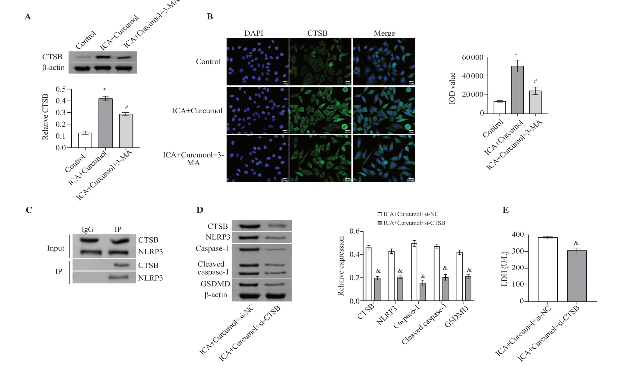
CTSB expression was significantly increased when PC3 cells were treated with ICA plus curcumol.Conversely,treatment with 3-MA significantly reduced CTSB expression in PC3 cells (Figure 5A-B).The CTSB was found to be associated with NLRP3 through a Co-IP assay (Figure 5C).After inhibition of CTSB expression,the levels of CTSB,NLRP3,caspase-1,cleaved-caspase-1,and GSDMD were significantly reduced (Figure 5D).Furthermore,the release of LDH was dramatically decreased in the ICA plus curcumol group(Figure 5E).These findings demonstrate that CTSB regulates ICA and curcumol-induced pyroptosis in PC3 cells through enhanced autophagy.

Figure 4.ICA plus curcumol induces PC3 cell pyroptosis by enhancing autophagy.(A-B) Caspase-1 and GSDMD expressions were observed utilizing the IF(magnification: ×400,scale bar=25 μm).(C) Western blot was used to analyze the NLRP3,caspase-1,cleaved-caspase-1,GSDMD,IL-1β,and IL-18 levels.(D)The detection of LDH concentration.*P<0.05 vs.control.#P<0.05 vs.ICA+curcumol.
4.Discussion
Prostate cancer is a common malignant tumor in males,and its incidence increases each year.This disease poses a serious threat to patients’ health[18].Firstly,the early symptoms of prostate cancer are often obscure and can easily be overlooked or mistaken for other common issues,resulting in delayed treatment and further disease deterioration.Secondly,prostate cancer can spread to surrounding tissues and organs,such as lymph nodes,bones,and lungs,which severely impacts patients’ quality of life and leads to more serious consequences.Furthermore,traditional treatments such as surgery and radiotherapy,although partially effective,can cause significant trauma and side effects to the body[19].Therefore,it is crucial to search for drugs that are more effective and have fewer side effects for prostate cancer treatment.This not only improves survival rates and quality of life for patients,but also provides more treatment options to manage this disease that threatens male health.

Figure 5.Inhibition of CTSB can suppress pyroptosis of PC3 cells induced by ICA and curcumol-enhanced autophagy.(A) Western blot was used to analyze the CTSB expression.(B) IF was used to detect the expression of CTSB (magnification: ×400,scale bar=25 μm).(C) The combination of CTSB and NLRP3 was detected using a Co-IP assay.(D) Western blot was used to analyze the CTSB,NLRP3,caspase-1,cleaved caspase-1,and GSDMD levels.(E) The detection of LDH concentration.*P<0.05 vs.control,#P<0.05 vs.ICA+curcumol,&P<0.05 vs.ICA+Curcumol+si-NC.
In recent years,there has been an increasing amount of research on natural anticancer drugs,and the toxicity and activity of these drugs cannot be studied without preclinicalin vitroresearch[20,21].Our study focused on the effects of ICA plus curcumol on PC3 cells.We found that ICA plus curcumol inhibited the growth of PC3 cells and enhanced autophagy through the mTOR pathway.In addition,we discovered that ICA plus curcumol induced pyroptosis by promoting autophagy,and this process was regulated by CTSB.
Previous studies have shown that natural products can activate the autophagy of prostatic cells,thereby inhibiting cell proliferation and relieving CRPC[22].ICA has been reported to inhibit the proliferation of various cancer cells,including breast cancer cells[23],lung adenocarcinoma cells[24],and lung cancer cells[7].However,the effects of ICA plus curcumol on PC3 cells have not been investigated.Since treatments for CRPC have mainly focused on the androgen receptor signal axis,it is more meaningful to study AR-negative CRPC cell PC3[25,26].Our experiments demonstrated that ICA plus curcumol effectively inhibited PC3 cell proliferation.Furthermore,colony formation experiments verified the inhibitory effects of ICA plus curcumol on PC3 cell proliferation,which were consistent with the effects of natural products[27].
LC3 is a crucial RNA-binding protein involved in the rapid degradation of mRNA during autophagy[28].In our study,the LC3 protein was also used as an important marker to assess the level of autophagy.The combination of drug ingredients and ICA enhanced mitochondrial autophagy[29] and ICA was found to regulate cell autophagy[30].Nevertheless,the effect of ICA plus curcumol on autophagy in PC3 cells has not yet been reported.In our study,after treatment with ICA plus curcumol,the levels of LC3 and beclin-1 were increased significantly,while the level of p62 was markedly decreased.These results were consistent with the previous literature[31] and confirmed our hypothesis that ICA plus curcumol effectively enhances autophagy in PC3 cells.
The mTOR pathway has been extensively studied in terms of its relationship with autophagy and its underlying mechanisms.The regulation of the PI3K/AKT/mTOR signaling pathway has been found to affect the proliferation,apoptosis,and autophagy of prostate cancer cells[32].Autophagy induction has been shown to inhibit the metastasis of prostate cancer through the ROS/AMPK/mTOR pathway[33].In addition,the mTOR-Foxo3a pathway has been found to induce autophagy-dependent apoptosis[34].Our study investigated the role of the mTOR pathway in ICA plus curcumolinduced autophagy.We found that ICA plus curcumol treatment resulted in increased levels of LC3-Ⅱ/LC3-Ⅰand beclin-1,as well as decreased levels of p62 and mTOR.These changes were reversed by overexpression of mTOR.Furthermore,the addition of 3-MA,an autophagy inhibitor,decreased the levels of autophagy and increased proliferation of PC3 cells.These findings suggest that ICA plus curcumol regulates the mTOR pathway to affect autophagy in PC3 cells.
Previous studies have shown that ICA can inhibit NLRP3 inflammasome-mediated pyroptosis in chondrocytes[35].Additionally,the use of 3-MA can suppress the activation of NLRP3 inflammasome and inhibit cell pyroptosis in HK-2 cells[16].Therefore,we investigated whether ICA plus curcumol could induce pyroptosis in PC3 cellsviaenhancing autophagy.Our previous research demonstrated the ability of ICA plus curcumol to initiate autophagy.CTSB,a component of the lysosomal cathepsin family,plays a role in both autophagy and apoptosis[36].The promotion of autophagy can release CTSB,further activating NLRP3 inflammasomes and causing cell pyroptosis[16].Our subsequent investigation indicated a noticeable increase in CTSB expression after treatment with ICA plus curcumol,which was then noticeably reduced by the application of 3-MA.Our study established that CTSB expression correlated with autophagy.In addition,activating CTSB in cells could elicit alternating incidents of pyroptosis and autophagy[37].The cytosolic CTSB expression and LDH release,as well as the NLRP3 inflammasome activation and pyroptosis,were related to autophagy[38].Our further investigation determined the role of CTSB in regulating autophagy and pyroptosis and revealed a significant decrease in pyroptosis-related proteins and markedly reduced LDH release after suppressing CTSB expression compared to treatment with ICA plus curcumol.Therefore,our results confirmed that ICA plus curcumol induces PC3 cell pyroptosis by enhancing autophagy,and CTSB may participate in regulating this process.
The correlation between pyroptosis and prostate cancer has been stated by some researchers[14].It is necessary to conduct further investigations to determine whether ICA plus curcumol affects prostate cancer treatment through its influence on pyroptosis.The NLRP3/Caspase-1/GSDMD pathway has been implicated in inducing pyroptosis in various malignancies,including triplenegative breast cancer[39],endometrial cancer[40],and abdominal aortic aneurysm[41].Our analysis has delved into the proteins that are associated with this pathway.Furthermore,the mechanisms of pyroptosis have been reported in the relevant literature.During the assembly of the NLRP3 inflammasome,caspase-1 is cleaved and activated,leading to the clearance of GSDMD and the formation of membrane pores,thus inducing pyroptosis[42].Additionally,the activation of the NLRP3 inflammasome stimulated the secretion of pro-inflammatory cytokine IL-1β/18 and triggered pyroptosis[43].Our findings align with these previous studies.Treatment with ICA plus curcumol significantly increased the expression levels of caspase-1,GSDMD,NLRP3,IL-1β,IL-18,LDH concentration,as well as pyroptosis in PC3 cells.The addition of 3-MA markedly decreased the levels of these proteins in the presence of ICA and curcumol.These results indicate that ICA plus curcumol induces pyroptosis in PC3 cells by enhancing autophagy.
Despite the promising results obtained in this study,certain limitations need to be acknowledged.Firstly,although ICA plus curcumol was found to enhance autophagy through the mTOR pathway and induce pyroptosis mediated by CTSB,the exact molecular mechanisms underlying these effects were not fully elucidated.Further research is needed to explore the specific signaling pathways and downstream targets involved in these observed effects,which will provide a more comprehensive understanding of the therapeutic potential of ICA plus curcumol for the treatment of prostate cancer.Additionally,this study primarily focused on the cellular and molecular changes induced by ICA plus curcumol treatment.Further investigations using animal models or clinical trials are needed to evaluate the efficacy and safety of ICA plus curcumol as a potential therapeutic agent for prostate cancerin vivoor in humans.Lastly,although this study examined the effects of ICA plus curcumol on autophagy and pyroptosis,other cellular processes and pathways involved in prostate cancer progression and treatment response were not investigated.Future studies should investigate the effects of ICA plus curcumol on other relevant pathways,such as apoptosis,cell cycle regulation,or angiogenesis,to provide a more comprehensive understanding of its overall therapeutic effects.
In conclusion,our findings indicate that the combination of ICA and curcumol has the potential to enhance autophagyviathe mTOR pathway and induce pyroptosis in prostate cancer cells,specificallyviaCTSB mediation.These results provide preliminary evidence to support further investigation on the therapeutic potential of ICA plus curcumol for treating CRPC.
Conflict of interest statement
The authors declare that they have no conflict of interest.
Funding
This study was supported by Natural Science Foundation of Hunan Province (No.2023JJ40511),Excellent Youth Project of Scientific Research Program of Hunan Education Department (No.22B0370),Project of Traditional Chinese Medicine Administration of Hunan Province (No.B2023034),Science and Technology Development Foundation of Beijing Hospital of Traditional Chinese Medicine Affiliated to Capital Medical University (No.LYYB202214),and Hunan Provincial Hygiene and Health Commission Health Research Project (No.W20243165).
Data availability statement
The data supporting the findings of this study are available from the corresponding authors upon request.
Authors’contributions
WS supervised,conceived and designed the study;XYW and WJX searched the literature.XYW,WJX,BNL,and TSS conducted the experiments and acquired the data.All authors contributed to data and statistical analyses.XYW and WJX prepared,WS edited,and all authors reviewed the manuscript.All authors have accepted responsibility for the entire content of this manuscript and approved its publication.
 Asian Pacific Journal of Tropical Biomedicine2024年2期
- Asian Pacific Journal of Tropical Biomedicine的其它文章
- Catalpa bignonioides extract improves exercise performance through regulation of growth and metabolism in skeletal muscles
- Hydrangea serrata extract exerts tumor inhibitory activity against hepatocellular carcinoma HepG2 cells via inducing p27/CDK2-mediated cell cycle arrest and apoptosis
- Benzydamine hydrochloride ameliorates ethanol-induced inflammation in RAW 264.7 macrophages by stabilizing redox homeostasis
- NUDT5 promotes the growth,metastasis,and Warburg effect of IDH wild-type glioblastoma multiforme cells by upregulating TRIM47
