Electroacupuncture alleviates ciliary muscle cell apoptosis in lens-induced myopic guinea pigs through inhibiting the mitochondrial signaling pathway
Zhao-Hui Yang, Jia-Wen Hao, Jin-Peng Liu, Bo Bao, Tu-Ling Li, Qiu-Xin Wu,Ming-Guang He, Hong-Sheng Bi,, Da-Dong Guo
1Shandong University of Traditional Chinese Medicine, Jinan 250014, Shandong Province, China
2Affiliated Eye Hospital of Shandong University of Traditional Chinese Medicine, Shandong Provincial Clinical Medical Research Center of Optometry and Adolescent Low Vision Prevention and Control, Jinan 250002, Shandong Province, China
3State Key Laboratory of Ophthalmology, Zhongshan Ophthalmic Center, Sun Yat-sen University, Guangzhou 510060, Guangdong Province, China
4Shandong Provincial Key Laboratory of Integrated Traditional Chinese and Western Medicine for Prevention and Therapy of Ocular Diseases, Shandong Academy of Eye Disease Prevention and Therapy, Medical College of Optometry and Ophthalmology, Shandong University of Traditional Chinese Medicine, Jinan 250002, Shandong Province, China
Abstract
● KEYWORDS: myopia; electroacupuncture; ciliary muscle;apoptosis; mitochondrial signaling pathway; guinea pigs
INTRODUCTION
Myopia is one of the common diseases affecting eye health.It is estimated that half of the world’s population will suffer from myopia by 2050[1].The prevalence of myopia among young people in East Asia is as high as 80%to 90%, and it has become the leading cause of blindness[2].With China being the most populous country in the world, the prevalence of myopia is of high attention and importance[3].As the investigation continues, researchers have found that myopia is related to environmental and genetic factors[4].Among environmental factors, the high intensity of close work and less duration of outdoor exercise seem to be highly correlated with the occurrence and development of myopia[5-6].Once high myopia is diagnosed, there will be a genetic tendency[7].In addition, vision problems can seriously affect people’s physical health leading to a reduced sense of well-being in life.Therefore, it is crucial to investigate the pathogenesis of myopia and control its development of myopia.
Currently, the pathogenesis of myopia is still unclear.Some studies have found that the regulation of the ciliary muscle may play an important role in the development of myopia[8-9].The ciliary muscle is composed of three types of muscle fibers,including longitudinal, radial, and circular fiber bundles[10].The role of the circular portion is to increase the thickness of the lens, while the longitudinal and radial portions can inhibit this effect.However, the change in ciliary muscle thickness of myopia is controversial.Baileyet al[11]found that thickened ciliary muscle leads to poor contractile responses and thus induces accommodative dysfunction in an early stage, indicating that the thickened ciliary muscle may affect its traction force on the lens[9,12].However, a recent trial found that emmetropic eyes had thicker anterior and thinner posterior ciliary muscles than myopic eyes.
Guinea pigs are the best model animals for studying myopia because of their similar longitudinal muscle fiber tissues and the strong power of the eye’s lens to humans[13].Apoptosis refers to the process of cell death controlled by genes to maintain the stability of the internal environment.Apoptosis is initiated by internal or external stimuli and caused by two different pathways: internal pathway-mitochondrial mediated and external pathway-death receptor[14].Mitochondrial division and fusion are involved in the internal pathway of cell apoptosis[15].Mitochondrial fission protein dynaminrelated protein 1 (DRP1) is a key mediator of mitochondrial fission.DRP1 connected with mitochondrial outer membrane protein (FIS1) and mitochondrial fission factor (MFF) is recruited to the mitochondrial fission site.Then DRP1 directly participates in cell apoptosis by controlling the permeability of the mitochondrial outer membrane[16-17].It was reported that the retinal endothelial cells grown in 30 mmol/L high glucose (HG) showed overexpression of DRP1, and reducing the expression of DRP1 can prevent HG-induced apoptosisin vitro[18].However, the effect of myopia on DRP1 in the ciliary muscle is still unclear.
Electroacupuncture (EA) is a technical skill combining acupuncture and electrical pulse equipment that is widely used in clinical treatments.Studies have shown that acupuncture can relax muscle tensions caused by various diseases, such as ischemic stroke[19]and spastic cerebral palsy[20].It was found that acupuncture can up-regulate the expression of antiapoptotic gene B-cell leukemia/lymphoma (BCL-2), downregulate the expression of apoptotic gene BCL2-associated X protein (BAX), and reduce granulosa cell apoptosis[21].The therapeutic effect of acupuncture on myopia has also been reported[22].Nevertheless, the underlying mechanism is still unclear.
To explore the effect of EA on myopia and to elucidate the underlying mechanism, we established an experimental myopic model in guinea pigs, isolated the ciliary muscle tissues from the guinea pigs after different treatments to measure the changes in DRP1, cleaved cysteine aspartate protease 3 (Caspase3/P17/P19), apoptotic protease activator 1(APAF1), and the adenosine triphosphate (ATP) contents.Based on the relevant experiments, we explored the role of the mitochondrial apoptosis pathway mediated by DRP1 in the ciliary muscle of guinea pigs with lens-induced myopia (LIM),investigated the effect of EA on the development of myopia,clarified whether EA can improve the function of ciliary muscles to inhibit the development of myopia by affecting the mitochondrial apoptotic signaling pathway.Our results will provide new insights into the treatment of myopia with EA.
MATERIALS AND METHODS
Ethical ApprovalThe present study was approved by the Experimental Animal Ethics Review Committee of Affiliated Hospital of Shandong University of Traditional Chinese Medicine (Approval code: AWE-2022-055) and was conducted according to the Association for Research in Ophthalmology Statement for the Use of Animals in Ophthalmic and Vision Research (ARVO).
AnimalsIn this study, 2-week-old healthy guinea pigs (Cavia porcellus, male, 110-130 g, the English short hair stock) were purchased from Jinan Jinfeng Experimental Animal Company(Jinan, China).The refraction and axial length (AL) of guinea pigs were measured before enrollment to exclude spontaneous myopia, cataract, and other eye diseases.Subsequently, the eligible guinea pigs were randomly divided into normal control(NC) group, LIM group, LIM+EA group, and LIM+SHAM group.All animals were housed at a temperature of 24℃-28℃in a 12-hour light/12-hour dark circulation environment (8:30 to 20:30) and were given fresh vegetables twice a day and purified water adequately.
Experimental Myopic Induction and Electroacupuncture TreatmentThe guinea pigs in the NC group did not receive any intervention, whereas the animals from LIM,LIM + SHAM, and LIM+ EA groups were covered with a-6.0 D lens on the right eyes while the left eyes received no intervention as self-control[23].Animals in the LIM + EA group received electrical pulse stimulation at bilateral acupoints (i.e.,LI4, EX-HN5)[23-24], while those in the LIM + SHAM group were treated with sham points which were close to the lateral side of the “degenerated tail” (Figure 1).The acupuncture needle was penetrated approximately 3 mm and was connected to the electrical stimulator for EA.EA intervention was conducted for 30min between 8:30a.m.and 9:30a.m.every day.The parameters were set as continuous wave electrical pulses (0.1ms duration), frequency of 2 Hz, and intensity of 2 mA[25].The guinea pigs in each group received 1, 2, or 4wk treatments.During experiments, once the lenses fell off, they would be wiped with 75% alcohol and stuck immediately to ensure consistent visual qualities of all guinea pigs in the study.
Detection of Ocular ParametersThe refractions of guinea pigs in each group were measured at 1, 2, and 4wk.Before measurements, guinea pigs in each group were acclimated in the dark room for 30min, and cyclopentolate hydrochloride eye drops (Alcon, Geneva, Switzerland) were dropped into the conjunctival sac three times at an interval of 5min[26].After 35min of eye dropping, a streak retinoscope (Suzhou 66 Vision Technology Co., Ltd, China) for refractive examination was used to determine the refraction parameters.The working distance was set to 50 cm and the final refraction was determined by the spherical column method.Two eyes were measured at least three times, and the average value was the final experimental result.The AL was determined by A-scan ultrasonography (CineScan, Quantel Medical, France).We dropped proparacaine hydrochloride (Alcon Geneva,Switzerland) in the eyes before the measurements.The A-scan parameter was as follows: 1557.5 m/s in the anterior chamber, 1723.3 m/s in the lens, and 1540 m/s in the vitreous chamber[27].The detector was wiped with 75% alcohol cotton and then stuck vertically and gently to the apex of the guinea pig cornea for 10 consecutive measurements.After removing the largest and smallest error data, the average data were regarded as the final result.The entire experiment process was completed by the eligible optometrist.
Histopathological Staining and TUNEL StainingAfter 4-week treatments, the right eyes of the guinea pigs in each group were isolated for hematoxylin-eosin (H&E) staining.First, the right eye was washed in sterile cold phosphate buffer saline (PBS) and placed in 4% polyformaldehyde for 24h, then the eyeballs were dehydrated, embedded with paraffin, and sectioned into 8 mm to observe the changes in the structure and physiological morphology of the ciliary muscle[28].Regarding the TUNEL assay, the paraffin sections were routinely dewaxed, washed with PBS three times, incubated with protease K at 37℃ for 22min, and treated with membrane breaking solution.Next, the TUNEL kit was used to perform the TUNEL assay according to the instructions.The sections were re-stained with DAPI solution, and the apoptotic cells in the ciliary muscle were observed with the fluorescence microscope (Axio Vert.A1, Zeiss, Germany).

Figure 1 Experimental flow chart NC: Normal control; LIM: Lensinduced myopia; SHAM: Sham acupoint; EA: Electroacupuncture;qPCR: Real-time quantitative polymerase chain reaction; ATP:Adenosine triphosphate; H&E: Hematoxylin and eosin staining; TUNEL:Terminal-deoxynucleoitidyl transferase mediated nick end labeling.
Measurements of Adenosine TriphosphateWe measured the ATP contents in the ciliary muscle of the right eyes in each group.We set the ciliary muscle into a 2 mL grinding tube and added cold distilled water at a ratio of 1:9, and then sonicated on the ice with a sonicator.Subsequently, the solutions were centrifuged at 5000 rpm for 10min at 4℃ to collect the supernatants, and the BCA protein assay kit (Beyotime Biotechnology, Shanghai, China) was used to determine the protein concentration of the supernatants with 96-well plates(Nest, Wuxi, China).Further, the ATP contents were measured with an ATP content kit (Nanjing Jiancheng Bioengineering Institute, Nanjing, China) according to the instructions.Finally, the absorption values of the samples at 636 nm were determined by an ultraviolet spectrophotometer (UNICO,Shanghai, China).
Quantification of mRNA LevelCiliary muscles after 1-,2-, and 4-week treatments were extracted from 1 mm behind the corneal limbus of the eye.Then the samples were frozen in liquid nitrogen immediately, and stored at -80℃.Before measurements, the ciliary muscle was weighed and the total RNAs were extracted using a modified tissue/cellular RNA rapid extraction kit (SparkJade Science Co., Ltd., Jinan, China).Subsequently, the cDNA of each sample was synthesized using the HiScript II Q RT SuperMix for quantitative polymerase chain reaction (qPCR) (+gDNA wiper) (Vazyme Biotech Co.,Ltd., Nanjing, China).The expression of DRP1, Caspase3, and APAF1 was determined by a Light Cycler 480 II Instrument(Roche, Switzerland) and was corrected by glyceraldehyde 3-phosphate dehydrogenase (GAPDH).The mRNA relative expression of DRP1, Caspase3, and APAF1 in ciliary muscle was quantitatively analyzed by the 2-△△CTmethod.The primer sequences are listed in Table 1.
Western Blot AnalysisTo determine the change of the molecules related to the mitochondrial signaling pathway, we used Western blot analysis to define the DRP1, Caspase3, and APAF1 protein levels of the ciliary muscle in each group.The ciliary muscle was isolated, then added RIPA buffer cracking solution containing PMSF at a ratio of 10 mg:100 μL, ground with an electric homogenizer.Subsequently, the samples were centrifuged at 5000 rpm for 6min (NEST, Wuxi, China).The supernatant was used to measure the protein concentration with the BCA protein assay kit (Beyotime Biotechnology, Shanghai,China).In the present study, 6% sodium dodecyl sulfatepolyacrylamide gel electrophoresis (SDS-PAGE) was used to measure APAF1, 12% SDS-PAGE was used to measure DRP1 and Caspase3/P17/P19.DRP1 (dilution 1:1800; Wanlei Bio, Shenyang, China), Caspase3/P17/P19 (dilution 1:800;Proteintech, Wuhan, China), and APAF1 (dilution 1:800;Wanlei Bio) primary antibodies were incubated with the polyvinylidene difluoride (PVDF) membrane at 4℃ overnight.Then the PVDF membrane were incubated with secondary antibodies against DRP1 (dilution 1:1800), Caspase3/P17/P19(dilution 1:800), and APAF1 (dilution 1:800) for 1h at 4°C.Finally, the FUSION-FX7 imaging system (Vilber Lourmat,Marne-la-Vallée, France) was used to visualize the membrane with DAB (Sigma-Aldrich, St.Louis, MO, USA) and the results were quantified with fusion CAPT software (Vilber Lourmat, Marne-la-Vallée, France).
Statistical AnalysisSPSS Statistics 23.0 (64-bit IBM,Chicago, IL, USA) software was used for significant analysis.The pairedt-test was performed for the eye parameters of different intervention periods.In this study, one-way ANOVA was employed for relative mRNA and protein expressions,respectively.In addition, one-wayANOVA was used to analyze the ATP contents.P<0.05 was considered a significant difference.
RESULTS
Changes in Refraction and Axial LengthThe refraction and AL of all guinea pigs were measured before enrollment.There was no significant difference between any of the two groups.However, after myopia induction, the refraction in the LIM group was significantly decreased compared with those of the NC group, whereas AL in the LIM group was significantly increased compared with those of the NC group at 1wk (LIMvsNC, refraction:P<0.01; Figure 2A; AL:P<0.01;Figure 2B), 2wk (LIMvsNC, refraction:P<0.001; Figure 2A;AL:P<0.001; Figure 2B) and 4wk (LIMvsNC, refraction:P<0.001; Figure 2A; AL:P<0.001; Figure 2B).By contrast,the refraction in the LIM+EA group was increased compared with those of the LIM group after EA intervention, and AL in the LIM+EA group was decreased compared with those of the LIM group after EA intervention at 1wk (LIM+EAvsLIM,refraction:P<0.05; Figure 2A; AL:P<0.01; Figure 2B), 2wk(LIM+EAvsLIM, refraction:P<0.01; Figure 2A; AL:P<0.01;Figure 2B), and 4wk (LIM+EAvsLIM, refraction:P<0.001;Figure 2A; AL:P<0.001; Figure 2B), respectively.

Table 1 Primer sequence of the target genes
Electroacupuncture Alleviates Apoptosis of Ciliary Muscle CellsTo explore the effect of EA on the development of myopia in ciliary muscles, we performed H&E staining after a 4wk treatment in each group (Figure 3).The results showed that the ciliary muscle fibers of the guinea pigs in the NC group were arranged orderly with normal structures, whereas those in the LIM group and LIM+SHAM group guinea pigs were significantly disordered, while the fibers in the LIM+EA group were improved compared with those of the LIM group.To study the change of apoptosis in ciliary muscles after different treatments for 4wk, we conducted TUNEL staining(Figure 4).The result showed that apoptotic cells presented green fluorescence, and the nuclei presented blue fluorescence.The green fluorescent intensity increased significantly in both LIM and LIM+SHAM groups; however, the green fluorescent intensity reduced significantly after EA treatment.
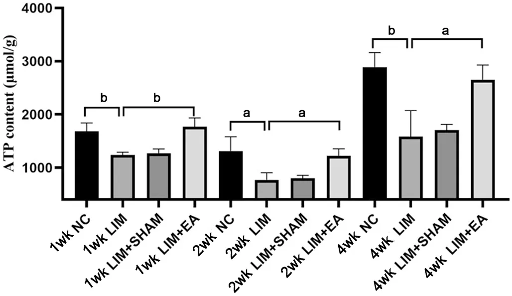
Electroacupuncture Elevates ATP Content in Ciliary Muscle CellsTo determine experimental myopia on ATP content, we measured the ATP contents in ciliary muscles in each group after myopia induction for 1, 2, and 4wk.As shown in Figure 5, the ATP contents in both LIM and LIM+SHAM guinea pigs decreased significantly after myopia induction for 1 (LIMvsNC,P<0.01), 2 (LIMvsNC,P<0.05), and 4wk(LIMvsNC,P<0.01), respectively.However, the ATP contents in LIM+EA guinea pigs increased after EA treatment for 1(LIM+EAvsLIM,P<0.01), 2 (LIM+EAvsLIM,P<0.05), and 4wk (LIM+EAvsLIM,P<0.05), respectively (Figure 5).

Figure 2 Changes in refraction and AL after different treatments The refraction (A) and AL (B) in the right eyes were compared with those in the NC, LIM, LIM+SHAM, and LIM+EA groups (mean±SD) at 1, 2, and 4wk.aP<0.05, bP<0.01, and cP<0.001.AL: Axial length; NC: Normal control;LIM: Lens-induced myopia; SHAM: Sham acupoint; EA: Electroacupuncture.

Figure 3 Histological morphology of ciliary muscles after 4wk different treatments NC: Normal control; LIM: Lens-induced myopia; SHAM:Sham acupoint; EA: Electroacupuncture.
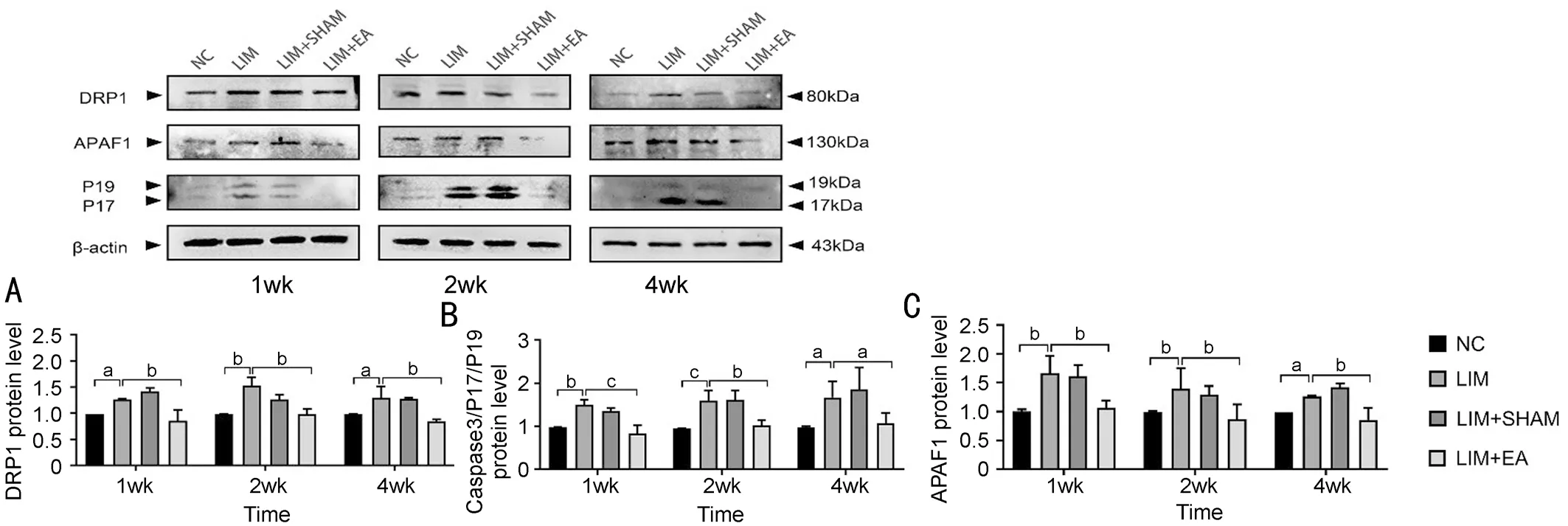
Electroacupuncture Ameliorates Apoptosis of Ciliary Muscle CellsTo investigateDRP1,Caspase3, andAPAF1gene expression in the ciliary muscles in each group, we measured theDRP1,Caspase3, andAPAF1expression at mRNA levels after 1, 2, and 4wk treatments.Results of qPCR showed thatDRP1,Caspase3, andAPAF1gene expression in LIM and LIM+SHAM groups was elevated at 1 (DRP1: LIMvsNC,P<0.05; Caspase3: LIMvsNC,P<0.001; APAF1: LIMvsNC,P<0.01), 2 (DRP1: LIMvsNC,P<0.05; Caspase3: LIMvsNC,P<0.01; APAF1: LIMvsNC,P<0.05), and 4wk (DRP1: LIMvsNC,P<0.01; Caspase3: LIMvsNC,P<0.05; APAF1: LIMvsNC,P<0.05), respectively; whereas those in LIM+EA group decreased at 1 (DRP1: LIM+EAvsLIM,P<0.05; Caspase3:LIM+EAvsLIM,P<0.01; APAF1: LIM+EAvsLIM,P<0.01),2 (DRP1: LIM+EAvsLIM,P<0.001; Caspase3: LIM+EAvsLIM,P<0.01; APAF1: LIM+EAvsLIM,P<0.01), and 4wk(DRP1: LIM+EAvsLIM,P<0.01; Caspase3: LIM+EAvsLIM,P<0.01; APAF1: LIM+EAvsLIM,P<0.01), respectively (Figure 6).The trend of DRP1, Caspase3/P17/P19, and APAF1 protein expression is consistent with that of DRP1, Caspase3/P17/P19,and APAF1 gene expression.That is to say, myopia induction could elevate the expression of DRP1, Caspase3/P17/P19,and APAF1 (P<0.05); however, after EA treatment, the DRP1, Caspase3/P17/P19, and APAF1 expression significantly decreased (P<0.05).Meanwhile, compared with the levels of the LIM group, the DRP1, Caspase3/P17/P19, and APAF1 levels in the LIM+SHAM group had no significant change (Figure 7).

Figure 5 Measurement of the ATP contents in ciliary muscles in the NC, LIM, LIM+SHAM, and LIM+EA groups after 1, 2, and 4wk treatments Compared with the NC group, the ATP contents in the LIM group reduced significantly, whereas EA treatment could elevate the ATP content in LIM+EA guinea pigs.aP<0.05, bP<0.01.NC: Normal control; LIM: Lens-induced myopia; SHAM: Sham acupoint; EA: Electroacupuncture; ATP:Adenosine triphosphate.

Figure 6 The gene levels of DRP1, Caspase3, and APAF1 in the ciliary muscle of guinea pigs were investigated by qPCR at 1, 2, and 4wk in the NC, LIM, LIM+SHAM, and LIM+EA groups aP<0.05, bP<0.01, and cP<0.001.DRP1: Dynamin-related protein 1; APAF1: Apoptotic protease activator 1; NC: Normal control; LIM: Lens-induced myopia; SHAM: Sham acupoint; EA: Electroacupuncture.

Figure 7 Western blot analysis of DRP1, Caspase3, and APAF1 Relative levels of the proteins DRP1 (A), Caspase3 (B), and APAF1 (C) in guinea pigs in the NC, LIM, LIM+SHAM, and LIM+EA groups at 1, 2, and 4wk.aP<0.05, bP<0.01, and cP<0.001.DRP1: Dynamin-related protein 1; APAF1:Apoptotic protease activator 1; NC: Normal control; LIM: Lens-induced myopia; SHAM: Sham acupoint; EA: Electroacupuncture.
DISCUSSION
In this study, we found that the levels of mitochondrial apoptotic signaling pathway-related molecules elevated, the ATP levels were significantly reduced, and mitochondrial function was impaired in ciliary muscle cells in experimental myopic guinea pigs.However, EA can significantly reduce the levels of mitochondrial apoptotic signaling pathway-related molecules and increase ATP levels, thereby inhibiting the activation of the mitochondrial apoptotic signaling pathway and delaying myopia progression.We demonstrated that EA effectively alleviated the expression of DRP1, APAF1, and Caspase3/P17/P19 and increased ATP levels, indicating that EA can improve some mitochondrial functions, ameliorate cell apoptosis in the ciliary muscle tissues, restore cell physiological function, and ultimately delay the development of myopia by enhancing the regulatory role of the ciliary muscles.
To date, the evidence regarding the association between myopia and ciliary muscle morphology remains inconclusive.Previous studies found that children suffering from myopia had thicker ciliary muscles than normal children, and this change may lead to insufficient contraction of the ciliary muscle and explain the regulation lag found in myopic children[29].However,Buckhurstet al[30]found a lack of correlation between ciliary muscle thickness and AL.A recent study indicated that emmetropic eyes had thicker anterior (up to 1.4 mm from the scleral spur) and thinner posterior (1.4 to 4.5 mm from the scleral spur) ciliary muscles than myopic eyes[8].In addition,Puckeret al[31]found that form deprivation-induced myopia(FDM) maintained for 7d in guinea pigs leads to atrophy of ciliary muscle cells.However, the specific mechanism of occurrence is not yet clear.Researchers speculate that myopia caused by form deprivation may be related to the shorter length and thinner ciliary muscle thickness.This contradicts the initial clinical observation that children with myopia have thicker ciliary muscles.Although the current research findings do not provide a clear understanding of the relationship between myopia and ciliary muscle morphology, they demonstrate that abnormal morphology and structure of the ciliary muscle may play a significant role in the progression of myopia.
According to DRP1, research has mainly focused on cardiovascular diseases.DRP1 is considered the primary protein involved in mitochondrial fission in mitochondria[17].Mitochondrial fragmentation and remodeling caused by DRP1 may lead to mitochondrial dysfunction, ultimately resulting in cellular apoptosis[32].Studies have shown that DPR1 plays an important role in smooth muscle remodeling[33], and the ciliary muscle of guinea pigs is mainly composed of longitudinal smooth muscle.Touvieret al[34]found that the overexpression of DRP1 can induce a reorganization of the mitochondrial network and impairs muscle development.Excessive activation of DPR1 can lead to increased permeability of the mitochondrial outer membrane, obstructing mitochondrial function, and then cytochrome-C was released from the mitochondria, and apoptosis was initiated.Previously, Puckeret al[31]found that 7d of visual deprivation inhibited the ciliary muscle growth,leading to axial myopia.Nevertheless, no reports suggest a connection between the ciliary muscle and cell apoptosis.
EA plays an essential role in the treatment of various diseases.Researchers found that acupuncture has become an important way to treat myopia in children[35-36].Acupuncture and moxibustion can alleviate eye hypoxia and are conducive to the recovery of myopia.Ciliary muscle belongs to smooth muscles and has the general properties of muscles.Researchers confirmed that acupuncture can help relieve the fatigue of muscles and improve muscle injury caused by exercise[37].In addition, the ciliary muscles are mainly innervated by dense cholinergic parasympathetic nerves[38].EA stimulation can lead to nerve excitation and change the local blood flow[39].These findings suggest that EA may play an important role in regulating the physiological function of ciliary muscle after myopia.EX-HN5 and LI4 points have been reported frequently to control the development of myopia[40].EX-HN5 point is located around the eye.EA stimulation can dredge the eye meridians and regulate blood flow.Acupuncture at the LI4 point can promote Qi and blood circulation, activate the activities of the temporal and occipital lobes of the brain,and promote the recovery of vision.Therefore, we combined EX-HN5 with LI4 to observe the effect of EA on myopia.Moreover, to validate the efficacy of acupoints, we also added the pseudoacupoint invention as the SHAM group.The SHAM group received non-acupoint stimulation, and the sham acupoint was set at a point that is near the outside of the“degenerated tail” of the gluteus muscle and was far from the traditional meridians.
In this study, we conducted relevant experiments to confirm that EA intervention effectively controls myopia development.We found that the activation of mitochondrial signaling pathway in the ciliary muscles of guinea pigs in the LIM group led to apoptosis of ciliary muscle cells.At the same time, EA treatment significantly inhibited the activation of mitochondrial apoptosis pathway after myopia induction.After 1, 2, and 4wk of myopia induction, the expression of DRP1 significantly increased, and the ATP content significantly decreased.After 1, 2, and 4wk of EA intervention, the expression of DRP1 was significantly reduced compared to the LIM group, while the ATP content was significantly increased.Mitochondria are centrally implicated in tissue injury due to the function of supplying ATP to the tissue, regulating oxidative metabolism during the pathologic process, and contributing to apoptotic cell death[41].DPR1 is a marker molecule for judging mitochondrial dysfunction which participates in mitochondrial fission and can form helical oligomers to wrap and divide the outer membrane of mitochondria.It is confirmed that the activation of DPR1 leads to mitochondrial fragmentation and promotes cell apoptosis[42].The lack of expression of DRP1 leads to a decrease in the release of cytochrome C in the mitochondrion[43].It is reported that EA can regulate the expression of DRP1, promote the production of ATP, and contribute to the recovery of mitochondrial function after polyhedral injury[44].It was found that acupuncture can also improve cellular homeostasis by regulating cytoskeleton remodeling and cell repair according to selecting acupoints such as EX-HN3 and EX-HN5[45].Therefore, we speculated that EA might alleviate the development of myopia by inhibiting the expression of DRP1, promoting ATP production,and improving cell metabolism.
Currently, there have been no reports on myopia leading to apoptosis of ciliary muscle cells.Our study found for the first time that after myopia induction, the expression of Caspase3/P17/P19 and APAF1 increased and after acupuncture treatments, the expression decreased.TUNEL staining showed that the number of apoptotic cells in the LIM+EA group was significantly lower than in the LIM group.APAF1 is the central component of apoptotic bodies and a multidomain protein.After the permeability of the outer membrane of mitochondria increases, APAF1 combines with cytochrome-C released from mitochondria and then activates the effector factors Caspase3,Caspase 6, or Caspase 7 to perform cell death functions[46-47].Cleaved Caspase3 is the active form of Caspase3 and is the main cleaving enzyme that promotes cell apoptosis[48].After activation of Caspase3, the endonuclease is activated, resulting in DNA fragmentation, and destruction of nuclear protein and cytoskeleton, protein cross-linking, and expression of phagocytic ligand[49].Based on the experimental results, we speculated that the activation of Caspase3 and APAF1 in the ciliary muscle in the LIM group leads to cell apoptosis.At the same time, EA can reduce cell apoptosis by decreasing the expression of Caspase3 and APAF1, improve the contraction function of the ciliary muscle, and enhance its regulatory role.Nevertheless, there are still limitations in our current research.It is still unknown whether stimulating EX-HN5 and LI4 acupoints will affect other apoptotic pathways besides mitochondrial apoptotic signaling pathway.The role of other reported related acupoints in delaying myopia development remains to be investigated.In this study, we put forward a new idea to explore the regulatory function of the ciliary muscle and provide direction in treating myopia.
In summary, experimental myopia can lead to the activation of DRP1 and elevate the levels of Caspase3 and APAF1.The high level of cleaved Caspase3 and APAF1 will activate the mitochondrial apoptosis pathway, leading to ciliary muscle cell apoptosis and dysfunction of the ciliary muscle.The mitochondrial dysfunction of the ciliary muscle in experimental myopic guinea pigs will further exaggerate myopia.EA can suppress the activation of DRP1, inhibit mitochondrial apoptosis pathway, and delay the development of myopia by increasing cell metabolism and restoring the regulatory function of ciliary muscles.Taken together, our findings show that the mitochondrial apoptosis pathway of the ciliary muscle is activated in experimental myopia and EA intervention can reduce cell apoptosis and restore the physiological function of the ciliary muscle, providing new insights for the treatment of myopia with EA.
ACKNOWLEDGEMENTS
Foundations:Supported by National Key R&D Program of China (No.2021YFC2702103; No.2021YFC2702100);the National Natural Science Foundation of China(No.82104937); the Key R&D Program of Shandong Province(No.2019GSF108252).
Conflicts of Interest: Yang ZH,None;Hao JW,None;Liu JP,None;Bao B,None;Li TL,None;Wu QX,None;He MG,None;Bi HS,None;Guo DD,None.
 International Journal of Ophthalmology2023年12期
- International Journal of Ophthalmology的其它文章
- Dynamic tear meniscus parameters in complete blinking:insights into dry eye assessment
- Effects of diquafosol sodium in povidone iodine-induced dry eye model
- Morroniside ameliorates lipopolysaccharide-induced inflammatory damage in iris pigment epithelial cells through inhibition of TLR4/JAK2/STAT3 pathway
- Role of reactive oxygen species in epithelial-mesenchymal transition and apoptosis of human lens epithelial cells
- De novel heterozygous copy number deletion on 7q31.31-7q31.32 involving TSPAN12 gene with familial exudative vitreoretinopathy in a Chinese family
- A pedigree with retinitis pigmentosa and its concomitant ophthalmic diseases
