Effectiveness of hip upslip correction method on ambulation and back pain in patients with lumbar discopathy:a case report
Osama Alshana
1Physiotherapy Department,Nasser Medical Complex,Khan Younis P9439028,Palestine.
Abstract Background: Low back pain can be caused by a variety of accidents,ailments,or diseases,the most common of which is a back muscle problem or tendon injury.The intensity of pain varies from mild to severe.Lower back pain usually improves with rest,pain medications,and physical therapy (PT).Injections of cortisone and manual treatments such as osteopathic or chiropractic manipulation can help reduce pain and speed up the healing process.Some back injuries and disorders necessitate surgery.Methods:A multidisciplinary team evaluation was applied.The patient received medications for a prolonged period that lasted more than 4 to 6 weeks without improvement.Seven sessions of conservative and physical therapy manipulation were applied over a period of two weeks to correct the lower body mechanics and realign the body posture properly.Results:The neglected adolescent patients responded well to physical therapy sessions.The patient can walk with normal body posture and practice his activities of daily life without obstacles.The right up-slip hip was corrected successfully,and both posterior iliac spine joints were functional after the intervention.The patient expressed his satisfaction with physical therapy treatment.Conclusion:Corrections of body mechanics in patients with discopathy should be taken into consideration during treatment.
Keywords: discopathy;chronic low back pain;manipulation;hip upslip
Introduction
The term “sacroiliac joint dysfunction” is used to describe sacroiliac joint pain (SI joint).The sacroiliac joint's abnormal motion,such as hyper-or hypomobility,or misalignment,are the usual causes.For 15% to 30% of persons with mechanical low back pain,the sacroiliac joint condition is a significant source of discomfort.The dysfunction of the SI joint is different from sacroiliitis.While sacroiliac joint dysfunction is a disorder brought on by inappropriate motion or slight mispositioning of the SI joint,sacroiliitis is unique to an inflammatory process existing in the SI joint,and the pain felt is a direct result of those inflammatory processes.Physicians and physiotherapists frequently fail to recognize the difficult-to-diagnose illness known as sacroiliac joint syndrome which mainly could lead to posture abnormality such as hip upslip [1].
Hip upslip is one form of posture abnormalities which mainly reflected in the walking and back pain.Manipulation for sacroiliac joint and specifically for hip upslip is significantly improved in patients with hip upslip [2].
Chronic low back pain (CLBP),defined as pain that lasts more than three months,is the leading cause of work-related impairment in those under 45 years old [3,4].CLBP is thought to be caused by lumbar degenerative disc degeneration in many cases [5].Lumbar spine disc,herniation is a potentially overlooked differential diagnosis for children and adolescents with low back and leg pain.Relatively common in adults,increasingly degenerative illnesses of the lumbar spine are becoming more prevalent as the population ages,the most common of which is lumbar disc herniation [6,7].
Surgery is considered for those patients with CLBP who do not respond to conservative treatment.According to studies,only about 3% of all patients treated for disc herniation were under the age of 20[8].Kurihara and Kataoka[7] observed that 15% of low back surgery patients in Japan were children or adolescents.
Despite multiple systematic reviews and prospective randomized studies comparing spinal surgical to nonsurgical treatment,the outcomes are still ambiguous [9,10].
Currently,CLBP is treated with a combination of activity adjustment,medication,cognitive therapy,and physical therapy[11-13].
In this study,we describe a patient with lumber discopathy with abnormal posture during normal ambulation to explore its natural course,diagnosis,and physiotherapy session procedure.
Case report
The patient signed informed consent form.The study was approved by Nasser medical complex ethics committee (2022NMC166).
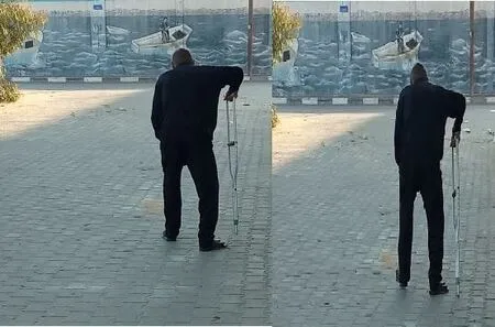
A 61-year-old male presented to the physiotherapy department with a complaint of chronic back pain at the right buttock,and posterior thigh pain.The onset of pain was sudden,there was no reported injury or fall.Even though the symptoms had been present for nine weeks,they were aggravated after daily fishing.The pain was described as a dull ache that spread distally along the posterior aspect of the right leg to the knee,with "numbness" sensations.Moreover,Sitting and coughing increased the severity of pain,but supine reclining and standing helped to relieve it.likewise,the patient reported no bowel or bladder dysfunction.the patient was practicing his daily activities regularly.After sleeping,the patient experienced acute back discomfort.Physician-prescribed medications,such as analgesics and anti-inflammatory drugs,were used to control the pain episodes.These medications' effect was temporary during the day,and the patient stopped working due to his pain.After the physician assessment,the patient was referred to the physiotherapy department after finishing his medication prescription.Examination revealed a healthy,slim,and tall old man in moderate discomfort during standing with abnormal body posture shifting to right side to avoid the pain (Figure 1).
The patient walked into the outpatient physiotherapy hall,unable to straighten his right leg,and stood with right lateral flexion.In addition to that,the patient was utilizing one axillary crutch to stabilize his body during ambulation with his right hand.In forward flexion,his lumbar spine's range of motion was severely limited.On the right,heel walking was impossible.The lower limbs' reflexes and sensory assessments were unremarkable.Straight leg raising was 25 degrees on the right,causing only leg pain,with crossover pain in the left buttock when the right leg was raised.The right hamstring muscle was found to be tight.Regarding trunk range of motion,it was observed severe range of motion restrictions in thoracic and lumber spine regions (trunk flexion 35,extension 5,lateral flexion 5 to left and 0 to right,and rotation was 10 to the right side and 0 to the left side)
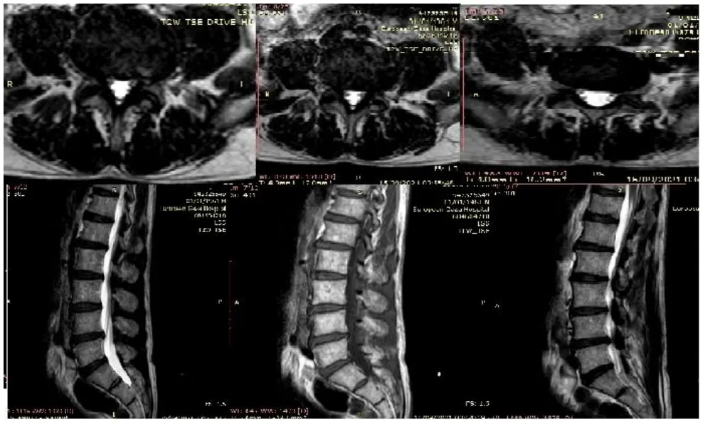
Magnetic resonance imaging findings confirmed a diagnosis of L3-L4 left posterolateral disc bulging impacting the thecal sac and left nerve root based on MRI images acquired in different planes and pulse sequences without IV contrast.Furthermore,L4-L5 disc bulging affects the thecal sac and both nerve roots.Similarly,a posterior disc bulge of L5-S1 affects the thecal sac and both nerve roots,with the bulging being larger on the right.Muscle spasms cause the typical lumbar spine lordosis to be lost (Figure 2).
Physiotherapy treatment sessions at the beginning consisted of transcutaneous electrical nerve stimulation (TENS) (intensity:25W/cm2,duration: 15 mins) applied to the lower back for pain control,soft tissue therapy for the lumbosacral area,and stages of Mackenzie extension exercises in a prone position three sessions per week.The pain exacerbated over the next weeks and the patient was referred for a neurosurgical consultation.One week following the consultation,the pain severity lessened with drugs.After the neurological consultation,the physiotherapy department staff noticed abnormal changes in the mechanical body posture and decided to improve and rectify the detected improper posture.The symmetry of both anterior superior iliac spines (ASIS) was altered,with the left ASIS being higher than the right (Figure 1) (upslip).The left posterior iliac spine (PSIS) was immobile throughout the clinical examination and during normal motion.Hip symmetry is achieved using the upslip hip corrective technique by the following procedure,pulling out the ilium inferior with a thrust,pulling on the leg at the ankle the restoration of both PSIS hypomobility and hip muscle workouts as well.From the side-line,manipulation of the left lower hip joint in an axial distraction and internal rotation position was used to correct the postural changes in the closed chain position.The therapist holds the distal leg with stabilization of the trunk,the therapist performed internal rotation with adduction and then thrust (Figure 3).
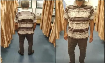
During follow-up,there were significant improvements in the trunk's range of motion.The patient reported being pain-free and unrestrained throughout daily activities at the six-week follow-up(Figure 4).Furthermore,the body trunk's range of motion was unrestricted.Ultimately patient returned to his activity’s daily living and fishing.

Figure 1 Pre-session abnormal body posture

Figure 2 Different MRI images in axial and sagittal views

Figure 3 Upslip-pelvic position and correction

Figure 4 Post-intervention of pelvic upslip correction
Discussion
The low back pain could affect the sacroiliac joint and sacrum nutation during flexion and extension of the hip.clinically,there are clusters of special tests that provide the highest diagnostic accuracy for sacroiliac dysfunction,these tests are sacroiliac (SI) gapping,sacroiliac compression,thigh thrust test (P4),sacral thrust,Gaenslen’s test,these 5 tests were clustered together as a group and at least 3/5 had a positive finding and illustrated a high diagnostic accuracy.The optimal technique for treating sacroiliac joint(SIJ)Syndrome involves a multidisciplinary approach,and it is typical to incorporate modalities like manipulation in the program.Exercise therapy and manual therapy make up the conservative course of treatment.During treatment,it is crucial to identify and deal with the root reasons for dysfunction [14].
Another technique for correcting hip upslip with rotation consists of a prone position,and the patient lies on his back with the therapist standing opposite the side to be manipulated.The patient places his hands behind his head and the therapist then moves the patient passively into side bending to end range toward the target side.The therapist then delivers a quick thrust to the ASIS in a posterior and inferior direction [15].The purpose of this technique is to manipulate an iliac anterior rotation displacement sacroiliac joint dysfunction[15].
For a hypermobile SI joint,the sacroiliac belt is recommended,and it should be worn continuously for six to twelve weeks.The belt should be used in combination with physical exercises and manual therapy in the case of joint dysfunction and muscle imbalance.Once the patient's lumbopelvic musculature is under better control,the belt may be taken off.To help support and stabilize the pelvis,the sacroiliac belt should be placed at the superior aspect of the PSIS[16].
Conclusion
Manipulation should be taken into consideration during the treatment of hip upslip.Furthermore,a differential diagnosis of low back pain in the adolescent age group and inquiry might aid in therapy decision-making.
 Clinical Research Communications2022年4期
- Clinical Research Communications的其它文章
- A study of ultrasonography for the treatment of Qingre Liangxue decoction on blood-heat psoriasis
- Meta-analysis of the efficacy and safety of Shenkang injection combined with Western medicine in the treatment of chronic glomerulonephritis
- Effect of Xingnao-Jianshen granules in treating AIS patients: study protocol for a non-randomized controlled intervention trial
- POEMS syndrome characterized by peripheral neuropathy as the first symptom: a rare case report
- Not just a sponge:novel functions of circRNAs in cholangiocarcinoma
