Retinal ganglion cell protection by hop-flower extract as a novel neuroprotective strategy for glaucoma
Tomoko Hasegawa, Hanako Ohashi Ikeda
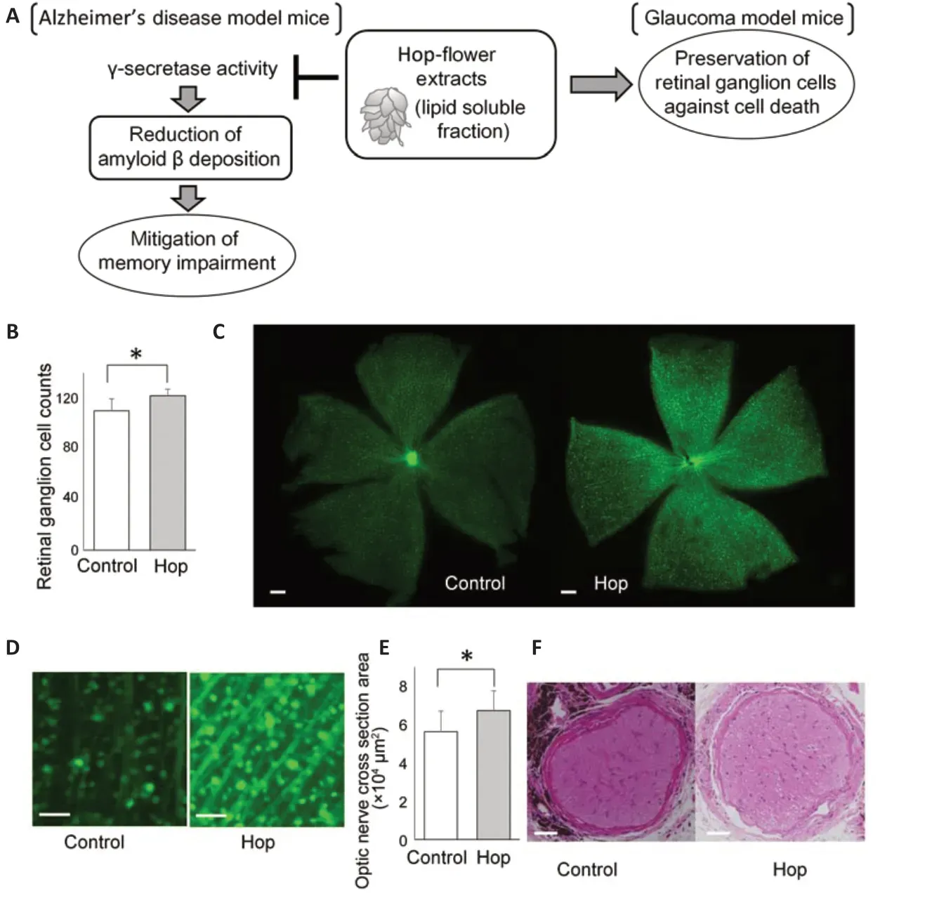
In glaucoma, a leading cause of blindness, retinal ganglion cells are progressively damaged. Intraocular pressure reduction is the only established treatment for glaucoma (Collaborative Normal-Tension Glaucoma Study Group, 1998; Vass et al., 2007). However, in some cases, visual field loss progresses despite sufficiently reduced intraocular pressure (Collaborative Normal-Tension Glaucoma Study Group, 1998; Killer and Pircher, 2018). While intraocular pressure and age are known risk factors for glaucoma (Ernest et al., 2013), the underlying mechanisms of glaucoma progression are not fully understood. Many factors that may influence glaucoma progression, including myopia and blood flow impairment, have been investigated (Marcus et al., 2011; Ernest et al., 2013). A factor that may be related to glaucoma is the concurrent occurrence of Alzheimer’s disease, which is caused by the accumulation of amyloid β (Aβ) in the brain. Glaucomatous retinal changes in patients with Alzheimer’s disease have been reported (Wang and Mao, 2021). Other studies have reported Aβ accumulation in animal models of glaucoma with ocular hypertension (Guo et al., 2007; Ito et al., 2012). Moreover, Aβ induces apoptotic retinal ganglion cell death (Guo et al., 2007). Considering that Aβ induces retinal ganglion cell death, reducing Aβ accumulation and consequently preventing retinal ganglion cell death may be a therapeutic strategy for glaucoma (Figure 1A).
Effects of hop-flower extract in Alzheimer’s disease model mice:A thorough screening of more than 1600 plant extracts showed that hop-flower extract (lipid-soluble fraction) inhibits γ-secretase (Sasaoka et al., 2014). It also inhibits Aβ production in cultured cells and decreases Aβ deposition in the brain of Alzheimer’s disease model mice, in which the C-terminal fragment of the human amyloid precursor protein with the Indiana mutation (V717F) is expressed in neuronal cells (Sasaoka et al., 2014). Hop-flower extract mitigated memory impairment in Alzheimer’s model mice without deleterious side effects during lifelong administration (Sasaoka et al., 2014).
Retinal ganglion cell death prevention by hop-flower extract in glaucoma model mice:Aβ induces retinal ganglion cell death (Guo et al., 2007). Therefore, we investigated whether hop-flower extract prevents retinal ganglion cell death in glaucoma.
We used glutamate-aspartate transporter (GLAST) knockout mice as a glaucoma model (Harada et al., 2007). GLAST knockout mice, which manifest chronic retinal ganglion cell death and optic nerve degeneration without elevated intraocular pressure, have been used as glaucoma models with normal intraocular pressure. Retinal ganglion cells are damaged more severely in GLAST-/-mice than in GLAST+/-mice (Harada et al., 2007).
Administration of hop-flower extract to GLAST-/-mice was started on postnatal day 7. Retinal thickness was examined using spectral domain-optical coherence tomography (Hasegawa et al., 2020). Hop-flower extract was administered intraperitoneally from postnatal day 7 until 2 months of age and orally afterward. The ganglion cell complex, composed of the retinal nerve fiber layer, retinal ganglion cell layer, and inner plexiform layer, was thicker in 8- and 12-week-old GLAST-/-mice that received hop extract than in non-treated mice. The thickness of the outer retinal layer, composed of the outer nuclear layer, photoreceptor myoid zone, photoreceptor ellipsoid zone, and outer segment layer, did not show significant differences between mice that received hop-flower extract and non-treated mice (Hasegawa et al., 2020).
To evaluate the effect of hop-flower extract over a longer period, hop-flower extract was administered orally to GLAST+/-mice starting from 1 month of age until 18 months of age (Hasegawa et al., 2020). Mice that were orally administered the hop-flower extract hadad libitumaccess to water containing 1 g/L of hopflower extract, which resulted in a daily uptake of approximately 0.2 g/kg of the extract. GLAST knockout mice were crossbred with Thy1-CFP mice, whose retinal ganglion cells express cyan fluorescent protein (Feng et al., 2000), to generate GLAST+/-:Thy1-CFP mice. Retinal ganglion cells were imagedin vivousing a scanning laser ophthalmoscope and imaged on retinal flat-mount after enucleation. The retinal ganglion cell numbers counted with the scanning laser ophthalmoscope and the retinal flat-mount were significantly higher in 12-month-old GLAST+/-mice receiving hop-flower extract than in nontreated mice (Hasegawa et al., 2020) (Figure 1B-D). The optic nerve was thicker in 18-month-old GLAST+/-mice that received hop-flower extract than in non-treated mice (Hasegawa et al., 2020) (Figure 1EandF). Immunostaining of the optic nerve of 18-month-old GLAST+/-mice did not show any difference in Aβ deposition between mice that received hop-flower extract and non-treated mice (Hasegawa et al., 2020).

Figure 1|Neuroprotective effects of hop-flower extract in neurodegenerative diseases and suppression of retinal ganglion cell death by hop-flower extract in glaucoma model mice.
These results show that hop-flower extract attenuated retinal ganglion cell death in a mouse model of glaucoma through mechanisms other than Aβ reduction. However, the underlying mechanisms remain to be elucidated.
Conclusion:We showed that hop-flower extract was effective against retinal ganglion cell death in a mouse model of glaucoma. Our findings may provide a potential novel therapeutic strategy for glaucoma.
The present study was supported in part by research grants from the Ministry of Health, Labour and Welfare of Japan (to HOI).
Tomoko Hasegawa, Hanako Ohashi Ikeda*
Department of Ophthalmology and Visual Sciences, Kyoto University Graduate School of Medicine, Kyoto, Japan (Hasegawa T, Ikeda HO)Research Fellow of Japan Society for the Promotion of Science, Tokyo, Japan (Hasegawa T)
*Correspondence to:Hanako Ohashi Ikeda, MD, PhD, hanakoi@kuhp.kyoto-u.ac.jp.
https://orcid.org/0000-0001-9572-8659(Hanako Ohashi Ikeda)
Date of submission:March 30, 2021
Date of decision:May 19, 2021
Date of acceptance:July 6, 2021
Date of web publication:October 29, 2021
https://doi.org/10.4103/1673-5374.327344
How to cite this article:Hasegawa T, Ikeda HO (2022) Retinal ganglion cell protection by hopflower extract as a novel neuroprotective strategy for glaucoma. Neural Regen Res 17(6):1267-1268.
Copyright license agreement:The Copyright License Agreement has been signed by both authors before publication.
Plagiarism check:Checked twice by iThenticate.
Peer review:Externally peer reviewed.
Open access statement:This is an open access journal, and articles are distributed under the terms of the Creative Commons Attribution-NonCommercial-ShareAlike 4.0 License, which allows others to remix, tweak, and build upon the work non-commercially, as long as appropriate credit is given and the new creations are licensed under the identical terms.
- 中国神经再生研究(英文版)的其它文章
- The importance of fasciculation and elongation protein zeta-1 in neural circuit establishment and neurological disorders
- Promoting axon regeneration in the central nervous system by increasing PI3-kinase signaling
- Microglial voltage-gated proton channel Hv1 in spinal cord injury
- Liposome based drug delivery as a potential treatment option for Alzheimer’s disease
- Retinal regeneration requires dynamic Notch signaling
- All roads lead to Rome — a review of the potential mechanisms by which exerkines exhibit neuroprotective effects in Alzheimer’s disease

