
Correlation between NF-κB/TNF-αpathway and atrial fibrillation
2022-08-20 04:53:48NingXuJunJieLengZhuoYaYaoBiTangPinFangKangHengZhang
Journal of Hainan Medical College 2022年12期
Ning Xu, Jun-Jie Leng, Zhuo-Ya Yao, Bi Tang, Pin-Fang Kang, Heng Zhang
Department of Cardiology,The First Affiliated Hospital of Bengbu Medical College,Bengbu 233000,China
Keywords:Atrial fibrillation NF-κB TNF -α Inflammatory mechanism
ABSTRACT Objective: To detect the levels of TNF-α and NF-κB in serum and NF-κB in peripheral blood lymphocytes of patients with atrial fibrillation.To investigate the regularity and significance of its changes in patients with atrial fibrillation. Methods: Patients with atrial fibrillation hospitalized in our hospital from January 2020 to December 2021 were selected,including paroxysmal AF group (PAF group, n=75) and non-paroxysmal AF group (nPAF group, n=60), and healthy subjects were selected as the control group (CON group, n=107).Routine examination and color doppler echocardiography were improved after admission.The expression levels of TNF-α and NF-κB in AF and CON groups were detected by ELISA. The expression of NF-κB in peripheral blood lymphocytes of AF and CON groups was detected by Western blot.To study the relationship between TNF-α, NF-κB and atrial fibrillation. Results: The levels of TNF-α and NF-κB in AF group were higher than those in CON group, and the differences were statistically significant (P<0.05). nPAF group was higher than PAF group, the difference was statistically significant (P < 0.05).The levels of TNF-α, NF-κB, LVEDD and LVEDV in PAF group were higher than those in CON group,but lower than those in nPAF group, the difference was statistically significant (P < 0.05).The levels of LVEF and FS in PAF group were lower than CON group, but higher than nPAF group, with statistical significance (P < 0.05).Pearson analysis showed that TNF-α, NF-κB were positively correlated with LVEDD and LVEDV in atrial fibrillation group compared with normal group (P<0.05), was negatively correlated with LVEF and FS (P<0.01).Conclusion:The levels of TNF-α and NF-κB in serum and NF-κB in peripheral blood lymphocytes were significantly increased in patients with atrial fibrillation. The occurrence and development of atrial fibrillation were related to NF-κB and TNF-α.
1. Introduction
Atrial fibrillation (AF) is the most common arrhythmia, and according to statistics, there are more than 30 million atrial fibrillation patients worldwide [1-3].Despite advances in the treatment of atrial fibrillation, it remains a major cause of stroke,heart failure, and sudden death [4-6].At present, substantial progress has been made in the understanding of the pathophysiology of AF, but the etiology and pathogenic factors of AF, including its underlying mechanism, are not fully understood.Therefore,biomarkers are needed to provide a new experimental basis for the diagnosis and treatment of atrial fibrillation.
Currently, autonomic nerve remodeling, atrial electrical remodeling and atrial structural remodeling are considered to be the main mechanisms of atrial fibrillation [7].Previous studies have confirmed that the incidence and prognosis of AF are related to the level of inflammatory biomarkers in serum [8-9].Studies have shown [10] that inhibition of NF-κB/TNF-αsignaling pathway can improve the susceptibility to atrial fibrillation.
Tumor necrosis factor -α(TNF-α) and nuclear factor -κB(N F-κB) are the most characteristic inflammatory signal transduction pathways.TNF-α is an endogenous inflammatory mediator involved in a variety of cellular processes, including activation of genes involved in inflammatory and immunomodulatory responses,proliferation, growth inhibition and cell death [11-12].NF- к B is a redox sensitive transcription factor, which can inhibit the transcription of myocardial Na+ channels and participate in the regulation of ion channels and transcription factors of AF during oxidative stress [13].
Due to the fact that TNF-α and NF-κB have been widely studied in animal trials of AF, but few clinical studies have been carried out, and the mortality and morbidity of AF are still increasing gradually, new ideas for the diagnosis and treatment of AF with biomarkers are still needed.The purpose of this study was to investigate the relationship between NF-κB and TNF-α levels of major participants in classic inflammatory signaling pathways and the disease of patients with atrial fibrillation, and to provide some new ideas for clinical diagnosis, severity evaluation and possible treatment of atrial fibrillation.
2. Objects and methods
2.1 Subjects
A total of 135 patients hospitalized with atrial fibrillation in our hospital from January 2020 to December 2021 were included in the study.All patients with atrial fibrillation were diagnosed according to the 2014 American Heart Association (AHA)/American College of Cardiology (ACC)/American Heart Rhythm Society (HRS) Atrial fibrillation Guidelines [14] and confirmed by electrocardiogram (ECG) or dynamic electrocardiogram (Holter)monitoring.Inclusion criteria: Clinical data were complete and atrial fibrillation was divided into paroxysmal atrial fibrillation(PAF) and non-paroxysmal atrial fibrillation (nPAF) based on occurrence and duration.Diagnostic criteria: PAF was defined as af lasting less than 7 days and cured on its own or with intervention.Exclusion criteria: patients with hypertrophic cardiomyopathy,severe infection, pacemaker implantation, cardioversion, other types of arrhythmia, various malignant tumors, chronic kidney disease,acute ST-segment elevation myocardial infarction, and pulmonary embolism.135 patients with AF were divided into paroxysmal af and non-paroxysmal af, of which 75 were paroxysmal af and 60 were non-paroxysmal AF. At the same time, 107 patients without a history of AF were selected as the normal control group. This study was approved by the Ethics Committee of our hospital (2019KY023),and all subjects signed informed consent.
2.2 Specimen collection and preparation
After fasting from water for 8 hours, 5ml peripheral venous blood was taken from all subjects and put into heparin anticoagulant test tube, centrifuged at 3000 r/min for 10min, then supernatant was absorbed and stored in refrigerator at -80℃ for later use.After the serum was extracted, PBS with the same dose as the serum was pumped to mix the lower tissues.Add the mixed lower layer of tissue to the same dose of lymphocyte separator (this process requires adherent and slow addition so that the mixed lower layer of tissue is suspended on the lymphocyte separator).Centrifugation at 2000 r/min for 20min, four layers were obtained, from top to bottom,including plasma, lymphocyte, lymphocyte separator and red blood cell.The middle lymphocyte layer was added to the 15ml centrifuge tube, balanced to 10ml with normal saline, and centrifuged at 4000 r/min for 5min. The upper layer of normal saline was removed, and lymphocyte cryo-storage solution was added, then pumped and mixed evenly, and transferred to the cryo-storage tube, which was stored in the refrigerator at -80℃ for future use.
2.3 Serum TNF-α and NF-κB activity were detected
The serum levels of TNF-αand NF-κB were measured using HumanTNF-αand NF-κB (Shanghai feather) after melting.The serum levels of TNF-α and NF-κB were detected using HumanTNF-αand NF-κB (Shanghai yudo) after melting the specimens. The OD values of TNF-α and NF-κB were obtained by ELISA method according to the kit instructions.
2.4 Western Blot was used to detect the expression of NFκB in peripheral blood lymphocytes of patients with atrial fibrillation and normal controls
The protein concentration in peripheral blood of patients with atrial fibrillation and normal control group was measured by BCA method, and 10% SDS-PAGE electrophoresis (80V 120min) was performed.PVDF membrane transfer (200mA 100min); Skim milk was sealed at room temperature for 2 h; Primary antibody (1:1000)was incubated at room temperature for half an hour;4 ℃ refrigerator overnight; Secondary antibody (sheep vs. rabbit 1:8000) was incubated at room temperature for 1 h. The ECL luminescence kit emits light and the gel imaging system captures the image.
2.5 Statistical Treatment
Measurement data were expressed as mean ± standard deviation,and comparison between the two groups was performed by T test.Counting data were expressed in percentage ratio andχ² test was used. The correlation of TNF-αand NF-κB with LVEF, LVEDD,LVEDV and FS was analyzed by Pearson method. The above data were analyzed by SPSS25.0. Graphad PRISm8 software was used to analyze Western Blot data.
3. Results
3.1 Comparison of baseline data between the two groups
There was a significant difference in age between the two groups(P < 0.05), suggesting that age was related to atrial fibrillation. There was no statistical significance in gender, history of hypertension,history of diabetes, blood routine examination, liver and kidney function and other related indexes between the two groups (P>0.05)(Table 1).
3.2 The comparison of TNF-α, NF-κB and the parameters related to cardiac ultrasonic central function between atrial fibrillation group and normal group
Compared with normal control group, the concentrations of TNF-α and NF-κB, LVEDD and LVEDV in atrial fibrillation group were increased, while LVEF and FS were decreased, the differences were statistically significant (P< 0.05) (Table 2).
3.3 Comparison of TNF-α, NF-κB and central function parameters in patients with paroxysmal atrial fibrillation and non-paroxysmal atrial fibrillation
Compared with paroxysmal af group, TNF-αand NF-κB concentration, LVEDD and LVEDV were increased in nonparoxysmal af group, while LVEF and FS were decreased, the difference was statistically significant (P< 0.05) (Table 3).
3.4 Correlation analysis of TNF-α, NF-κB and cardiac ultrasound central function indexes
Pearson correlation analysis showed that TNF-α and NF-κB were positively correlated with LVEDD and LVEDV higher in atrial fibrillation group than in normal group (r=0.163,0.182,0.167,0.144,P< 0.05), were negatively correlated with LVEF and FS (r=-0.733,-0.716, -0.312, -0.314, P< 0.01).

Table 1 Comparison of baseline data between the two groups of subjects
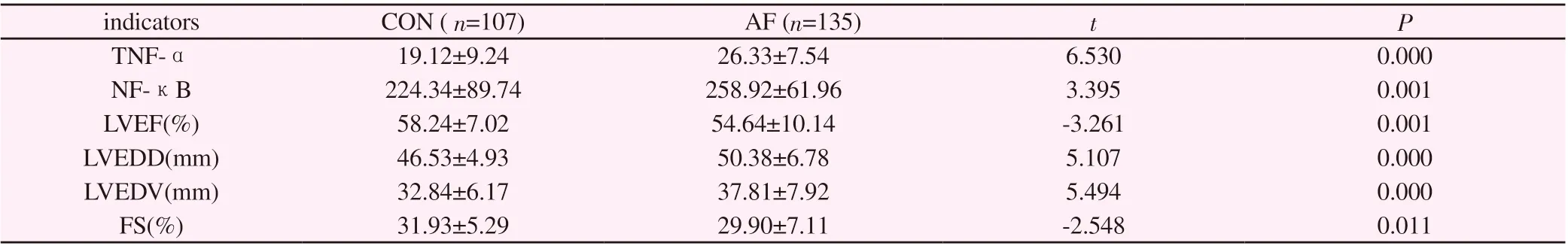
Table 2.Comparison of TNF-α and NF-κB with LVEF, LVEDD, LVEDV and FS in the two groups

Table 3 Comparison of TNF-α and NF-κB with LVEF, LVEDD, LVEDV and FS in patients with different atrial fibrillation types

Table 4.Correlation analysis of TNF-α, NF-κB with LVEF, LVEDD, LVEDV and FS
3.5 Comparison of NF-κB protein expression in peripheral blood lymphocytes of atrial fibrillation group and control group.
The expression of NF-κB protein in PAF group was higher than that in CON group and lower than that in nPAF group, with statistical significance (P < 0.05) (Figure 1).

Figure1 A:NF-κB protein bands in peripheral blood lymphocytes of the 3 groups; B: Compared with CON group, *P < 0.05; Compared with PAF group, #P < 0.05
4. Discussion
Atrial fibrillation is a complex disease involving many factors.The presence of atrial fibrillation increases the mortality of patients with cardiovascular diseases and reduces their quality of life [15].Current studies have shown that structural remodeling and electrical remodeling are the two main mechanisms for the persistence of atrial fibrillation, and atrial fibrosis in structural remodeling is closely related to inflammatory response [16].Previous studies have also found that inflammatory cell infiltration was observed in atrial tissue of patients with atrial fibrillation [17], and inflammatory response was found to accompany the occurrence and development of atrial fibrillation [16,18].Therefore, the occurrence and development of atrial fibrillation involves various mechanisms such as atrial fibrosis and inflammatory response.
Multiple studies [19-21] have shown that the increased incidence of atrial fibrillation is associated with the activation of NF-κB,a member of the transcription factor protein family.Ye et al. [10]suggested that squirrel (5, 7-dihydroxyflavone) can inhibit NFκB/TNF-α signaling and reduce the level of pro-inflammatory cytokines, which may help to improve the susceptibility to atrial fibrillation.It has also been found [22] that colchicine can reduce the susceptibility to atrial fibrillation in rats by inhibiting inflammatory react-mediated atrial fibrosis.In conclusion, the present study hypothesized that down-regulation of NF-κB/TNF-αsignaling pathway and reduction of inflammatory response could improve atrial fibrosis in patients with AF to a certain extent.Therefore,studying the expression of NF-κB and TNF-α in patients with atrial fibrillation may provide a new idea for the diagnosis and treatment of atrial fibrillation.
Nuclear factor-κB(NF-κB) is one of the key factors in regulating gene transcription and is characterized by immediate transcription[23].Previous experiments have also shown [24] that in alloxan induced diabetic rats, the occurrence of AF is related to the activation of NF-κB signaling pathway, resulting in pro-inflammatory state,myocardial hypertrophy and fibrosis.Studies have shown [24,25]that hyperglycemia can cause collagen fiber accumulation and interstitial fibrosis in myocardial cells, thereby promoting structural remodeling, and atrial remodeling in diabetes mellitus is partially attributed to activation of NF-κB/TGF-β signaling pathway.In this study, there was no statistical difference in the prevalence of diabetes between the AF group and the control group, so the influence of cardiac remodeling caused by diabetes on the experimental results was excluded.In addition, the serum NF-κB concentration in patients with AF was higher than that in the control group, and that in patients without nPAF was higher than that in the PAF group,suggesting that NF-κB gradually increases with the increase of the severity of AF.In this study, the concentration of NF-κB protein in peripheral blood lymphocytes was determined, and the trend of WB protein concentration was consistent with the ELISA results.It is suggested that the increase of NF-κB has certain reference value for the existence, occurrence and development of atrial fibrillation.NF-κB, as a key node in the inflammatory pathway, is expected to identify therapeutic targets for atrial fibrillation by inhibiting NF-κB [26].Previous studies [10,27,28] have shown that activated NF-κB enters the nucleus and upregulates transcription of target genes - tumor necrosis factor-α, leading to the occurrence of atrial fibrillation.
TNF-α is a key mediator and regulator of the immune response in mammals under healthy and disease conditions, regulating the development of the immune system, cell survival signaling pathways, cell proliferation and regulating metabolic processes [6].Tumor necrosis factor -αis closely related to the occurrence and maintenance of atrial fibrillation, mainly manifested in the following two aspects [29] :① Tumor necrosis factor -αcan promote atrial fibrosis and atrial dilation, change the expression of connexin Cx43,and lead to heteroconduction;② TNF-αcan also directly change the calcium ion processing in cardiomyocytes, providing a substrate for the occurrence of atrial fibrillation.Therefore, TNF-αcan trigger atrial fibrillation and provide theoretical support for the deterioration of cardiac function caused by atrial fibrillation.In this study, the level of serum TNF-αin patients with atrial fibrillation was higher than that in the normal control group, indicating that the increase of serum TNF-αconcentration is related to the occurrence of atrial fibrillation.Serum levels of TNF-αin PAFand nPAF were measured.It was found that TNF-αexpression was different in the two types of AF, and increased gradually with the increase of the severity of AF, which was statistically significant.These results suggest that the increase of serum TNF-αconcentration is related to the occurrence and development of atrial fibrillation.
In addition, through the analysis of cardiac color doppler ultrasound indexes of subjects in this study, it was found that the quantitative indexes of cardiac fibrosis such as LVEDD and LVEDV in patients with AF were higher than those in the control group, and increased with the severity of AF.In this study, we further investigated the relationship between the expression of NF-κB/TNF-αsignaling pathway and the quantitative indexes and hemodynamics of cardiac fibrosis, and found that serum TNF-αand NF-κB levels were positively correlated with LVEDD and LVEDV in patients with atrial fibrillation, while negatively correlated with LVEF and FS.Therefore,we hypothesized that activation of NF-κB/TNF-αpathway leads to excessive inflammatory response and aggravation of ventricular remodeling, which increases LVEDD, LVEDV and other indicators,while intensification of cardiac remodeling leads to deterioration of cardiac function and decrease of LVEF, FS and other indicators [30].In conclusion, the levels of TNF-α and NF-κB increased in patients with atrial fibrillation and gradually increased with the severity of atrial fibrillation.In the future clinical work, on the one hand, targeted detection of TNF-α and NF-κB levels can be used to evaluate the early diagnosis and severity of atrial fibrillation,which can significantly reduce the occurrence of stroke, heart failure and other complications, and have a positive impact on the prognosis of patients. On the other hand, the inhibition of NF-κB/TNF-α signaling pathway can also provide a new theoretical basis for the development of new drugs and personalized therapy for patients with atrial fibrillation.
Xu Ning: Carried out experiments, sorted out data and wrote papers. Zhang Heng, Kang Pinfang, TANG Bi: Design the experiment and make key corrections to the content of the paper. Yao Zhuoya and Leng Junjie participated in the experimental content and data collection.
 Journal of Hainan Medical College2022年12期
- Journal of Hainan Medical College的其它文章
- Research progress of Cassytha filiformis L
- Research on anti-pancreatic cancer mechanism of Codonopsis codonopsis based on network pharmacology
- Risk factors for infection with multidrug-resistant organisms in diabetic foot ulcer patients: A systematic review and meta-analysis
- Effectiveness of Shengmai Injection on angina pectoris based on realworld propensity score method
- Construction and validation of prognostic model of hepatocellular carcinoma based on epigenetic factors
- Preparation and characterization of hemihydrate calcium sulfate-calcium hydroxide composite bone repair materials