Picosecond time-resolved X-ray ferromagnetic resonance measurements at Shanghai synchrotron radiation facility
Xia Yang· Jie-Feng Cao · Jun-Qin Li · Fang-Yuan Zhu ·Rui Yu · Jian He· Zi-Long Zhao · Yong Wang · Ren-Zhong Tai
Abstract An experimental picosecond time-resolved X-ray ferromagnetic resonance (TR-XFMR) apparatus with a time resolution of 13 ps (RMS) or 31 ps (FWHM)was constructed and demonstrated in the 07U and 08U1A soft X-ray beamlines at the Shanghai Synchrotron Radiation Facility(SSRF)using pump-probe detection and X-ray magnetic circular dichroism (XMCD) spectroscopy. Element and time-resolved ferromagnetic resonance was excited by continuous microwave phase-locking of the bunch clock within the photon beam during synchrotron radiation and was characterized by detecting the magnetic circular dichroism signals of the elements of interest in the magnetic films. Using this equipment, we measured the amplitude of the element-specific moment precession during ferromagnetic resonance (FMR) at 2 GHz in a single Ni81Fe19 layer.
Keywords Ferromagnetic resonance · Time resolution ·Pump-probe · Synchrotron radiation
1 Introduction
In 1988,Fert et al.and Gru¨nberg et al.discovered a new physical effect, the giant magnetoresistance effect (GMR)[1, 2], which indicated that two basic properties of the electron, the charge and spin, could carry information.Study of spintronics emerged and developed rapidly[3,4].The research and development of spintronics are inseparable from the microscopic characterization of materials and devices. FMR is a widely used technique for determining the fundamental parameters of thin magnetic films.It has been used to detect magnetic permeability,saturation magnetization, damping factor, and interlayer exchange coupling [5–7]. Thus, it is essential in studying functional materials and spin-based electronic devices in some applications. However, conventional FMR has some limitations;it can only obtain the static response of a multilayer film structure, cannot distinguish alloy sublattice information without ambiguity, and cannot resolve spin dynamics in multilayer samples.A unique measurement that directly reveals the microscopic origin of magnetic interactions[8–11] is required to develop innovative near-modern magnetic materials and devices. Picosecond TR-XFMR can directly detect spin dynamical processes to indicate the spin current transfer in complex magnetic multilayers.
Compared to conventional FMR technology,picosecond TR-XFMR in synchrotron radiation facilities is a powerful technique with unique advantages for studying magnetization dynamics such as spin-transfer torque,spin–orbit torque, and spin current [12–18]. Similar magnetization dynamics concepts and experimental apparatus have been developed in the 4-ID-C beamline of the Advanced Photon Source (APS) [19] at the Argonne National Laboratory, with approximately 60 ps (FWHM) for the photon bunch length, and in the I06 beamline at the Diamond Light Source [20], with approximately 35 ps (FWHM) for the photon bunch length. Synchrotron radiation [21–23]has an element-specific resolution capability, allowing mapping of the spin dynamical processes of each ferromagnetic layer. Synchrotron radiation has a time structure that can be applied to detect spin dynamical processes on a picosecond time scale. Synchrotron radiation can provide X-rays in any polarization direction.Combined with X-ray magnetic linear dichroism (XMCD) [24, 25], the magnetism of ferromagnetic materials can be determined. The magnetism of antiferromagnetic materials can be determined by X-ray magnetic linear dichroism (XMLD).Picosecond TR-XFMR measurements in a synchrotron radiation facility can directly measure the spin precession of different elemental magnetic moments, instead of relying on indirect means such as measuring a spin–orbit torque driven by spin precession [26], the second harmonic optical effect[27]caused by the spin current,or the inverse spin Hall effect(ISHE)[28,29],preventing effects such as magnetic order in non-magnetic interfaces.Picosecond TRXFMR measurements allow time-resolved spectroscopy measurements of magnetization dynamics to investigate the nature of magnetism, including dipole field strength measurement, magnetocrystalline anisotropy behavior,two-step demagnetization behavior, interlayer exchange coupling, and spin scattering.
In this study, we successfully demonstrated the first experimental apparatus for picosecond TR-XFMR at the SSRF in China. Under a constant bias field (HB) and a microwave field (Hrf), ferromagnetic materials were excited to generate spin precession. The phases of the microwave and photon bunch clocks were synchronized using a series of electronic devices. Left- and right-circularly polarized X-rays were used to detect the components of the magnetic moment parallel to the direction of the photon beam. The experimental apparatus was equipped with a timing system, an XFMR electronic circuitry system, and microwave excitation. Using this apparatus, representative TR-XFMR spectra were tested at the Ni L3edges in an Ni81Fe19stripe in the 07U and 08U1A soft X-ray beamlines at the SSRF. The experimental results demonstrate that spin precession of the magnetic moment can be detected using this experimental apparatus, providing a powerful technique for studying spin-based devices. This experimental method provides a research platform for many scientific researchers.
2 Experimental setup
The picosecond time-resolved X-ray ferromagnetic resonance method is a ‘‘pump-probe’’ time-resolved method in the phase-shifting mode. Synchrotron radiation is pulsed electromagnetic radiation with pulse time structure. A continuous-wave microwave excites the physical materials. X-ray pulses are used to measure physical phenomena.Using the periodic time structure of the SSRF,the dynamics of electron spin precession can be measured repeatedly [30–32]. During measurements with the same delay time, consecutive X-ray pulses with an interval of 2 ns detect the magnetic momentum at the same precession angle.Information at the precession angle is obtained when the accumulated magnetic circular dichroism signal is sufficient. In the next measurement, signals at another precession angle in the cycle are measured by manipulating the phase shift between the microwave excitation and X-ray pulses. The complete dynamics of electron spin precession with time can be obtained by arranging the measurement data at different precession angles in the cycle.

Fig. 1 (Color online) Experimental apparatus for TR-XFMR measurement at SSFR
In the TR-XFMR system,a continuous-wave microwave magnetic field is applied to excite the ferromagnetic material into a steady precession state around a DC magnetic field (more accurately, around the effective field).With XMCD, the spin dynamical processes of individual elements in multilayer films can be measured. When the eigenfrequency of the magnetic material is consistent with the microwave frequency,the spin precession amplitude of the magnetic moment is the largest, and ferromagnetic resonance occurs. The instantaneous component of magnetization along the direction of the photon beam in the spin precession is sampled by X-ray pulses [33]. Figure 1 shows a photograph of the TR-XFMR experimental apparatus, containing the X-ray, coplanar waveguide(CPW), magnet, motor platform with a two-dimensional linear motor, sample, and photodiode (PD). HBwas oriented with the orientation of the CPW center conductor produced by the electromagnet. The orientation of Hrfwas perpendicular to the DC magnetic field within the same plane of the CPW. The sample was deposited near the shorted end of the CPW, to be near where the continuouswave microwave magnetic field is strongest. The X-ray pulse was incident at an angle of approximately 45° with respect to the CPW plane, perpendicular to the DC magnetic field. Incident X-ray pulses were focused on the sample deposited on the CPW center conductor to avoid non-uniform continuous-wave microwave excitation. The spot size was 300 × 60 μm2(FWHM) at the CPW center conductor (smaller than the sample). The fluorescence intensity was recorded using a soft X-ray-sensitive standard photodiode (OptoDiode Corp., SXUV100) mounted above the sample.

Time-resolved measurements were performed in multibunch mode. The operation modes of electron beam injection from a linear accelerator to the storage ring at the SSRF are single-bunch mode and multi-bunch mode[36–38]. For single-bunch mode, only one electron beam runs in the storage ring, and the injected electron beam must be filled in the same high-frequency bucket. The beam current is approximately 20 mA. In multi-bunch mode, there are as many as 720 high-frequency buckets with an interval of 2 ns in the storage ring.When all highfrequency buckets in the storage ring are fully filled, the beam current is approximately 260 mA. In multi-bunch mode, the beam current is higher, and the experimental signal is more intense [39]. There is no need for fast detectors to monitor each X-ray pulse.
There are two signal detection methods: in-reflection and in-transmission. For in-transmission mode, time-resolved XFMR spectra were measured as the luminescence yield of the luminescent substrate.The sample deposited on top of the substrate was mounted over a hole in the central conductor of the CPW. For in-reflection mode, time-resolved XFMR spectra were measured as the fluorescence yield directly excited from the sample. This mode was selected for sample characterization in TR-XFMR measurements.
The timing system, XFMR electronic circuitry system,and microwave excitation devices are the key elements for stroboscopic measurements. For the timing system, phaselocking and adjustable time delay are achieved between the microwave and X-ray pulses. For an XFMR electronic circuitry system, the microwave frequency can be multiplied to the GHz band. The microwave power is flexibly modulated and can be monitored in real time. The microwave power can be tuned up to 1.11 W. For microwave excitation devices,we designed a CPW that can convert the current to a microwave and provide the sample with a homogeneous and stable microwave field. These three systems are introduced in the following section.
2.1 Timing system
For time-resolved measurement, it was necessary to clarify the adjustable time delay between the X-ray pulse and the microwave. A digital delay generator uses an event-timing system. Rapid development of high-speed serial communication and field-programmable gate array(FPGA)technology has advanced the event timing system,which has become preeminent in large-scale accelerator facilities, especially at the SSRF [40–43]. The main advantage of an event-timing system is that the triggers and clocks can be simultaneously transmitted to optical fiber networks, as shown in Fig. 2. The optical fiber network signal is fanned out from the main timing system at the synchrotron radiation facility. Thus,all triggers and clocks can be provided regardless of where the timing system optical fiber networks arrive.

Fig. 2 Logic structure of event timing system
The logic diagram of the event-timing system consists of an event generator (EVG), event receiver (EVR), and optical fiber networks [44, 45]. The parallel clock for the frame is known as the event clock. In the EVG, the event clock originates from the storage ring bunch clock. In the EVR, the trigger is decoded from the event code, and the recovered event clock is extracted from the data frame. In this manner, all triggers and clocks in the EVR can be synchronized with the storage ring bunch clock in the EVG.
The EVG receives the photon bunch clock signal and generates all sequence trigger pulse event codes and convolutional frequencies [44]. These messages are transmitted to all events through a single-stage or multistage equallength multimode fiber fan-out network receiver. After passing the same length of the optical fiber networks, all EVRs synchronously receive the timing information sent by the EVG and generate the corresponding delayed pulse output.
In our experiment, the digital delay generator manipulated phase shifting between the microwave and X-ray pulses with a step resolution of 5 ps, sufficiently smaller than the 12 ps (RMS) pulse width of the photon bunches.Based on the time delay between the X-ray pulses and microwave radio frequency, projection of the magnetization can be obtained at the same phase in the steady precession. The time-varying projection of an elemental moment can be measured using a variable time delay sweeping scheme.
2.2 XFMR electronic circuitry system
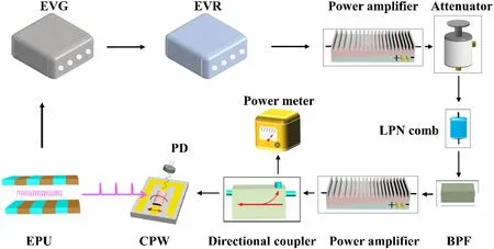
The magnetic moment of magnetic thin films precesses in the GHz band.The frequency of the photon bunch within the storage ring was 499.654 MHz. The microwave frequency was multiplied by the bunch clock frequency into the GHz band. A microwave magnetic field harmonically generated by microwave current via a CPW is required; it must be strong enough to excite the spin precession of the magnetic film. The microwave excitation power must be modulated and amplified.We used electronic equipment to perform these functions, illustrated in Fig. 3.
The microwave signal is elicited by the high-frequency clock signal of the accelerator through a single-mode fiber[30, 34, 45]. A diagram of the TR-XFMR experimental apparatus developed to generate this radio frequency signal is shown in Fig. 4. At the SSRF, the storage-ring bunch clock generates an X-ray pulse. The signal of the highfrequency clock is set in a customized timing system. The phase shift between the microwave and X-ray pulses can be flexibly adjusted. After passing through the timing system,a bunch clock signal is introduced into the amplifier. The amplified signal is introduced into a low-phase noise comb generator (LPN comb) through an attenuator. The attenuator can reduce the power of the signal to protect the LPN comb and adjust the power of the bunch clock signal. The harmonic spectrum of an X-ray pulse frequency input signal was generated (simultaneously with the synchrotron). After frequency multiplication, the signal was fed into a tunable, narrow bandpass filter (BPF)to select a microwave frequency. A BPF with a frequency of 2 GHz was used for stroboscopic measurements. To monitor the microwave frequency power in real time, a final amplified input signal was directly applied to a directional coupler.The final amplified input signal was fed into the CPW via the auxiliary port of the directional coupler.
Using the TR-XFMR measurements, a series of harmonic microwave frequencies from an X-ray pulserepetition frequency input signal and the power of the microwave excitation were obtained.The microwave radio frequency can be multiplied to GHz range. Microwave radio power can be monitored in real time and flexibly modulated.

Fig. 3 (Color online) Timeresolved electronic device in landscape

Fig. 4 (Color online)Schematic of TR-XFMR setup developed to generate microwave frequency signal
2.3 Microwave excitation system
Broadband coplanar waveguides are commonly used in ferromagnetic resonance techniques.A coplanar microstrip transmission line is typically applied during sample preparation. A CPW was constructed using a thin center conductor, a dielectric slab, and two ground plates. The center conductor and two ground plates were deposited on the same surface of the dielectric slab [46]. The typical CPW structure simplifies connection of external components, enabling electronic device integration.
In the CPW,the microwave magnetic field propagates in standard TEM mode [47]. Circularly polarized microwave frequency magnetic fields are generated at the surface of a thin metallic film.The microwave magnetic field is parallel to the CPW plane and perpendicular to the center conductor. To improve the homogeneity and stability of the microwave magnetic field exciting the sample, a magnetic film is deposited on the surface of the center conductor using photolithography and magnetron sputtering technology instead of sample-mounted on a CPW using an insulating adhesive [48].

Fig. 5 (Color online) Diagram and photograph of CPW
Figure 5 shows a diagram of the CPW model. To transmit microwave currents to the CPW, a standard subminiature A (SMA) was connected to the CPW using conductive silver paste. The CPW was designed using Ansoft HFSS software [49]. To ensure efficient microwave circuit transmission, the CPW characteristic impedance was set as 50 Ω.A copper film with a thickness of 120 nm was sputtered onto high-resistance silicon with a thickness of 500 ± 25 μm.The center conductor width and dielectric space width on each side of the center conductor were 200 and 116 μm, respectively, as shown in Fig. 5. The total length of the center conductor was 7.38 mm. The CPW head was expanded to match the SMA center pin. A dead short was formed by terminating the dielectric spacing at the CPW end to increase the microwave magnetic field.Given an input excitation of 30 dBm (1 W), a magnetic field Hrf~1 mT was obtained from the HFSS result.
3 Sample preparation
For TR-XFMR measurement, the CPW was fabricated using photolithography and RF magnetron sputtering. The patterning of the Cu signal line was performed on a highresistance silicon substrate using a 99.99% pure Cu target.The base pressure of magnetron sputtering was 5 × 10⁻⁸Torr. The Cu layer was deposited at an Ar pressure of 3 × 10⁻³Torr at a sputtering rate of 0.375 nm/s. The coplanar waveguide fabricated on a high-resistance silicon substrate achieved a microwave loss of less than 2 dB/cm in the tested 2 GHz band. The Ni81Fe19film was deposited near the CPW shorted end, forming a polycrystalline layer with DC UHV magnetron sputtering at an Ar pressure of 3.5 × 10⁻³Torr.The base pressure of magnetron sputtering was 4 × 10⁻⁸Torr. The thickness of the Ni81Fe19magnetic layer was 40 nm.The growth rate of the Ni81Fe19film was 0.067 nm/s. The thicknesses of the Cu and Ni81Fe19layers were determined from X-ray reflectivity measurements.
4 Experimental results
In studying the spin dynamics of magnetic materials,the relative change in magnetization is usually measured by XMCD [50]. XMCD is characterized by dichroism. Magnetic materials have different absorptions of left-and rightcircularly polarized X-rays caused by different densities of spin-up and spin-down electrons. The absorption edge of an element can be obtained by tuning the incident X-ray energy. When spin dynamics are excited by a microwave magnetic field, the magnetization component along the direction of the X-ray pulse changes, and the X-ray absorption intensity changes accordingly.
Before the TR-XFMR experiment, static X-ray absorption spectra(XAS)were obtained from the absorption edge of the Ni element using the total fluorescence yield (TFY)method to select the photon energy of the element, as shown in Fig. 6. The saturation effects in the TFY mode may affect the XMCD spectra at both L2,3edges, which may change the ratio between L3and L2in the XMCD sum rules and prevent application of sum rule analysis.

Fig. 6 Experimental XAS spectra at Ni L2,3 edge of Ni81Fe19
Under a bias field and microwave excitation, spin precession is driven in the sample. When a series of HBis applied to the sample, the magnetic moment is tuned.Depending on the series of delay times, the X-ray absorption intensity is measured as the spin precession amplitude. The X-ray absorption intensity changes according to a sinusoidal law. The time-resolved XFMR measurements of Ni magnetic moment precession in the Ni81Fe19layer are shown in Fig. 7, confirming that timeresolved XFMR spectra can be detected by our apparatus.
At a certain microwave excitation frequency and power,the spin precession of the magnetic moment can be adjusted by varying HB. The microwave excitation field drives spin precession at GHz frequencies in the thin magnetic films. For our measurement, a microwave radio frequency of 2 GHz was used to excite the Ni81Fe19film to produce precession. A high-power (0.6 W) microwave excitation field was used to ensure that a larger spin precession amplitude was recorded.
Time-dependent projection of spin precession was achieved by tuning the phase shift at a constant bias field.Each of the scans was confirmed at a series bias field from 2.5–7.5 mT. For the same bias field, phase shifting with a step resolution of 20 ps was used. Each precession curve collected a total of 20 data points. To optimize the signalto-noise ratio, each data point was represented by the average value of 20 sample fluorescence signals at the same phase shift. As expected for spin dynamical processes, the XMCD signals of the magnetic moment amplitude exhibited a sinusoidal distribution. The solid lines in Fig. 7 were fitted. The solid points in different colors represent the experimental data. All TR-XFMR measurements were performed at room temperature.
5 Conclusion
We set up a picosecond time-resolved X-ray ferromagnetic resonance apparatus with a time resolution of 13 ps(RMS)or 31 ps(FWHM)at the SSRF.Using this system,a series of higher harmonic microwave frequencies were driven at multiples of the synchrotron radiation photon bunch clock. A phase shift of up to 5 ps was achieved between the microwave and X-ray pulses. For our pumpprobe experiments, the step resolution for phase shifting was chosen as 20 ps,sufficient to resolve a spin precession process with a period of 500 ps. With the electronic circuitry system, the continuous-wave microwave power that excites spin precession can be adjusted up to 1.11 W. The continuous-wave microwave frequency can be adjusted within the GHz range. The designed CPW was properly integrated in this experimental setup and provided a stable and uniform microwave magnetic field of up to 1 mT.
Furthermore, we successfully detected the spin precession of the Ni magnetic moment in an Ni81Fe19sample at 2 GHz in different magnetic fields on the Ni L3X-ray absorption edge energy by synchronizing and manipulating the phase shift. The experimental results demonstrate that the platform can excite spin precession at GHz frequencies in thin magnetic films.The amplitude and phase of the spin precession can be detected and verified on a picosecond timescale using this setup.These findings indicate that this setup is a powerful tool for exploring element-specific magnetization dynamics in magnetic multilayers. Continuous development of this technique will be instructive for other magnetic systems and complex magnetic dynamic processes in the future.
The picosecond TR-XFMR method was successfully applied in the BL07U and BL08U1A end stations at the SSRF. Based on the same ‘‘phase shift’’ time resolution principle and other synchrotron radiation experimental methods, time-resolved scanning transmission X-ray microscopy and time-resolved photoemission electron microscopy will be considered at the SSRF.A temperaturevarying system is being installed in the cavity to upgrade the experimental platform.
Open Access This article is licensed under a Creative Commons Attribution 4.0 International License, which permits use, sharing,adaptation,distribution and reproduction in any medium or format,as long as you give appropriate credit to the original author(s) and the source,provide a link to the Creative Commons licence,and indicate if changes were made.The images or other third party material in this article are included in the article’s Creative Commons licence,unless indicated otherwise in a credit line to the material. If material is not included in the article’s Creative Commons licence and your intended use is not permitted by statutory regulation or exceeds the permitted use, you will need to obtain permission directly from the copyright holder. To view a copy of this licence, visit http://creativecommons.org/licenses/by/4.0/.
Author contributionsAll authors contributed to the study conception and design. Material preparation, data collection, and analysis were performed by Xia Yang,Jie-Feng Cao,and Jun-Qin Li.The first draft of the manuscript was written by Xia Yang, and all authors commented on previous versions of the manuscript. All authors read and approved the final manuscript.
 Nuclear Science and Techniques2022年5期
- Nuclear Science and Techniques的其它文章
- Spatial resolution and image processing for pinhole camera-based X-ray fluorescence imaging: a simulation study
- Nonrecursive residual Monte Carlo method for SN transport discretization error estimation
- Density fluctuations in intermediate-energy heavy-ion collisions
- Isospin effects on intermediate mass fragments at intermediate energy-heavy ion collisions
- Identification of anomalous fast bulk events in a p-type pointcontact germanium detector
- Free-radical evolution and decay in cross-linked polytetrafluoroethylene irradiated by gamma-rays
