
Solvent Synthesis and Characterization of a New Dinuclear Zinc(II) Complex Zn2(L)4(phen)2·H2O
2022-06-24 06:39LIChongyuLILixuanYUPeng
湖南师范大学自然科学学报 2022年3期
LI Chong-yu, LI Li-xuan, YU Peng
(School of Chemistry and Materials Science, Hunan Agricultural University, Changsha 410128, China)
Abstract A new dinuclear zinc (II) complex Zn2(L)4(phen)2·H2O was synthesized with zinc sulfate, p-chlorobenzoic acid (HL) and 1,10-phenanthroline(phen) in the mixed acetonitrile/water (5∶1 in volume) mixtures. Its crystalline pattern belongs to the monoclinic space group C2/c, with a=2.340 3(2) nm,b=1.560 59(15) nm,c=1.560 72(15) nm, β=121.040(10)°, Mr=1 131.40, V=4.883 9(8) nm3, Z=4, Dc=1.510 g·cm-3, F(000)=2 256, and μ(Mo Kα)=0.80. The final GOOF is 1.033, R1 is 0.037 3, and wR2 is 0.054 3. The Zn (II) ion is six-coordinated to give a distorted octahedral geometry. The complex remains thermally stable before 429 K and holds a strong fluorescence at around 419 nm.
Key words Zinc (II) complex; thermal stability; spectroscopic characterization
1 INTRODUCTION
Metal hydrolases in organism, which can be used for hydrolyzing protein, phosphate ester, DNA, etc., play an important role in biochemical reactions[1-4]. A lot of studies have shown that identifying the structure of the active sites and the catalytic mechanism of different functional metalloenzymes are the key issues of bioinorganic chemistry[5-8]. As zinc is an essential element to sustain all forms of life, it is of great significance to design and synthesize zinc complexes to simulate hydrolases and explore the corresponding reaction mechanism[9-11]. Herein, we report the synthesis of a new dinuclear zinc complex Zn2(L)4(phen)2·H2O usingp-chlorobenzoic acid (HL) and 1,10-phenanthroline (phen) as ligands. The information on the structure and function of this complexe with flexible cyclic carboxylic ligands[12]was acquired. Moreover, the optical performance and thermal stability of this complexe were preliminaried characterized.
2 EXPERIMENTAL
2.1 Chemical reagents and instruments
All chemicals were of analytical grade and used without further purification. A PE-2400(II) elemental analyzer was used to conduct C, H and N analyses and a F-7000 fluorescence spectrophotometer (from 200 to 700 nm) was adopted to obtain a fluorescence spectrum at room temperature. The IR spectra were obtained on a FT-IR spectrophotometer (ALPHA, Bruker) employing KBr disks. The thermogravimetric properties of crystals were measured by a TGA2 instrument (METTLER TOLEDO, Switzerland). All samples were heated under air atmosphere from 25 to 700 ℃ at a heating rate of 10 ℃·min-1.
2.2 Synthesis of the complex
Zinc sulfate (0.2 mmol, 0.057 2 g) and HL (0.4 mmol, 0.062 5 g) were added into a mixed solvent (30 mL) of acetonitrile and water (5∶1 in volume). The pH of reaction mixture was adjusted to 6.5—7.0 using dilute sodium hydroxide and phen was then added (0.2 mmol, 0.036 0 g). The mixture was stirred for an additional 15 h at 343 K. Afterwards, the mixture was stored at room temperature for diffusion. Yellow single crystals were obtained one month later, which was employed for further characterization. Yield: 50.20%. Anal. calcd.(%) for C52H34Cl4Zn2N4O9(Mr=1 131.40): C, 55.41; N, 4.94; H, 3.00; Found: C, 55.16; N, 4.94; H, 3.04. Main IR (KBr, cm-1): 550(w), 686(w), 726(m), 849(m), 1 062(m), 1 240(w), 1 279(m), 1 344(vs), 1 428(vs), 1 479(m), 1 622(vs), 2 942(s), 3 331(w).
2.3 Determination of crystal structure of samples
X-ray diffractionanalysis of the complex was carried out on a Bruker SMART APEX CCD area detector at 295(2) K using graphite-monochromatized Mo Kα(λ=0.071 1 nm) radiation. Direct methods were used to analyze the crystal structure, which was subsequently optimized by SHElXS-2014[13]and SHELXL-2014[13]programs using a full-matrix least-squares technique.Lpfactors were corrected and the empirical adsorption was adjusted. Anisotropic thermal parameters were applied to refine all non-hydrogen atoms. The final refinement was convergedwR2=0.054 3 andR=0.037 3 (w=1/[S2(F02)+(0.036 5P)2+5.323 4P], whereS=1.033,P=(Fo2+2Fc2)/3), (Δρ)max=0.512, (Δρ)min=-455 e·nm-3, and (Δ/σ)max=0.00).
3 RESULTS AND DISCUSSION
3.1 Crystal structure of the complex
The molecular structure of the complex is shown in Fig. 1. The packing diagram is given in Fig. 2. The selected bond lengths and bond angles are listed in Table 1.

Table 1 Partial bond angles (°) and bond lengths (nm) of the complex
The entire molecule of the complex composes of two zinc ions, four L-1groups, two phen molecules and one water molecule. The Zn (1) is surrounded by two nitrogen atoms (N (1) and N (2) from the phen molecule) and four oxygen atoms (O(5) from the water molecule and O(1), O(2A) and O(3) from the L-1anions), giving a distorted octahedral geometry. In the crystal structure of the binuclear zinc complex, each zinc atom is situated in the N2O4coordination environment, and all of oxygen atoms O(5) act as “bridges” between Zn(1) and Zn(1A) to form the familiar Zn-O-Zn structure. The Zn-N bond lengths range from 0.226 4 to 0.229 2 nm. The coordination mode of the carboxylic acid group is the monodentate with zinc atoms. The average distance of Zn-O(carboxyl)ranges from 0.214 0 to 0.217 6 nm, shorter than 0.226 3 nm of the bond length Zn-O(water). The Zn…Zn distance (throughLgroups and O (5) bridging) is 0.351 3 nm, which is close to the similar Zn (II) complexes (Zn4C112H138BrCl8N12O18Zn…Zn=0.347 6 nm, Zn4C112H138Cl9N12O18Zn…Zn=0.355 6 nm)[14], falling in the normal range. The bond angle of O/N-Zn-N/O ranges from 73.19(8)° to 174.25(7)°. In addition to this, as shown in Fig. 2, the shortest center distance between aromatic cycles of phen group is 0.399 6 nm greater than 0.370 0 nm, which indicates the existence of a stacking interaction (π-π stacking interaction)between the aromatic rings[15].
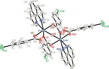
Fig. 1 Molecular structureof the title complex (A:1-x, y, 0.5-z)

Fig. 2 Packing diagram of the title complex in a cell (matr2)

Fig. 3 Spectra of the title complex and ligands (a: complex; b: HL; c: phen)
3.2 Spectroscopic characterization
Infrared spectrum of the title complex appears an absorption peak at 3 332 cm-1, belonging to the absorption peak of —OH in H2O. The anti-symmetrical and symmetrical stretching vibration absorption peaks appear at 1 623 and 1 345 cm-1, which are in contrast with the corresponding peaks of free ligands (1 687 and 1 593 cm-1). The characteristic peaks at 1 429, 850 and 727 cm-1are ascribed to the absorption peaks of phen ligand.
The solid-state emission spectra of the title complex and the free ligand at room temperature are shown in Fig.3 in the range of from 320 to 550 nm (the excitation wavelength 320 nm). As seen in Fig. 3, phen, HL and the title complex have characteristic emission peaks at 452, 413 and 419 nm, respectively. Compared with that of free ligand HL, the emission spectrum of the title complex has similar figure and characteristic emission peak, and its luminescence is stronger than that of free ligand HL, suggesting that luminescence of the title complex may be mainly attributed to the electronic transition in the intra-ligand HL[16].
3.3 Thermal Stability
TG-DTG results are depicted in Fig. 4. There are three weight-loss stages from ambient temperature to 700 ℃ as illustrated by the TG curve. The first stage occurs from 138 ℃ to 156 ℃ with the weight-loss of 1.50% (calcd 1.60%), attributed to the release of one water molecule. The second stage emerges from 156 ℃ to 300 ℃ with the weight-loss of 33.0% (calcd 32.8%) because of the loss of two phen molecules. The DTG thermal absorption peak appears at about 266 ℃, which corresponds to a strong thermal decomposition. The third stage occurs from 300 ℃ to 520 ℃ with the weight-loss of 51.5% (calcd 51.20%) as a result of the departure of four L-1molecules, which is consistent with the crystal structure. The final products is ZnO with the residual weight being 14.00% (calcd 14.40%).

Fig. 4 TG and DTG curves of the title complex
4 CONCLUSIONS
1) A new dinuclear zinc (II) complex [Zn2(L)4(phen)2]·H2O has been successfully synthesized usingp-chlorobenzoic acid and 1,10-phenanthroline as ligands and characterized.
2) The crystalline pattern of [Zn2(L)4(phen)2]·H2O can be indexed to the monoclinic space groupC2/c, witha=2.340 3(2) nm,b=1.560 59(15) nm,c=1.560 72(15) nm,β=121.040(10)°,Mr=1 131.40,V=4.883 9(8) nm3, andZ=4.
3) [Zn2(L)4(phen)2]·H2O remains thermally stable before 429 K and has a strong fluorescence at around 419 nm.
