The apoptotic effect of ozone therapy on mitochondrial activity of highly metastatic breast cancer cell line MDA-MB-231 using in vitro approaches
Merve Yıldırım, Selen Erkis¸i, Hzl Yılmz, Nz Ünsl, Elif ˙Inç, Yıldıry Tnrıver,Polen Koçk
a Department of Genetics and Bioengineering, Faculty of Engineering and Architecture, Yeditepe University, 26 A˘gustos Campus, Kayisdagi Cad., Kayisdagi, 34755,Istanbul, Turkey
b Infinity Regenerative Clinic, Thy Sitesi Yolu, Ulus, 34347, Istanbul, Turkey
c Department of Biomedical Engineering, Faculty of Engineering, Istinye University, Vadi Campus, Azerbaycan Cad., Sariyer, 34396, Istanbul, Turkey
Keywords:Ozone therapy Antioxidants Apoptosis Breast cancer
ABSTRACT Ozone (O3) gas is the triatomic state of oxygen and it is used as a disinfection agent due to its strong oxidizing effect,since its discovery in the mid-nineteenth century.Ozone therapy is also an alternative therapeutic approach for some diseases like circulatory disorders,AIDS,asthma,cardiovascular diseases,and certain types of cancer by increasing the oxygen levels in the blood by external addition of ozone to the body.In this study,the therapeutic potential of ozone therapy was examined by inhibiting the growth of breast cancer cells in a dose-dependent procedure. Ozone concentrations varying from 5 to 20 μg/ml were applied to the MDA-MB-231, human breast adenocarcinoma and HUVEC, human umbilical vein endothelium, cell lines, and MDA cells demonstrated an increased rate of death while its migration potential decreases. RT-PCR analysis showed mRNA expression levels of pro-apoptotic genes demonstrated higher folds in MDA cells after 10 μg/ml treatment. In the same context,Annexin V/PI and cell cycle analysis also concluded that ozone therapy causes apoptotic cell death on breast tumor cells.The use of ozone therapy for cancer treatment requires further and extensive research.However,this research has shown that ozone therapy is a promising source for cancer treatment in a way by inhibiting the proliferation of breast tumor cells.
1. Introduction
Ozone (O3) is a highly energetic and triatomic unstable form of oxygen in the atmosphere of normal oxygen gas.Since it has an extremely strong oxidizing effect, it is very effective in removing microbes.Discovered in the mid-1800s, ozone was first used in 1856 to purify surgical instruments and operating rooms from microbes. Ozone which started to be used in the disinfection of drinking water in the late 1800s;was also used in World War I for the treatment of wounds,frostbite,and the effects of poisonous gases.1
Ozone therapy is a complementary medicine method that aims to increase the level of oxygen in the blood by giving oxygen-releasing substances to the body in different forms and different ways. In this method, which is also known by different names such as oxygen therapy, oxidative therapy, oxygenation therapy, hyperoxygenation,hydrogen peroxide therapy, the most frequently used forms of oxygen are; hydrogen peroxide (H2O2) and ozone (O3).2The method is referred to as ozone therapy since ozone is the chemically active form of oxygen. Although the method can be applied in different ways and forms, the most common form of application is to apply ozone to the body by mixing it with various gases and liquids.It can be given to the body under pressure through the rectum, vagina, or other cavities in the body, or by injection into the muscle or under the skin.Within the scope of an approach called autohemotherapy, the practitioner can apply ozone using a special device by adding it to some blood taken from the patient and returning it to the patient's body. Different types of oxygen therapy are introduced as complementary treatments for dozens of diseases, including certain types of cancer, emphysema,AIDS, arthritis, cardiovascular diseases, multiple sclerosis, and Alzheimer's disease.3
Ozone therapy is often preferred as an auxiliary treatment method in many diseases. Some of these can be listed as follows: circulatory disorders, cancer, eye diseases, bacterial and fungal infections. One of the methods that can be preferred as a complementary therapy in cancer patients is ozone therapy. Ozone therapy, which increases the activation of the immune system and supports the production of cancer-fighting cells,contributes positively to the general resistance of the body and helps cancer treatment.It also plays an important role in reducing the negative effects of chemotherapy due to its vitality.4On the basis of the belief of practitioners today who claim that ozone therapy is effective in cancer, Dr. Otto Warburg's theories on cancer are included. In the 1930s, he was awarded the Nobel Prize in 1931 for his research on respiratory enzymes.Otto Warburg discovered that cancer cells have a lower respiratory rate than other cells. From this,he deduced that cancer cells grow in low-oxygen environments, and thus increasing the oxygen level can damage or even kill them.5Ozone has been proven to have varying effects in cell cultures, depending on its dosage, direct action on specific tumor types, and, in such situations, enhancing the effects of chemotherapeutic and radiotherapeutic drugs.6A prestigious publication, Science, reported in 1980 that,depending on the concentration, ozone could significantly suppress the growth of distinct human cancer cells (breast, lung, and uterus)while having no effect on non-tumor cell lines.7Ozone was shown to have a stimulatory impact on 5-fluorouracil in colon and breast cancer cell lines, and the combination treatment of ozone and 5-fluorouracil showed success in cancer cells that had previously been resistant to the drug.8It was also demonstrated that ozone has a significant impact in neuroblastoma cell cultures, where it enhanced the effects of etoposide and cisplatin.9Besides enhancing the effects of therapeutic drugs, ozone was found to have a direct effect on increasing the necrosis in tumors while causing a reduction in the rate of tumor growth.10
Growth-stimulatory and differentiation signals, together with the activation of cell death pathways, have a significant impact on the growth of epithelial cells that are covering the breast ducts and lobules.Malfunction in these homeostatic mechanisms can cause uncontrolled growth of epithelial cells and,as a result,breast cancer.Breast cancer is the consequence of the complex interaction of several variables,including genetic, hormones, lifestyle, and environmental factors.11Women with specific mutations in tumor suppressor genes BRCA1 and BRCA2 or who have inherited a mutated BRCA1 allele from either parent are found to be more likely to develop breast cancer at an early age and may face more severe consequences of the disease.12Breast cancer is a hormone-dependent disease and its dependency is shown by the fact that women who do not have functional ovaries and have never undergone hormone replacement therapy (HR) do not acquire breast cancer. Since the exposure to progesterone and estrogen in men is lower than in women, the incidence of breast cancer in men is 150 times less than in women. Although the hormone levels decrease in older women, the lifetime accumulation of these hormones increases the diagnosis of breast cancer by 10%.13Environmental pollutants and commonly used chemicals such as polychlorinated biphenyls and Polycyclic aromatic hydrocarbons (PAHs) also disrupt the hormones and cause breast cancer.14These disruptors are involved in the activation of carcinogens and cause tumor formation increase in the mammary glands where certain genetic polymorphisms are present. Besides genetic and hormonal factors, lifestyle is also an important risk factor for developing breast cancer.Obesity is associated with all-cause mortalities including breast cancer. Studies demonstrate a direct correlation between fat intake and the incidence of breast cancer.15,16Thus,exercising regularly and maintaining body mass index (BMI) between 19 and 25 kg/m2is found to be decrease breast cancer by approximately 30%. Moreover, regular alcohol intake increases the breast cancer risk by 26%.17
Breast cancer,which is the most common and the most common cause of death in women in our country and the world, is a malignant tumor and constitutes approximately 30%of all cancers seen in women.Breast cancer in men is observed much less frequently(less than 1%of all breast cancers)compared to women.It ranks first among the ten most common cancers in the world and our country. While the incidence of breast cancer is 46.3 per hundred thousand worldwide, it is 92.6 for Northern European countries, 39.2 for East Asia, 38.3 for the United States, and 45.6 for our country.180,000 new cases are detected annually in Europe and 184,000 new cases per year in the United States (USA). In a year,approximately 18,000 women are diagnosed with breast cancer in our country.18
2. Material & method
2.1. Cell culture and treatment
Human breast adenocarcinoma cell line (MDA-MB-231, ATCC ®HTB-26™) and human umbilical vein endothelium cell line (HUVEC,ATCC ® CRL-1730™) were obtained from American Type Culture Collection (ATCC). Cell lines were cultured in High-Glucose Dulbecco’s Modified Eagle Medium (DMEM) supplemented with 10% (v/v)Fetal Bovine Serum (FBS) and 1% Penicillin-Streptomycin-Amphotericin in a highly humidified Nuaire NU5510/E/G incubator(RH 80%) at 37˚C with 95% (v/v) air and 5% CO2. Ozone (O3)treatment was performed in isotonic sodium chloride solution at 5-10-20 μg/ml concentrations.
2.2. Cytotoxicity assay
HUVEC and MDA-MB-231 cell lines were seeded in 96-well plates at a concentration of 5×10³cells/well.Cells were treated with ozone at 5-10-20 μg/ml concentrations for 24, 48, 72 h in a highly humidified incubator at 37˚C.After each 24 h,the old medium was discarded from the wells, and cells were incubated in freshly prepared MTS Reagent-PBS Glucose (1:10 v/v) mixture for an additional hour at 37˚C. After the incubation, plates were read at 490 nm using an Elisa Plate Reader.
2.3. Wound healing (scratch) assay
MDA and HUVEC cells were seeded as 3 × 10⁶cell/well into 6-well plates. The old medium was replaced with PBS and cells were scratched using a 1000-μl sterile pipette tip.Then,PBS was discarded and fresh medium containing 5-10-20 μg/ml ozone was added to the wells.Cells were incubated for 48 h in a highly humidified incubator at 37˚C.At each 24 h, wound closure was imaged with a light microscope equipped with a digital camera under a 5×objective.
2.4. Cell cycle analysis
Each cell line was seeded into 6-well plates at a concentration of 1×10⁵cell/well. After cells were attached to the surface, the medium was discarded and fresh medium containing different ozone concentrations was added to the wells.At 24 and 48thhours,cells were harvested and centrifuged cells were washed with PBS twice.Cells were fixed using 70%EtOH and incubated at-20˚C for 2 h.After incubation,cells were washed with PBS two times.Pellets were resuspended in PBS containing 20 μg RNase A and 0.1%Nonidet and incubated at 37˚C for 30 min.Propidium Iodide(PI)was addedtothe flowtubes andthe cell cyclewas analyzed usingthe Becton Dickinson FACS Calibur flow cytometer.
2.5. Annexin V-FITC/PI assay
1 × 10⁵cell/well MDA and HUVEC cells were seeded into 6-well plates. Treatment was done and cells were incubated at 37˚C for 48 h.At 24thand 48thhours,cells were dislodged with trypsin and centrifuged cells were washed with PBS. Cells were incubated in Annexin V buffer and FITC Annexin V according to the manufacturer’s protocol.Propidium Iodide was added to the flow tubes before the analysis and the apoptotic effect of treatments was analyzed using the Becton Dickinson FACS Calibur flow cytometer.
2.6. Real-Time PCR
Total RNA was isolated from ozone-treated MDA and HUVEC cell lines by using TRIzol Reagent. Reverse transcription of RNA to cDNA was performed using iScript™Reverse Transcription Supermix for RTqPCR Kit (Bio-Rad, USA). Gene expression levels of apoptotic marker genes Cas9, Cas3, p53, and Bcl-2, and reference gene GAPDH were analyzed using QuantiTect SYBR Green PCR Kit (Qiagen, France) with iCycler RT-PCR system(Bio-Rad,USA)according to the manufacturer’s protocol.
2.7. Statistical analysis
The one-way analysis of variance(ANOVA)using Graph-Pad Prism 9(GraphPad Software Inc.,San Diego,CA)was used for statistical analysis.p ≤0.05 values were considered significant statistically.
3. Result
3.1. Cytotoxicity assay
The effect of various concentrations (5–20 μg/ml) of ozone/PBS on MDA and HUVEC cells viability was determined at 24, 48, and 72 h(Fig.1).Considering the cell viability in MDA cells,it was observed that the cells were killed by approximately 23% at three different concentrations for the first day. When the cell viability at the 48thhour is examined, it is seen that the death rate decreases to 30%, and the maximum cell death are 50%at the third dose at the 72ndh.In the first day's MTS analysis of HUVEC cells,cell viability was increased by 19%at all 3 doses. In MTS analyses at 48 and 72 h for HUVEC cells, it was observed that the cell viability decreased to 14%at most,and this value is considered normal.
3.2. Wound healing (scratch) assay
The effect of ozone on the migration capacities of cell lines was evaluated by scratch assay with three different doses.As can be seen in Fig.2,ozone-treated HUVEC cells migrated and artificial wounds were mostly closed in each dose. The artificial wound of 20 μg/ml ozonetreated HUVEC cells was found to close faster than other doses after 48 h of incubation.Conversely,the scratched area of the ozone-treated MDA cells was slightly closed at the end of the incubation period.
3.3. Apoptotic effect of ozone
Annexin V-FITC/PI assays were used to validate the apoptotic impact of ozone on MDA cells in order to further understand the cell cycle arrest and cell death process(Fig.3).Treatment of MDA cells with 5,10,and 20 μg/ml of ozone resulted in about 7%apoptotic cell death after 24 h when 20 μg/ml of ozone is applied.In neither 24 nor 48 h treatments,there was no substantial necrosis. The apoptotic cell death ratio in MDA cells elevated 14% after 48 h. For the HUVEC cell line which was used as a healthy cell line, there were no significant apoptotic cell deaths.Apoptotic cell ratio is increased in HUVEC cells from 0.50% to 0.58%after 48 h.
3.4. Cell cycle analysis
Flow cytometry was used to investigate the influence of ozone on cell cycle progression in MDA and HUVEC cells in order to determine what was causing the growth inhibition. Ozone treatment for 24 h indicates that even though no significant result was found at a 5 μg/ml dose for the G2/M phase,percentages of the G2/M phase were increased at 10 μg/ml and 20 μg/ml compared to control (Fig. 4). Although a slight decrease can be seen at the S phase and G0/G1 phase for each dose, there is no significant cell distribution.
3.5. Real-Time PCR
Gene expression levels of ozone-treated cell lines were examined using RT-PCR. After the ozone treatment, expression levels of proapoptotic genes, p53, Cas9, and Cas 3, and anti-apoptotic gene Bcl-2 were analyzed in both MDA and HUVEC cell lines by normalizing the expression levels with the housekeeping gene GAPDH.HUVEC cells were found to express almost twofold in the Bcl-2 anti-apoptotic gene while demonstrating lower expression in pro-apoptotic genes.As can be seen in the results, MDA cells demonstrated higher folds in all pro-apoptotic genes than the pro-apoptotic gene, Bcl-2. Also, there was a remarkable three to fourfold increase in all pro-apoptotic genes expressed in the 20 μg/ml ozone-treated MDA cells.
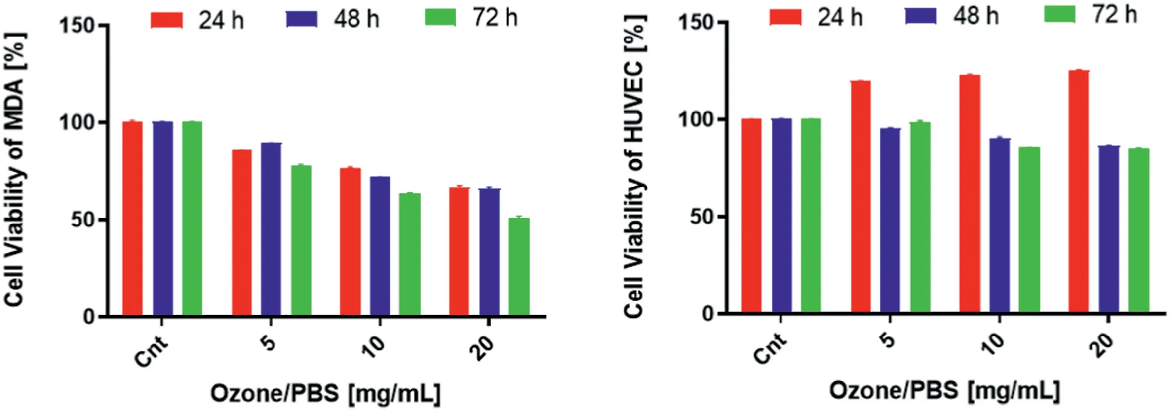
Fig. 1. Viability and proliferation of human breast adenocarcinoma cells (MDA) and human umbilical vein endothelium cells (HUVEC) were observed in various concentrations (5–20 μg/ml) of ozone/PBS after MTS assay was performed 24-, 48- and 72-h incubation (37 ˚C, 5% CO2) in DMEM high glucose medium supplemented with 10%FBS.The absorbance value of nontreated control cells was compared to 100 to determine cell death. The absorbance value obtained from NC: negative control, standard growth medium treated cells were assigned to 100% to compute the percentage of cell viability. The data are presented as the mean ± SD.
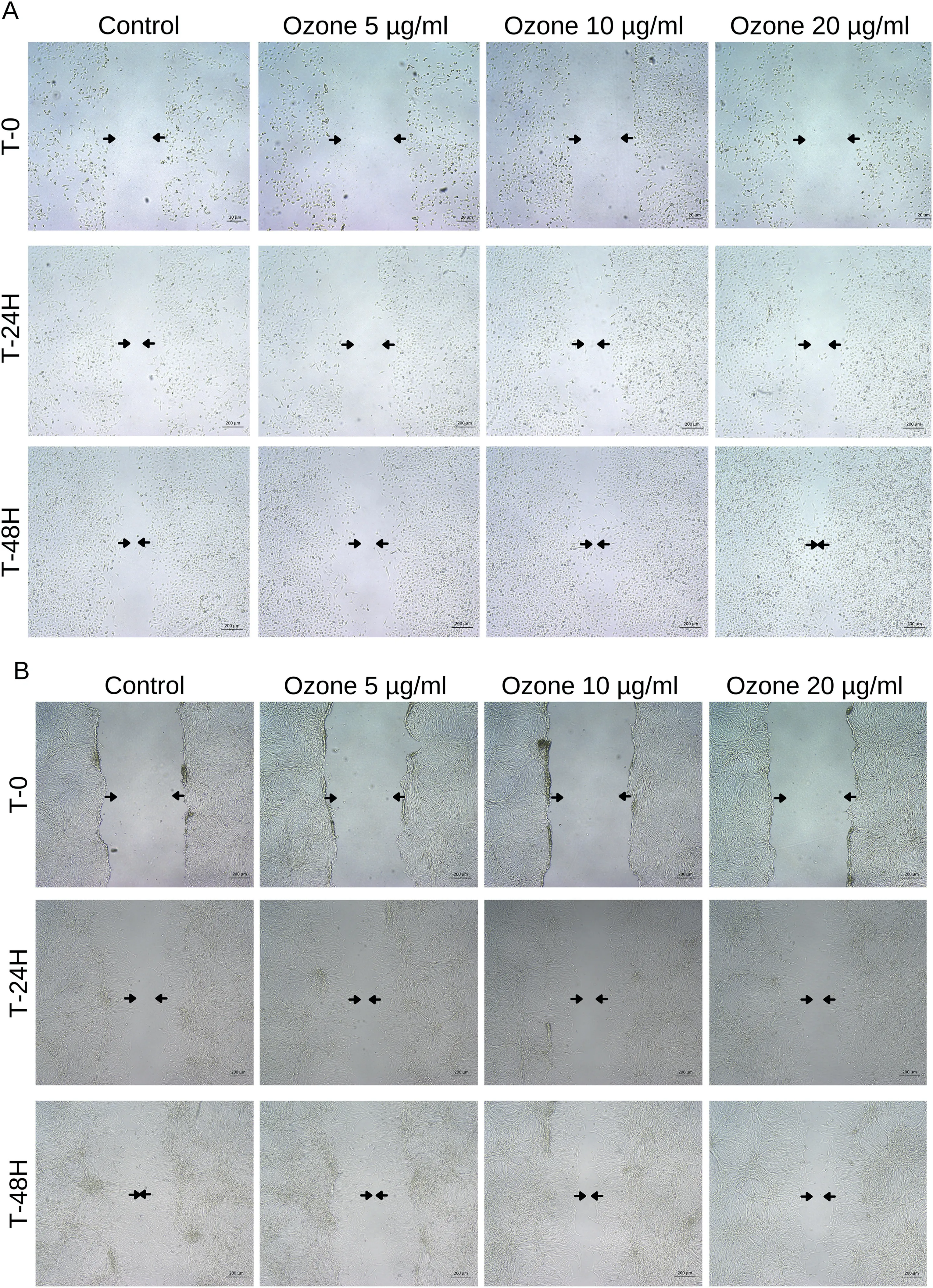
Fig.2. Microscope images at 5×magnification to evaluate cell migration in vitro in the scratch assay using A)HUVEC and B)MDA cell lines.Cells were treated with 5, 10, and 20 μg/ml ozone, and images of the artificial wound were taken at 0- , 24- , and 48-h for each cell line.
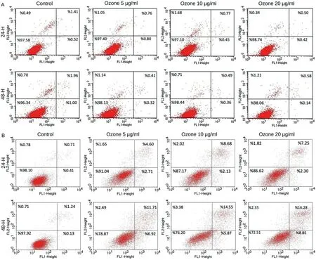
Fig.3. Apoptosis was detected in an AnnexinV/PI containing test on A)HUVEC and B)MDA cell line after treatment with 5,10,and 20 μg/ml of ozone for 24-and 48-h. Quantifications of early and late apoptosis were presented. The data are presented as the mean ± SD.
4. Discussion
The ozone therapy method used in the field of health has been used in many types of research for cancer treatment from past to present,and Dr.Otto Warburg's theory is that cancer cells multiply in a low-oxygen environment, so high oxygen levels will damage and kill them. In light of the effects described for ozone therapy by experimental and clinical studies,we intended to further evaluate the cytotoxicity effect that ozone yields on breast cancer and human umbilical vein endothelium cells, as well as to assess the impact that it has on the cancer treatment process.
The findings of this study show that ozone can be used as an antimetastatic procedure as well as an adjuvant in the treatment of cancer patients.Ozone therapy has the ability to alter the complex tumor process at specific points.First,it has been shown to have an influence on oxygen metabolism and oxygenation regulation, such as in the usage of the aerobic pathway for energy production and re-establishing normal metabolic activities while managing lactic acidosis. Then, by considerably and continuously boosting oxygen availability and microcirculation in the tumor, it may be possible to halt tumor growth and prevent metastatization. Second, the therapy's minor and temporary oxidative stress increase the production of cellular antioxidant enzymes that can protect against persistent oxidative stress. In addition, the immune system is modulated by ozone,allowing for the regeneration of the immune response against cancerous cells.19–21Ozone has been shown to suppress the growth of human cancer cells in culture, implying that cancer cells have a compromised defense system against ozone damage.7,22
In the current study, we demonstrated that ozone treatment could induce an anti-cancer effect on breast cancer cell lines(MDA).In Fig.1,cell viability results of breast cancer cells at different concentrations of ozone were observed to decrease significantly in the MTS analysis on the third day compared to the control, and the cell viability in the HUVEC cell line increased on the first day of MTS,while the viability decreased by 14% on the other days. This decrease was considered normal. Also,looking at the RT-PCR results in Fig. 5, the increase in pro-apoptotic genes in breast cancer cells and the decrease in the anti-apoptotic gene Bcl-2 show that the results are as expected and ozone is effective on cancer. In addition, when RT-PCR results in HUVEC cell lines are examined,it is seen that pro-apoptotic genes decrease and anti-apoptotic genes increase,and this result supports that ozone is effective in cancer.In the same context,the results of the wound healing assay also support the RT-PCR and MTS results considering that the wound closure of HUVEC cells was more than the MDA cells.
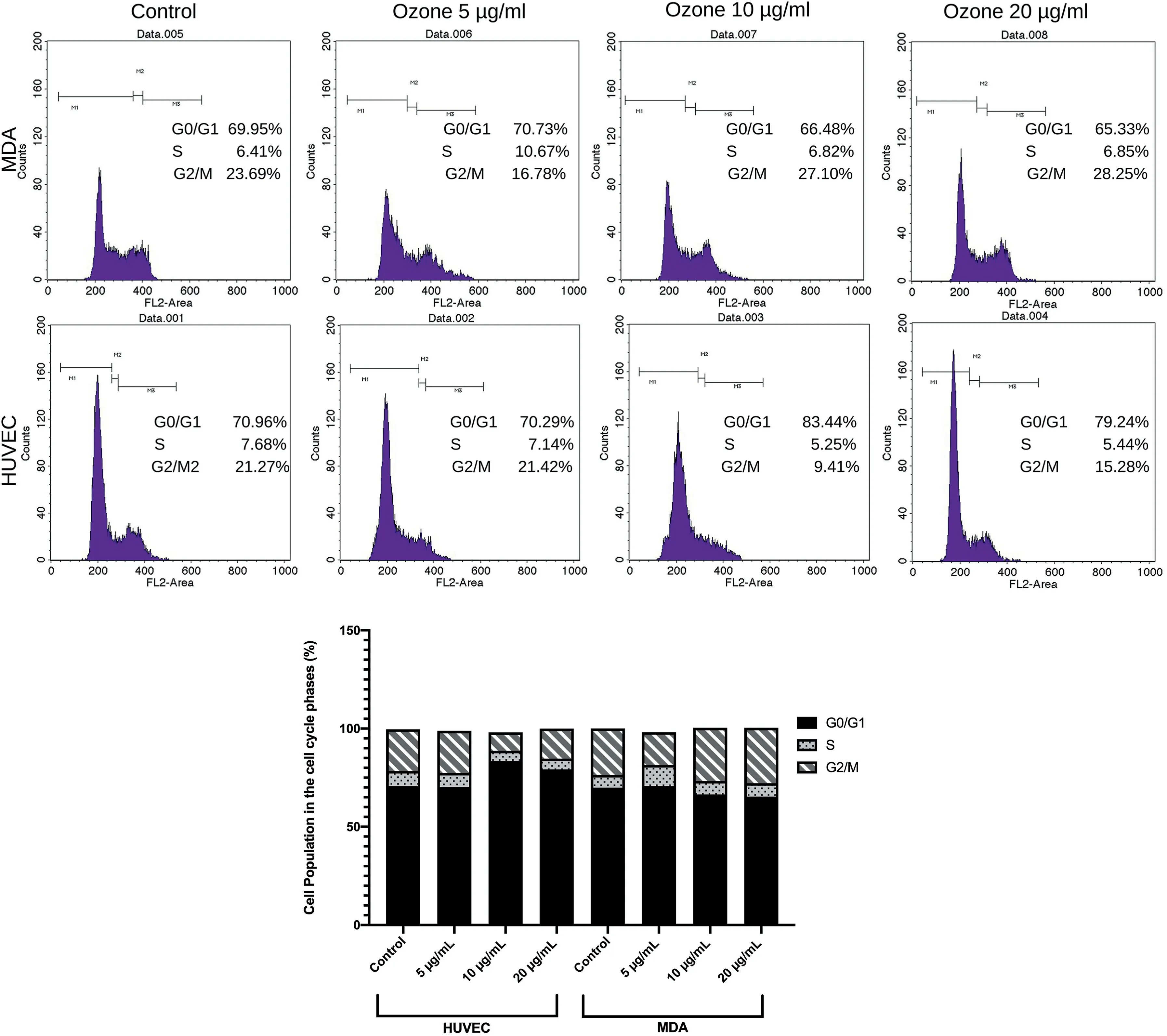
Fig.4. After treatments with ozone for 24h,A)cell cycle distributions and B)representative bar graph of MDA and HUVEC cell lines.Cell populations treated with 5,10, and 20 μg/ml of ozone were examined at G0/G1, S, and G2/M phases. The mean and standard deviation are used to express data. The data are presented as the mean ± SD.
Besides cell toxicity and gene expression analysis,in order to examine the apoptotic effect and cell cycle analysis were examined.When cells are arrested in the G2/M phase, apoptosis and autophagy are increased.23For this situation,the cell cycle should be analyzed for each 3,6,and 9thhour. However, our results did not indicate significant cell distribution for the G2/M phase.The ozone treatment did not elicit a significant cell cycle arrest in HUVEC cells which explains why ozone treatment had a little anti-proliferative impact.Annexin-V/PI staining test was performed for each cell line after three different doses of an ozone treatment to investigate cell cycle arrest and the way of cell death.At all the periods,MDA cells treated with 20 μg/ml of ozone showed apoptotic activity.Unlike MDA cells,the number of apoptotic cells in HUVEC cells remained non-significant in comparison to control cells.
In conclusion,the evidence reported here clearly suggests that ozone has therapeutic effects on MDA cancer cells while having little to no cytotoxicity on normal HUVEC cells. On the other hand, our findings revealed that ozone treatment with a specific dose can be effective for apoptosis and also can enhance apoptotic gene expression.
Declaration of competing interest
The authors declare that the paper has been submitted solely to the Journal of Interventional Medicine and that is not concurrently under consideration for publication in any journal. The authors declare that they have no conflict of interest.Thank you for your consideration.
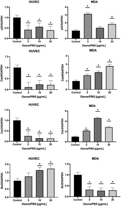
Fig. 5. Expression level analysis of Cas9, Bcl-2, p53, and Cas3 genes of 5, 10, and 20 μg/ml ozone-treated MDA and HUVEC cell lines. Gene expression levels were analyzed by using GAPDH as the negative control. The data are presented as the mean ± SD. Results of one-way ANOVA multiple comparisons for each group are presented at n = 3, *p < 0.01.
 Journal of Interventional Medicine2022年2期
- Journal of Interventional Medicine的其它文章
- Advances in the interventional therapy of hepatocellular carcinoma originating from the caudate lobe
- The underlying molecular mechanism of intratumoral radiofrequency hyperthermia-enhanced chemotherapy of pancreatic cancer
- Preparation and investigation of a novel iodine-based visible polyvinyl alcohol embolization material
- Intrahepatic flow diversion prior to segmental Yttrium-90 radioembolization for challenging tumor vasculature
- Safety and efficacy of transcatheter arterial embolization for management of refractory hematuria of prostatic origin
- Irreversible electroporation versus radiofrequency ablation for malignant hepatic tumor: A prospective single-center double-arm trial
