Computed tomography-based radiomic to predict resectability in locally advanced pancreatic cancer treated with chemotherapy and radiotherapy
lNTRODUCTlON
Pancreatic cancer(PC)is an aggressive disease,with increasing incidence and mortality rates,and a 5-year survival of less than 10%[1,2].At the time of diagnosis,roughly one-third of patients present with locally advanced PC(LAPC),typically due to extensive involvement of peripancreatic vessels(
,celiac axis,superior mesenteric artery and vein,portal vein,common hepatic artery),that precludes surgical resection[3].Nowadays,multiagent chemotherapy regimens,including gemcitabine plus nanoparticle albumin-bound(nab)-paclitaxel and 5-fluorouracil,leucovorin,irinotecan,plus oxaliplatin(FOLFIRINOX),represent the standard of care for LAPC,able to significantly improve survival compared to mono-chemotherapy schedules[4-6].In parallel,the integration of dose-escalated radiation therapy(RT)approaches to intensive chemotherapy regimens have suggested the possibility to maximize the benefits of the oncological treatment[7,8].Technological innovations,such as the use of stereotactic techniques,advanced organ motion management solutions,and accurate image guidance before treatment delivery,have allowed to safely deliver ablative doses to LAPC while sparing the surrounding critical organs at risk(OARs)[9,10].In addition,different series reported that a multimodal approach,including systemic induction therapy followed by(chemo-)radiotherapy,might represent an effective therapeutic option for LAPC,potentially improving the oncological outcome and the probability of surgical resection,at the price of acceptable postoperative complication rates[11-14].Thus,there is a strong need to determine the resectability of LAPC for treatment decision-making.
After neoadjuvant treatment,the therapeutic decision whether to perform surgical exploration is typically based on a multidisciplinary and multiparametric evaluation that includes patients- and tumour-specific features.Cross-sectional imaging at restaging is essential to rule out disease progression that would contraindicate surgery and drive the surgical strategy if radical resectability is considered feasible.Indeed,several studies have reported that computed tomography(CT)misestimates the resectability of LAPC after neoadjuvant treatment[15-17].After RT,CT cannot discriminate between posttherapy fibrosis,locoregional oedema,inflammatory changes,and viable tumour,thus underestimating the histological response[18-20].
Weeks later, with a joyful11 burst of energy, Timmy sprinted12 towards me. He proudly pulled the bandage off to show me the result if his work. See, he said, It s growing back really good! A year later, Timmy came to say good-bye to me. He and his family were moving to another neighborhood. Timmy s finger was completely healed. It was round and padded just as any index finger should be. Only a fine hairline scar remained.
Radiomics is gaining more and more popularity for the possibility of decoding crucial information underneath medical imaging.Unlike conventional CT image analysis,radiomics can use imaging data for the high-throughput extraction of large numbers of quantitative features,able to offer additional information related to tumour phenotype,its heterogeneity,and microenvironment,as well as posttreatment changes[21].Additionally,this information can be used to create accurate,reliable,and efficient predictive models throughout the application of machine learning algorithms[22].To date,only a few studies have investigated the application of the radiomics framework to CT imaging to obtain potential biomarkers predictive and prognostic for treatment response and survival in PC treated with chemotherapy and radiotherapy[23-27].More recently,the possibility of using CT radiomic biomarkers as predictors of resectability in oesophageal cancer[28]and thymic malignancies[29]has been investigated.
This study aimed to explore,for the first time,whether a CT-based radiomic model could assess LAPC patients' resectability after neoadjuvant treatment,including induction chemotherapy followed by ablative RT.
MATERlALS AND METHODS
Study design
Clinical,radiological,laboratory,surgical,and pathology data of LAPC patients receiving intensive induction chemotherapy followed by ablative RT from January 2017 to April 2020 were prospectively collected and retrospectively evaluated.The Institutional Review Board(IRB)approved the prospective collection of patient data(PAD-R n.1101 CESC).The National Comprehensive Cancer Network(NCCN)classification was used to define LAPC[30].As described in the original published study[14],inclusion criteria for Risk Adapted Ablative Radiation Therapy(RAdAR)were:Histologically-proven pancreatic ductal adenocarcinoma,ECOG performance status < 2,at least 3 mo of chemotherapy(with gemcitabine/nab-paclitaxel or FOLFIRINOX),biochemical response,and absence of disease progression at restaging CT-scan after induction chemotherapy.
The tradition has grown and someday will expand even further with our grandchildren standing14 around the tree with wide-eyed anticipation watching as their fathers take down the envelope. Mike s spirit, like the Christmas spirit, will always be with us.
RT protocol and surgery
Details of the radiation treatment have previously been reported[14].Briefly,the RAdAR approach consisted of anatomy- and simultaneous integrated boost(SIB)-based dose prescription strategy.If anatomically and dosimetrically feasible,the first treatment choice was stereotactic ablative RT(SAbR).However,in the following cases,the(hypo-)fractionated ablative radiotherapy(HART)schedule was adopted:Tumour 6 cm in greatest dimension,nodal spread of disease that could not be included in the SAbR target volume,tumour adhesion/infiltration of the stomach or duodenum,and/or impossibility to achieve SAbR planning objectives(
,non-respect of OARs dose constraints).
Following induction chemotherapy,RT was delivered with SAbR,administering 30 Gy in 5 fractions to the tumour volume(PTVt)and 50 Gy SIB to the vascular involvement,or with HART prescribing 50.4 Gy in 28 fractions to the PTVt,with a vascular SIB of 78.4 Gy.Thus,an ablative biologically effective dose(BED
= 100 Gy)within the tumour was prescribed for both SAbR and HART.The RAdAR was delivered using RapidArc
Technology(Varian Medical Systems,Palo Alto,CA,United States)or TomoTherapy
System(Accuray,Sunnyvale,CA,United States).Daily on-line volumetric image-guided radiotherapy(cone beam or megavoltage CT)was performed before each treatment fraction.After restaging,a multidisciplinary team re-evaluated the patient and,in the absence of tumour progression,re-considered surgery if radical resection was deemed feasible;if not,patients were candidate for follow-up.
Then he recognized Gerda, and said, joyfully10, “Gerda, dear little Gerda, where have you been all this time, and where have I been?” And he looked all around him, and said, “How cold it is, and how large and empty it all looks,” and he clung to Gerda, and she laughed and wept for joy
Image acquisitions and tumour segmentation
For simulation CT,patients were immobilized in the supine position with arms over the head.After a scan without contrast,a tri-phase contrast-enhanced simulation CT scan was carried out,including an arterial,a pancreatic parenchymal,and a portal venous phase,using a 64-row scanner(Brilliance 64;Philips,Eindhoven,The Netherlands).A minimum 3-h fasting period was required for all patients before simulation CT.For contrast-enhanced imaging,a weight-based amount of iodinated nonionic contrast agent was used,with an automatic power injector at a flow rate of 2-3.5 mL/s.A bolus-tracking technique at standardized time was used for contrast-enhanced phases.CT images were acquired with a 64 mm × 0.625-1.25 mm collimation,with a reconstruction thickness of 2 mm.
To create and validate a predictive model to predict LAPC resectability,throughout the application of machine learning algorithms to planning CT-radiomic features.
As I cared for my patients, George was right alongside. I watched him spread holiday cheer as he became a guest to the patients who had no visitors that day. When trays arrived he knew who needed assistance and who needed to be fed. He read letters and cards to those whose eyes could no longer see the letters on a printed page. George’s powerful body and tender hands were always ready to help hold, turn, pull-up or lift a patient. He was a “gopher” who made countless15 trips to the supply room for the “needs of the moment.”
Radiomic feature extraction
The potential to predict surgical resection of LAPC,especially in patients still considered unresectable after induction chemotherapy,could drive to a more aggressive effort of conversion to surgery by the means of RT.For this purpose,the use of simulation CT for features' extraction appears particularly appropriate to add homogeneity,repeatability,robustness,and simplicity to the model under evaluation.The use of free-contrast planning CT scans was previously reported as a basis of the analysis to derive the textural features related to survival in LAPC treated with SAbR.Analysing data of 100 patients,the authors found a significant association between a clinical-radiomic signature and survival of LAPC in both training and validation sets(
= 0.01 and 0.05 and concordance index 0.73 and 0.75,respectively)[25].
Statistical analysis
Statistical analysis of clinical(tumour location and size,cancer antigen 19-9(CA19-9)value at diagnosis and after chemotherapy,clinical stage,chemotherapy regimen and radiation approach)and radiomic data was implemented in R(v3.6.3).For the analysis,patients surgically explored(
exploratory laparotomy after RT)but not resected were integrated into the non-resected group.The complete workflow of statistical analysis included a first step of variable selection and a training/validation step to find the model that better predicted the outcome.Subsequently the validated model was applied to the whole dataset.
Multivariate analysis was performed firstly on the training set that included 70% of the whole database.Clinical data and radiomic features were tested for their capability to predict surgical resection using Wilcoxon rank-sum test.Correlated features were identified using Pearson correlation considering a threshold of 0.9 and were removed for further analysis.
values of the remaining variables were corrected for multiple comparisons considering Bonferroni correction(
corrected <0.01).Multivariate LASSO regression analysis was performed to select relevant variables and build the model.The optimal lambda parameter was chosen as an average of the regularization parameter obtained from 50 times repetition of the 5-fold cross-validation process.
Then she crept to the stove, and began to sob31 and cry and to pour out her poor little heart, and said: Here I sit, deserted32 by all the world, I who am a king s daughter, and a false waiting- maid has forced me to take off my own clothes, and has taken my place with my bridegroom, while I have to fulfill33 the lowly office of goose-girl
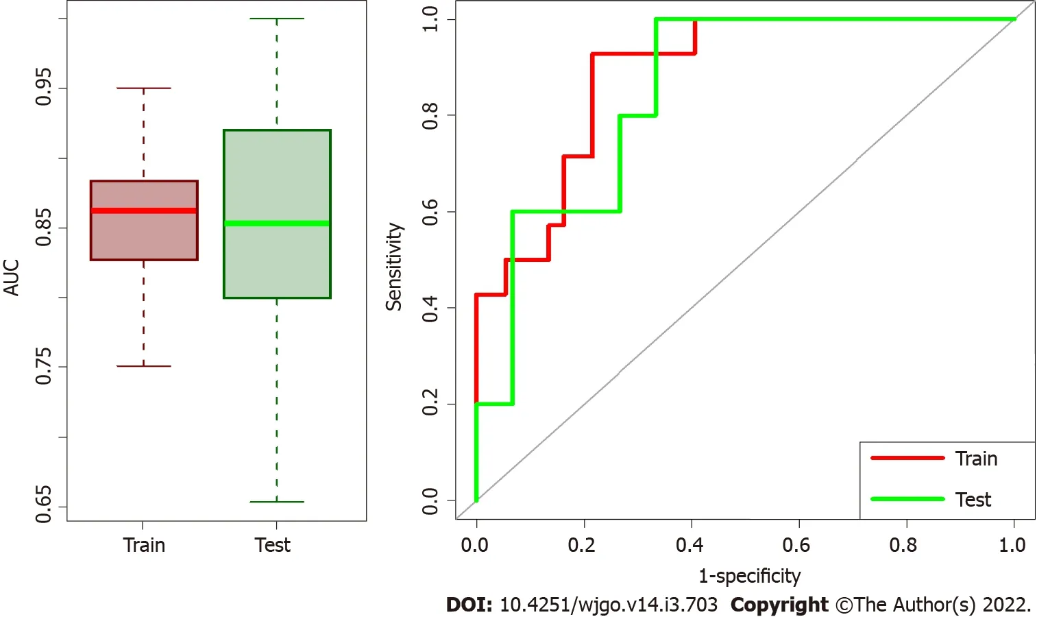
The model was then tested on the validation dataset,and corresponding area under the receiver operating characteristic curve(AUC)and their confidence intervals were computed.To improve the statistical significance and robustness of our analysis,all the steps were repeated 100 times.More precisely,among these repeated analyses,the solution that presents the median AUC was chosen as representative,and corresponding confidence intervals were computed throughout a bootstrap.The already tested model was re-trained on the whole dataset to obtain robust selected variables,and AUC was computed.To assess the predictive capability of our model,this was finally applied to the entire cohort to predict the surgical resection status and compute the OS for predicted surgery
no predicted surgery patients.The log-rank test was used to assess statistical significance for survival curves.
Jack my Hedgehog mounted his cock, and driving his pigs before him into the village, he let every one kill as many as they chose, and such a hacking13 and hewing14 of pork went on as you might have heard for miles off
RESULTS
Study population
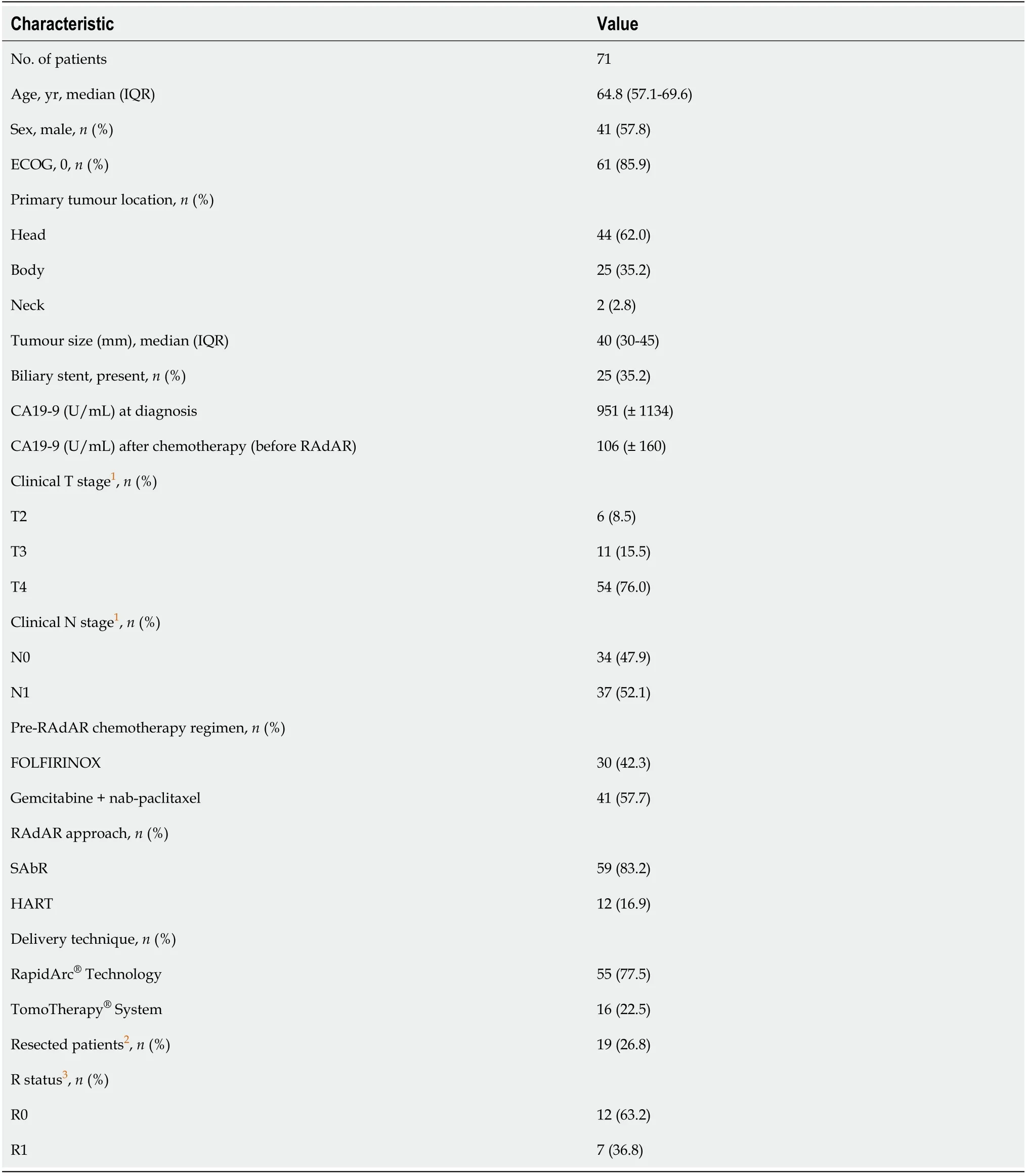
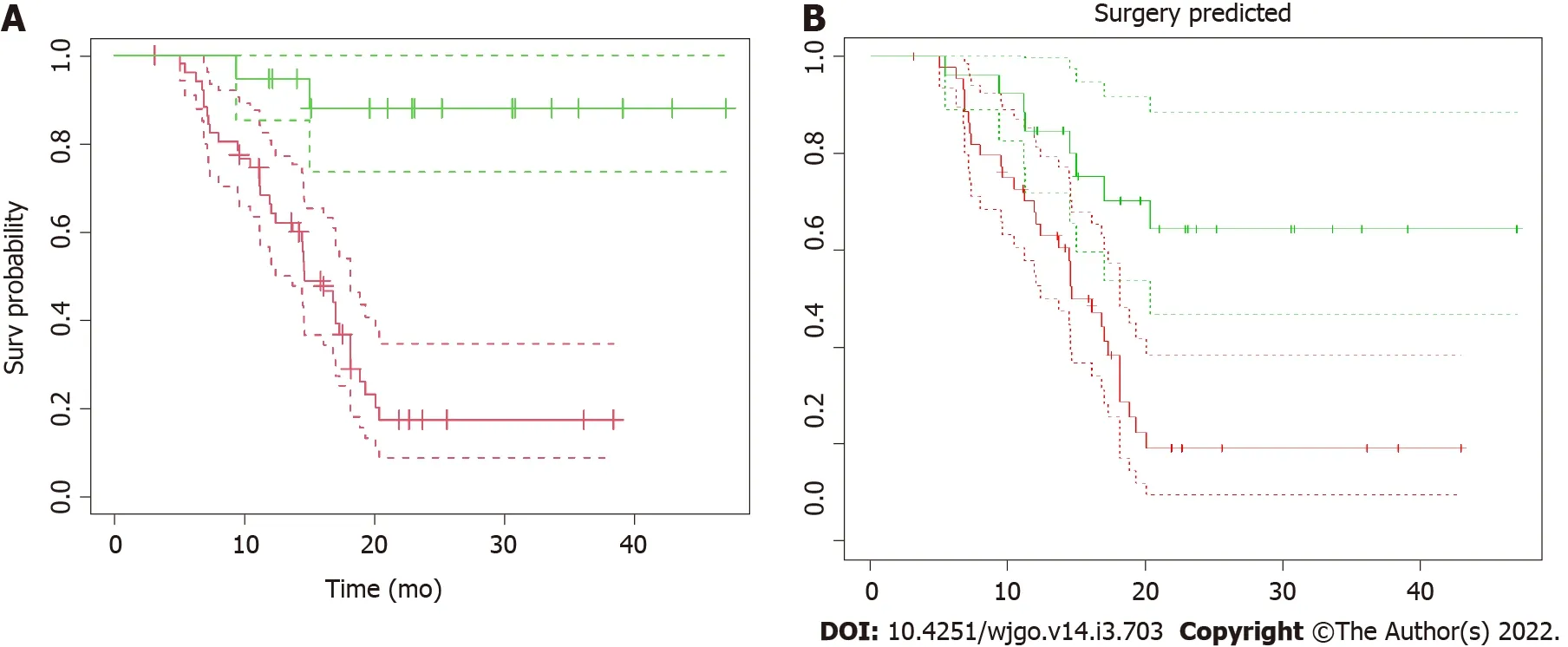
Seventy-one LAPC patients were included in the analysis.Baseline characteristics are outlined in Table 1.The median age was 65 years[interquartile range(IQR)57-69],and 57.8% of patients were male.The median period of induction chemotherapy was 6 mo(IQR 6-6 mo).SAbR was used in 59(83.1%)patients and HART in the remaining 12(16.9%).All patients completed the prescribed treatment.Thirty-two patients(45.1%)underwent exploratory laparotomy after RT,and 19(26.8%)patients ultimately received surgical resection,with a resection/exploration ratio of 59.4%.Postoperative 90-d mortality was nil.The median follow-up for the analysis was 15.0 mo(IQR 11.2-20.2 mo).Overall survival(OS)curves and their confidence intervals,estimated by Kaplan-Meier method as a function of surgical resection(resected
non-resected patients;
< 0.001),are shown in Figure 2A.
Prediction model for resectability
Median AUC for training and validation sets were 0.862(95%CI:0.792-0.921)and 0.853(95%CI:0.706-0.960),respectively.Box plots in Figure 3 summarized the AUC distributions for both datasets for all the 100 repetitions and the ROC curve of the median solution both for train and validation test.Among the 100 repetitions of the training process,the clinical variables were rarely selected from LASSO.On average,98% of the selected variables were radiomic features,indicating a higher predictive power of radiomic data with respect to clinical data.
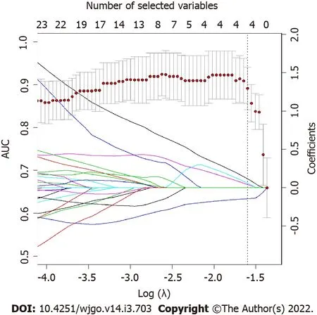
Applying the validated model on the entire dataset,4 features were selected from the LASSO regression.Figure 4 depicts the LASSO variable selection process and the AUC as a function of the lambda parameter.Four variables survived to LASSO regression in correspondence with the best lambda value(dotted line in Figure 4).Lambda was chosen as the value at one standard deviation after the value that maximises AUC.The selected features,the
value from the Wilcoxon test,and their adjusted
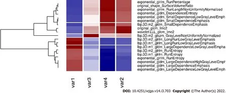
-value for Bonferroni correction are reported in Table 2.In addition,the slope of LASSO regression coefficients for each variable is shown.Starting from the selected variables,the correlationmatrix between these features and all other features with at least one Pearson correlation higher than 0.9 are reported in Figure 5,as a reference for external validation.
An important consideration of the current study is that clinical data was not significant to separate resected from not-resected patients and did not contribute to the building of the predictive model.This means that radiomic represents a fundamental tool to decode information that cannot be obtained from the direct observation of CT images or other clinical data alone[34].Notably,the “real” OS curves(Figure 2A)and “predicted” OS curves obtained applying the validated model to the entire database without considering the information of resectability status(Figure 2B)are comparable.This is an indirect validation of the model that can independently predict before RT whether the patient can be a candidate for surgical resection,leading to comparable OS curves.This finding is particularly relevant from a clinical point of view.Indeed,although recent retrospective series have suggested that RT can complement induction chemotherapy and improve LAPC resectability[11-13],the role of(chemo-)RT after systemic therapy in LAPC is still controversial[35].

AUC obtained for the model applied on the entire dataset is 0.944(95%CI:0.892-0.996)(Figure 6).OS curves and their confidence intervals,estimated by Kaplan-Meier method as a function of surgical resection(resected
non-resected patients)as predicted by the model,are shown in Figure 2B(
<0.001).
DlSCUSSlON
To the best of our knowledge,the present study is the first to propose a CT-based radiomics model to predict resectability in LAPC treated with intensive chemotherapy followed by ablative RT.The model was built,tested,and validated in a homogeneous cohort of LAPC patients,using clinical data and radiomic features extracted from the simulation-CT,and showed a reliable performance to predict surgical resection.
Surgical resection after neoadjuvant treatment is the main driver for improved survival in LAPC[11].However,the diagnostic performance of CT imaging to evaluate the residual tumour burden at restaging after neoadjuvant therapy is low,due to the difficulty in distinguishing neoplastic tissue from fibrous scar or inflammation[33].In this context,radiomics has gained popularity over conventional imaging as a complementary clinical tool capable of providing additional,unprecedented information regarding the intratumor heterogeneity and the residual neoplastic tissue,potentially serving in the therapeutic decision-making process.
In the present study,we identified four potential CT-based radiomic features(lbp.3D.m1_glrlm_LRLGE;wavelet.LLL_glcm_Imc2;lbp.3D.m
_glszm_GLLN;exponential_glrlm_RLNN)to build a CT radiomic model,which could be useful in predicting resectability in LAPC patients undergoing neoadjuvant therapy.Notably,the AUC obtained for the model was 0.94 if applied to the entire dataset.Radiomics has already been used in other contexts of surgical oncology,predicting resectability in oesophageal[28]and thymic cancers[29].
They were taller than mortal men, and the young man had a staff in his hand with golden wings on it, and two golden serpents twisted round it, and he had wings on his cap and on his shoes

Radiomic features were extracted from the VOI using PyRadiomics v3.0[31].Firstly,the VOI was resampled with an isotropic voxel of 2 mm × 2 mm × 2 mm,and HU were binned considering a width of 5 HU.The extracted features are defined according to Imaging Biomarker Standardized Initiative(IBSI)[32]and include first order statistics,shaped based both 2D and 3D,Gray Level Co-occurrence Matrix,Gray Level Run Length Matrix,Gray Level Size Zone Matrix,Neighbouring Gray Tone Difference Matrix,and Grey Level Dependence Matrix,as well as filtered features(logarithm exponential,gradient,LBP3D,and wavelet).A total of 1655 radiomic features were considered.
The analysis of the present study has some significant strengths.First,a three-step machine learning method was implemented.More precisely,the model was trained on a training subset of patients finding a robust AUC(median value:0.86);subsequently,this model was validated and confirmed on a smaller subset(30% of the total patients),confirming high AUC values(median value:0.85).Ultimately,it was re-applied on the whole database to extract the more significant features that contribute to the prediction,obtaining a high AUC value(0.94).Second,the statistical significance of all the steps gives robust and reliable predictions.Indeed,radiomic analysis and machine learning implementation are prone to lead to several errors and unreproducible results[36].To give strengths and add reproducibility to the analysis,all results were cross-validated or obtained throughout multiple repetitions and corrected for multiple testing.
This study had several limitations.First,the design was observational,with a retrospective analysis and a relatively small sample size.Second,the results may have been biased by the patient selection process since indications to chemotherapy,radiotherapy,and surgery were defined on a case-by-case basis by the Institutional board.The lack of external validation represents a third limit.An external multi-institutional validation may have been preferable.Finally,due to the limited number of events in the resected group,we were unable to perform an analysis on the impact of resection status(R0
R1)on survival.
A further frontier of the application of radiomics,not evaluated in the present study,is to analyse the change in radiomic features during or after treatment(the so-called delta radiomics)[37,38].The application of delta radiomic might further improve the ability of radiomics to provide predictive models for oncological outcomes and instruct clinical decisions.Furthermore,another potential application of radiomics in oncology is to realize prognostic and predictive models of the tumour pathological response(TRG)to neoadjuvant treatment[39-41].This information could lead to better identification of patients who may or may not benefit from preoperative approach,in order to allow for more effective treatment personalization.
In the analysis,all the features correlated with the most significant ones were rejected to maintain only those variables that explained more variance.All the significant features correlated with at least one of the selected features are shown in Figure 5.The names of correlated variables are provided,allowing other centres to reproduce the presented results.Further studies could find significant variables different to those reported in our study,but correlated with them,indicating they have the same underlying radiomic information.Lastly,the possibility to extract valuable information on planning CT without additional diagnostic methodologies adds simplicity and the possibility to apply this in the clinical practice.
My stomach suddenly heaved, and a bitter taste rose into my throat. I leaned over the rail and lost my lunch. I d never been so sick in all my life—and I was freezing! Goosebumps() stood out on my arms like grapefruits. Why hadn t I worn a jacket? Why had I even come? Who needed to go deep-sea fishing anyway? I suddenly realized I hated fish—especially the dead ones in the bait buckets. The stink4 of them filled my nose, my head—my stomach! Breakfast followed lunch.
CONCLUSlON
The present CT-radiomic model demonstrated a reliable performance to predict resectability in LAPC treated with induction chemotherapy followed by ablative RT.Radiomic information may complement clinical and morphological parameters in predicting surgical resectability.If further confirmed,the results of this study may allow integrating radiomic information into the pool of clinical and biochemical data to consider when a LAPC patient is candidate for surgical exploration after neoadjuvant therapy.
ARTlCLE HlGHLlGHTS
Research background
Radiomics is emerging as a promising tool in oncology,potentially improving,through the development of predictive and prognostic models,the therapeutic decision-making process.To date,however,few data are available regarding the use of radiomics in pancreatic cancer(PC).Since computed tomography(CT)misestimate the resectability of locally advanced PC(LAPC)after neoadjuvant treatment,the role of radiomics could be decisive to integrate traditional morphological parameters in predicting surgical resection.
Research motivation
To explore the potential role of CT-radiomic features to integrate clinical and morphological data to predict surgical resection in LAPC treated with neoadjuvant chemotherapy and radiotherapy.
Research objectives
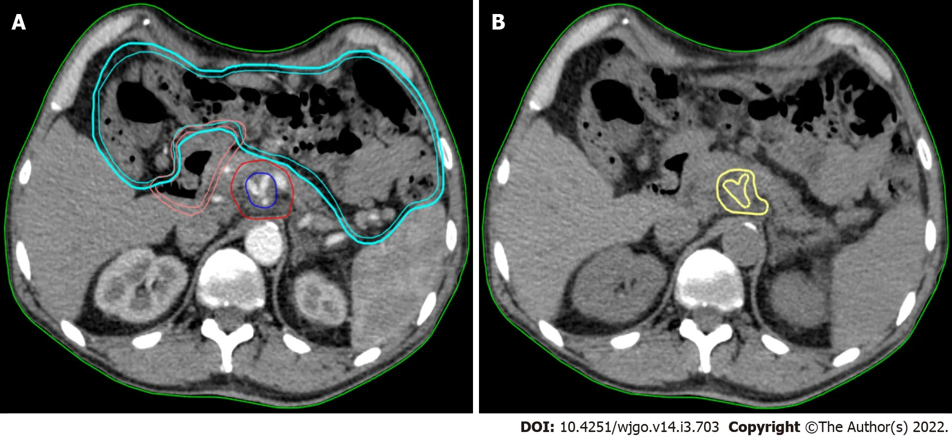
Texture analysis was performed using contrast-free simulation CT imaging.Tumour segmentation was performed using a software for medical image processing(MIM software;Mim Software Inc.,United States).The volume of interest(VOI)corresponded to the gross tumour volume(GTV)and was defined by a radiation oncologist with experience in PC and validated by a second radiation oncologist(Figure 1).The segmentation excluded vessels,biliary stent,calcifications,fiducial markers,or any other potential source of artifact from the GTV(VOI)(Figure 1).
Research methods
A total of 1655 radiomic features were extracted from planning CT inside the gross tumour volume.Resectability status predictive model was build starting from these radiomic features and clinical data.A first step of variable selection and a training/validation step to find the model that better predicted the outcome was adopted.Subsequently,the validated model was applied to the whole dataset.The discriminating performance of each model was assessed with the area under the receiver operating characteristic curve(AUC).
Research results
Seventy-one LAPC patients were included in the analysis.After neoadjuvant chemotherapy and radiotherapy,19(26.8%)patients underwent surgical resection.The training and validation steps resulted in a predictive model of resectability with a median AUC of 0.862(95%CI:0.792-0.921)and 0.853(95%CI:0.706-0.960),respectively.This model applied to the entire dataset allowed to select 4 radiomic features that predict the respectability status with an AUC of 0.944(95%CI:0.892-0.996).No clinical data contributed to the predictive model.
Husband, said the woman, have you caught nothing to-day? No, said the man, I did catch a Flounder, who said he was an enchanted prince, so I let him go again. Did you not wish for anything first? said the woman. No, said the man; what should I wish for? Ah, said the woman, it is surely hard to have to live always in this dirty hovel; you might have wished for a small cottage for us. Go back and call him. Tell him we want to have a small cottage, he will certainly give us that. Ah, said the man, why should I go there again? Why, said the woman, you did catch him, and you let him go again; he is sure to do it. Go at once. The man still did not quite like to go, but did not like to oppose his wife, and went to the sea.
Research conclusions
The present radiomic model could help predict resectability in LAPC treated with neoadjuvant therapy,suggesting a promising role in the context of a complex long-course downstaging and a challenging indication to surgery.
Research perspectives
The analysis of the change of radiomic features during or after treatment(delta radiomics)and the correlation with tumour response(
.
.,tumour regression grade)represent another intriguing application of radiomics that needs further exploration.
Then the hand of the person who has touched the bird will be held as in a vice9, and nothing will set it free, unless you touch it with this little stick which I will make you a present of
FOOTNOTES
Rossi G,Altabella L,and Simoni N designed the research;Rossi G,Benetti G,Rossi R,Venezia M,Paiella S,and Malleo G collected data;Rossi G and Simoni N analysed clinical and radiation data;Altabella L and Benetti G performed the radiomic features extraction,machine learning algorithm implementation,and statistical analysis;Rossi G,Altabella L and Simoni N wrote the manuscript;Benetti G,Rossi R,Venezia M,Paiella S,Malleo G,Salvia R,Guariglia S,Bassi C,Cavedon C,and Mazzarotto R reviewed the manuscript;All authors approved the final version of the manuscript.
The Institutional Review Board(IRB)approved the prospective collection of patient data,No.PAD-R n.1101 CESC.
All study participants or their legal guardian provided informed written consent about personal and medical data collection prior to study enrolment.
We have no financial relationships to disclose.
No additional data are available.
This article is an open-access article that was selected by an in-house editor and fully peer-reviewed by external reviewers.It is distributed in accordance with the Creative Commons Attribution NonCommercial(CC BYNC 4.0)license,which permits others to distribute,remix,adapt,build upon this work non-commercially,and license their derivative works on different terms,provided the original work is properly cited and the use is noncommercial.See:http://creativecommons.org/Licenses/by-nc/4.0/
Soon, I was following this routine every Sunday morning with almost religious zeal21. She never missed her rendezvous22 with me -- same place, same day, at precisely23 the same time, Seven o clock. Still, not a word was exchanged between us. I was too shy and she probably wanted to keep it this way -- a beautiful ethereal relationship -- a love so delicate that one wrong move might ruin everything. Meanwhile, I had developed a taste for fried pomfret -- quite surprisingly, considering that I had never eaten fish before.
Italy
Gabriella Rossi 0000-0002-3613-9229;Luisa Altabella 0000-0002-4816-6945;Nicola Simoni 0000-0003-4651-1757;Giulio Benetti 0000-0002-7070-0083;Roberto Rossi 0000-0002-8002-0495;Martina Venezia 0000-0001-5431-6420;Salvatore Paiella 0000-0001-6893-8618;Giuseppe Malleo 0000-0001-5467-2628;Roberto Salvia 0000-0002-3514-8473;Stefania Guariglia 0000-0002-3161-5345;Claudio Bassi 0000-0002-5416-329X;Carlo Cavedon 0000-0001-7710-4607;Renzo Mazzarotto 0000-0002-0122-1871.
AIRO;ESTRO.
Fan JR
Yeah, he said shyly. Well, last week, when I was sure the parole was coming through, I wrote her again. We used to live in Brunswick, just before Jacksonville, and there s a big oak tree just as you come into town. I told her that if she d take me back, she should put a yellow handkerchief on the tree, and I d get off and come home. If she didn t want me, forget it no handkerchief, and I d go on through.
Filipodia
Fan JR
1 Siegel RL,Miller KD,Jemal A.Cancer Statistics,2017.
2017;67:7-30[PMID:28055103 DOI:10.3322/caac.21387]
2 Rahib L,Smith BD,Aizenberg R,Rosenzweig AB,Fleshman JM,Matrisian LM.Projecting cancer incidence and deaths to 2030:the unexpected burden of thyroid,liver,and pancreas cancers in the United States.
2014;74:2913-2921[PMID:24840647 DOI:10.1158/0008-5472.CAN-14-0155]
3 Mizrahi JD,Surana R,Valle JW,Shroff RT.Pancreatic cancer.
2020;395:2008-2020[PMID:32593337 DOI:10.1016/S0140-6736(20)30974-0]
4 Maggino L,Malleo G,Marchegiani G,Viviani E,Nessi C,Ciprani D,Esposito A,Landoni L,Casetti L,Tuveri M,Paiella S,Casciani F,Sereni E,Binco A,Bonamini D,Secchettin E,Auriemma A,Merz V,Simionato F,Zecchetto C,D'Onofrio M,Melisi D,Bassi C,Salvia R.Outcomes of Primary Chemotherapy for Borderline Resectable and Locally Advanced Pancreatic Ductal Adenocarcinoma.
2019;154:932-942[PMID:31339530 DOI:10.1001/jamasurg.2019.2277]
5 Suker M,Beumer BR,Sadot E,Marthey L,Faris JE,Mellon EA,El-Rayes BF,Wang-Gillam A,Lacy J,Hosein PJ,Moorcraft SY,Conroy T,Hohla F,Allen P,Taieb J,Hong TS,Shridhar R,Chau I,van Eijck CH,Koerkamp BG.FOLFIRINOX for locally advanced pancreatic cancer:a systematic review and patient-level meta-analysis.
2016;17:801-810[PMID:27160474 DOI:10.1016/S1470-2045(16)00172-8]
6 Reni M,Zanon S,Balzano G,Nobile S,Pircher CC,Chiaravalli M,Passoni P,Arcidiacono PG,Nicoletti R,Crippa S,Slim N,Doglioni C,Falconi M,Gianni L.Selecting patients for resection after primary chemotherapy for non-metastatic pancreatic adenocarcinoma.
2017;28:2786-2792[PMID:28945895 DOI:10.1093/annonc/mdx495]
7 Chung SY,Chang JS,Lee BM,Kim KH,Lee KJ,Seong J.Dose escalation in locally advanced pancreatic cancer patients receiving chemoradiotherapy.
2017;123:438-445[PMID:28464997 DOI:10.1016/j.radonc.2017.04.010]
8 Krishnan S,Chadha AS,Suh Y,Chen HC,Rao A,Das P,Minsky BD,Mahmood U,Delclos ME,Sawakuchi GO,Beddar S,Katz MH,Fleming JB,Javle MM,Varadhachary GR,Wolff RA,Crane CH.Focal Radiation Therapy Dose Escalation Improves Overall Survival in Locally Advanced Pancreatic Cancer Patients Receiving Induction Chemotherapy and Consolidative Chemoradiation.
2016;94:755-765[PMID:26972648 DOI:10.1016/j.ijrobp.2015.12.003]
9 Toesca DAS,Ahmed F,Kashyap M,Baclay JRM,von Eyben R,Pollom EL,Koong AC,Chang DT.Intensified systemic therapy and stereotactic ablative radiotherapy dose for patients with unresectable pancreatic adenocarcinoma.
2020;152:63-69[PMID:32763253 DOI:10.1016/j.radonc.2020.07.053]
10 Reyngold M,O'Reilly EM,Varghese AM,Fiasconaro M,Zinovoy M,Romesser PB,Wu A,Hajj C,Cuaron JJ,Tuli R,Hilal L,Khalil D,Park W,Yorke ED,Zhang Z,Yu KH,Crane CH.Association of Ablative Radiation Therapy With Survival Among Patients With Inoperable Pancreatic Cancer.
2021;7:735-738[PMID:33704353 DOI:10.1001/jamaoncol.2021.0057]
11 Gemenetzis G,Groot VP,Blair AB,Laheru DA,Zheng L,Narang AK,Fishman EK,Hruban RH,Yu J,Burkhart RA,Cameron JL,Weiss MJ,Wolfgang CL,He J.Survival in Locally Advanced Pancreatic Cancer After Neoadjuvant Therapy and Surgical Resection.
2019;270:340-347[PMID:29596120 DOI:10.1097/SLA.0000000000002753]
12 Murphy JE,Wo JY,Ryan DP,Clark JW,Jiang W,Yeap BY,Drapek LC,Ly L,Baglini CV,Blaszkowsky LS,Ferrone CR,Parikh AR,Weekes CD,Nipp RD,Kwak EL,Allen JN,Corcoran RB,Ting DT,Faris JE,Zhu AX,Goyal L,Berger DL,Qadan M,Lillemoe KD,Talele N,Jain RK,DeLaney TF,Duda DG,Boucher Y,Fernández-Del Castillo C,Hong TS.Total Neoadjuvant Therapy With FOLFIRINOX in Combination With Losartan Followed by Chemoradiotherapy for Locally Advanced Pancreatic Cancer:A Phase 2 Clinical Trial.
2019;5:1020-1027[PMID:31145418 DOI:10.1001/jamaoncol.2019.0892]
13 Simoni N,Micera R,Paiella S,Guariglia S,Zivelonghi E,Malleo G,Rossi G,Addari L,Giuliani T,Pollini T,Cavedon C,Salvia R,Milella M,Bassi C,Mazzarotto R.Hypofractionated Stereotactic Body Radiation Therapy With Simultaneous Integrated Boost and Simultaneous Integrated Protection in Pancreatic Ductal Adenocarcinoma.
2021;33:e31-e38[PMID:32682686 DOI:10.1016/j.clon.2020.06.019]
14 Rossi G,Simoni N,Paiella S,Rossi R,Venezia M,Micera R,Malleo G,Salvia R,Giuliani T,Di Gioia A,Auriemma A,Milella M,Guariglia S,Cavedon C,Bassi C,Mazzarotto R.Risk Adapted Ablative Radiotherapy After Intensive Chemotherapy for Locally Advanced Pancreatic Cancer.
2021;11:662205[PMID:33959509 DOI:10.3389/fonc.2021.662205]
15 Jang JK,Byun JH,Kang JH,Son JH,Kim JH,Lee SS,Kim HJ,Yoo C,Kim KP,Hong SM,Seo DW,Kim SC,Lee MG.CT-determined resectability of borderline resectable and unresectable pancreatic adenocarcinoma following FOLFIRINOX therapy.
2021;31:813-823[PMID:32845389 DOI:10.1007/s00330-020-07188-8]
16 Ferrone CR,Marchegiani G,Hong TS,Ryan DP,Deshpande V,McDonnell EI,Sabbatino F,Santos DD,Allen JN,Blaszkowsky LS,Clark JW,Faris JE,Goyal L,Kwak EL,Murphy JE,Ting DT,Wo JY,Zhu AX,Warshaw AL,Lillemoe KD,Fernández-del Castillo C.Radiological and surgical implications of neoadjuvant treatment with FOLFIRINOX for locally advanced and borderline resectable pancreatic cancer.
2015;261:12-17[PMID:25599322 DOI:10.1097/SLA.0000000000000867]
17 Wagner M,Antunes C,Pietrasz D,Cassinotto C,Zappa M,Sa Cunha A,Lucidarme O,Bachet JB.CT evaluation after neoadjuvant FOLFIRINOX chemotherapy for borderline and locally advanced pancreatic adenocarcinoma.
2017;27:3104-3116[PMID:27896469 DOI:10.1007/s00330-016-4632-8]
18 Kim YE,Park MS,Hong HS,Kang CM,Choi JY,Lim JS,Lee WJ,Kim MJ,Kim KW.Effects of neoadjuvant combined chemotherapy and radiation therapy on the CT evaluation of resectability and staging in patients with pancreatic head cancer.
2009;250:758-765[PMID:19164113 DOI:10.1148/radiol.2502080501]
19 Cassinotto C,Cortade J,Belleannée G,Lapuyade B,Terrebonne E,Vendrely V,Laurent C,Sa-Cunha A.An evaluation of the accuracy of CT when determining resectability of pancreatic head adenocarcinoma after neoadjuvant treatment.
2013;82:589-593[PMID:23287712 DOI:10.1016/j.ejrad.2012.12.002]
20 Cassinotto C,Mouries A,Lafourcade JP,Terrebonne E,Belleannée G,Blanc JF,Lapuyade B,Vendrely V,Laurent C,Chiche L,Wagner T,Sa-Cunha A,Gaye D,Trillaud H,Laurent F,Montaudon M.Locally advanced pancreatic adenocarcinoma:reassessment of response with CT after neoadjuvant chemotherapy and radiation therapy.
2014;273:108-116[PMID:24960211 DOI:10.1148/radiol.14132914]
21 Lambin P,Leijenaar RTH,Deist TM,Peerlings J,de Jong EEC,van Timmeren J,Sanduleanu S,Larue RTHM,Even AJG,Jochems A,van Wijk Y,Woodruff H,van Soest J,Lustberg T,Roelofs E,van Elmpt W,Dekker A,Mottaghy FM,Wildberger JE,Walsh S.Radiomics:the bridge between medical imaging and personalized medicine.
2017;14:749-762[PMID:28975929 DOI:10.1038/nrclinonc.2017.141]
22 Giraud P,Giraud P,Gasnier A,El Ayachy R,Kreps S,Foy JP,Durdux C,Huguet F,Burgun A,Bibault JE.Radiomics and Machine Learning for Radiotherapy in Head and Neck Cancers.
2019;9:174[PMID:30972291 DOI:10.3389/fonc.2019.00174]
23 Ciaravino V,Cardobi N,DE Robertis R,Capelli P,Melisi D,Simionato F,Marchegiani G,Salvia R,D'Onofrio M.CT Texture Analysis of Ductal Adenocarcinoma Downstaged After Chemotherapy.
2018;38:4889-4895[PMID:30061265 DOI:10.21873/anticanres.12803]
24 Borhani AA,Dewan R,Furlan A,Seiser N,Zureikat AH,Singhi AD,Boone B,Bahary N,Hogg ME,Lotze M,Zeh HJ III,Tublin ME.Assessment of Response to Neoadjuvant Therapy Using CT Texture Analysis in Patients With Resectable and Borderline Resectable Pancreatic Ductal Adenocarcinoma.
2020;214:362-369[PMID:31799875 DOI:10.2214/AJR.19.21152]
25 Cozzi L,Comito T,Fogliata A,Franzese C,Franceschini D,Bonifacio C,Tozzi A,Di Brina L,Clerici E,Tomatis S,Reggiori G,Lobefalo F,Stravato A,Mancosu P,Zerbi A,Sollini M,Kirienko M,Chiti A,Scorsetti M.Computed tomography based radiomic signature as predictive of survival and local control after stereotactic body radiation therapy in pancreatic carcinoma.
2019;14:e0210758[PMID:30657785 DOI:10.1371/journal.pone.0210758]
26 Sandrasegaran K,Lin Y,Asare-Sawiri M,Taiyini T,Tann M.CT texture analysis of pancreatic cancer.
2019;29:1067-1073[PMID:30116961 DOI:10.1007/s00330-018-5662-1]
27 Parr E,Du Q,Zhang C,Lin C,Kamal A,McAlister J,Liang X,Bavitz K,Rux G,Hollingsworth M,Baine M,Zheng D.Radiomics-Based Outcome Prediction for Pancreatic Cancer Following Stereotactic Body Radiotherapy.
2020;12[PMID:32344538 DOI:10.3390/cancers12041051]
28 Ou J,Li R,Zeng R,Wu CQ,Chen Y,Chen TW,Zhang XM,Wu L,Jiang Y,Yang JQ,Cao JM,Tang S,Tang MJ,Hu J.CT radiomic features for predicting resectability of oesophageal squamous cell carcinoma as given by feature analysis:a case control study.
2019;19:66[PMID:31619297 DOI:10.1186/s40644-019-0254-0]
29 Batista Araujo-Filho JA,Mayoral M,Zheng J,Tan KS,Gibbs P,Shepherd AF,Rimner A,Simone CB 2nd,Riely G,Huang J,Ginsberg MS.CT Radiomic Features for Predicting Resectability and TNM Staging in Thymic Epithelial Tumors.
2021[PMID:33844992 DOI:10.1016/j.athoracsur.2021.03.084]
30 National Comprehensive Cancer Network.Pancreatic Adenocarcinoma Version 2.2015.NCCN Guidelines.(2015).[cited 10 April 2021].Available from:https://www.nccn.org/professionals/physician_gls/pdf/pancreatic.pdf
31 van Griethuysen JJM,Fedorov A,Parmar C,Hosny A,Aucoin N,Narayan V,Beets-Tan RGH,Fillion-Robin JC,Pieper S,Aerts HJWL.Computational Radiomics System to Decode the Radiographic Phenotype.
2017;77:e104-e107[PMID:29092951 DOI:10.1158/0008-5472.CAN-17-0339]
32 Zwanenburg A,Vallières M,Abdalah MA,Aerts HJWL,Andrearczyk V,Apte A,Ashrafinia S,Bakas S,Beukinga RJ,Boellaard R,Bogowicz M,Boldrini L,Buvat I,Cook GJR,Davatzikos C,Depeursinge A,Desseroit MC,Dinapoli N,Dinh CV,Echegaray S,El Naqa I,Fedorov AY,Gatta R,Gillies RJ,Goh V,Götz M,Guckenberger M,Ha SM,Hatt M,Isensee F,Lambin P,Leger S,Leijenaar RTH,Lenkowicz J,Lippert F,Losnegård A,Maier-Hein KH,Morin O,Müller H,Napel S,Nioche C,Orlhac F,Pati S,Pfaehler EAG,Rahmim A,Rao AUK,Scherer J,Siddique MM,Sijtsema NM,Socarras Fernandez J,Spezi E,Steenbakkers RJHM,Tanadini-Lang S,Thorwarth D,Troost EGC,Upadhaya T,Valentini V,van Dijk LV,van Griethuysen J,van Velden FHP,Whybra P,Richter C,Löck S.The Image Biomarker Standardization Initiative:Standardized Quantitative Radiomics for High-Throughput Image-based Phenotyping.
2020;295:328-338[PMID:32154773 DOI:10.1148/radiol.2020191145]
33 Cassinotto C,Sa-Cunha A,Trillaud H.Radiological evaluation of response to neoadjuvant treatment in pancreatic cancer.
2016;97:1225-1232[PMID:27692675 DOI:10.1016/j.diii.2016.07.011]
34 Limkin EJ,Sun R,Dercle L,Zacharaki EI,Robert C,Reuzé S,Schernberg A,Paragios N,Deutsch E,Ferté C.Promises and challenges for the implementation of computational medical imaging(radiomics)in oncology.
2017;28:1191-1206[PMID:28168275 DOI:10.1093/annonc/mdx034]
35 Hammel P,Huguet F,van Laethem JL,Goldstein D,Glimelius B,Artru P,Borbath I,Bouché O,Shannon J,André T,Mineur L,Chibaudel B,Bonnetain F,Louvet C;LAP07 Trial Group.Effect of Chemoradiotherapy
Chemotherapy on Survival in Patients With Locally Advanced Pancreatic Cancer Controlled After 4 Months of Gemcitabine With or Without Erlotinib:The LAP07 Randomized Clinical Trial.
2016;315:1844-1853[PMID:27139057 DOI:10.1001/jama.2016.4324]
36 Rizzo S,Botta F,Raimondi S,Origgi D,Fanciullo C,Morganti AG,Bellomi M.Radiomics:the facts and the challenges of image analysis.
2018;2:36[PMID:30426318 DOI:10.1186/s41747-018-0068-z]
37 Nasief H,Zheng C,Schott D,Hall W,Tsai S,Erickson B,Allen Li X.A machine learning based delta-radiomics process for early prediction of treatment response of pancreatic cancer.
2019;3:25[PMID:31602401 DOI:10.1038/s41698-019-0096-z]
38 Cusumano D,Boldrini L,Yadav P,Casà C,Lee SL,Romano A,Piras A,Chiloiro G,Placidi L,Catucci F,Votta C,Mattiucci GC,Indovina L,Gambacorta MA,Bassetti M,Valentini V.Delta Radiomics Analysis for Local Control Prediction in Pancreatic Cancer Patients Treated Using Magnetic Resonance Guided Radiotherapy.
2021;11[PMID:33466307 DOI:10.3390/diagnostics11010072]
39 Li ZY,Wang XD,Li M,Liu XJ,Ye Z,Song B,Yuan F,Yuan Y,Xia CC,Zhang X,Li Q.Multi-modal radiomics model to predict treatment response to neoadjuvant chemotherapy for locally advanced rectal cancer.
2020;26:2388-2402[PMID:32476800 DOI:10.3748/wjg.v26.i19.2388]
40 Yuan Z,Frazer M,Zhang GG,Latifi K,Moros EG,Feygelman V,Felder S,Sanchez J,Dessureault S,Imanirad I,Kim RD,Harrison LB,Hoffe SE,Frakes JM.CT-based radiomic features to predict pathological response in rectal cancer:A retrospective cohort study.
2020;64:444-449[PMID:32386109 DOI:10.1111/1754-9485.13044]
41 Yang Z,He B,Zhuang X,Gao X,Wang D,Li M,Lin Z,Luo R.CT-based radiomic signatures for prediction of pathologic complete response in esophageal squamous cell carcinoma after neoadjuvant chemoradiotherapy.
2019;60:538-545[PMID:31111948 DOI:10.1093/jrr/rrz027]
 World Journal of Gastrointestinal Oncology2022年3期
- World Journal of Gastrointestinal Oncology的其它文章
- lnflammatory bowel disease-related colorectal cancer:Past,present and future perspectives
- Barrett’s esophagus:Review of natural history and comparative efficacy of endoscopic and surgical therapies
- Gut and liver involvement in pediatric hematolymphoid malignancies
- Pathological,molecular,and clinical characteristics of cholangiocarcinoma:A comprehensive review
- Clinical significance of molecular subtypes of gastrointestinal tract adenocarcinoma
- Evolving roles of magnifying endoscopy and endoscopic resection for neoplasia in inflammatory bowel diseases
