
Basic Study CircRNA_0084927 promotes colorectal cancer progression by regulating miRNA-20b-3p/glutathione S-transferase mu 5 axis
2021-10-12 08:46FengLiuXiaoLiXiaoYuJingLiuRuoHuiXuWenJunZhouHanChenXuAiGuangZhaoYangXianXuYanQiDangGuangJi
World Journal of Gastroenterology 2021年36期
Feng Liu, Xiao-Li Xiao, Yu-Jing Liu, Ruo-Hui Xu, Wen-Jun Zhou, Han-Chen Xu, Ai-Guang Zhao, Yang-Xian Xu,Yan-Qi Dang, Guang Ji
Abstract BACKGROUND Colorectal cancer (CRC) is the third most common cancer and the second most common cause of cancer-related death worldwide.The 5-year survival rate of patients with early-stage CRC could reach 90%, but it is very low in patients with advanced-stage CRC.Recent studies have shown that circular RNAs play important roles in regulating the migration and invasion of CRC cells.AIM To elucidate the role of circRNA_0084927 (circ_0084927) in the migration and invasion of CRC cells and its underlying mechanism.METHODS Clinical tissue samples and cells were collected, and the expression of circ_0084927 was detected by quantitative polymerase chain reaction (qPCR).The diagnostic performance of circ_0084927 was assessed by receiver operating characteristic curve analysis.The role of circ_0084927 in CRC cell proliferation,migration, and invasion was determined using cell counting kit-8 assay, wound healing assay, and transwell assay, respectively.The regulatory relationship among circ_0084927, miRNA-20b-3p (miR-20b-3p), and glutathione S-transferase mu 5 (GSTM5) was identified using databases, luciferase reporter assay, qPCR,and Western blot analysis.AKT-mTOR signaling was also verified after circ_0084927 knockdown or miR-20b-3p mimic treatment.RESULTS The expression of circ_0084927 was significantly increased in CRC tissues and cells, and it was higher in advanced-stage CRC compared with early-stage CRC.The area under the curve (AUC) of circ_0084927 was 0.806 [95% confidence interval (CI): 0.683-0.896].In addition, the AUC was 0.874 (95%CI: 0.738-0.956) in patients with advanced-stage CRC and 0.713 (95%CI: 0.555-0.840) in those with early-stage CRC.Knockdown of circ_0084927 inhibited the migration and invasion of HCT116 cells.Moreover, circ_0084927 was found to act as a sponge of miR-20b-3p.MiR-20b-3p activation reduced the circ_0084927 level, whereas miR-20b-3p inhibition increased the circ_0084927 level.But the effect was not found after circ_0084927 mutation.In addition, miR-20b-3p expression in CRC patients was also reduced and negatively correlated with circ_0084927 expression.The function of circ_0084927 in HCT116 cells with circ_0084927 knockdown was rescued by miR-20b-3p.Moreover, GSTM5 expression was significantly decreased after overexpressing miR-20b-3p or inhibiting circ_0084927, but its expression was rescued when circ_0084927 and miR-20b-3p were both inhibited.Finally, AKTmTOR signaling was markedly regulated by circ_0084927 and miR-20b-3p.CONCLUSION The expression of circ_0084927 is significantly increased in CRC and higher in advanced-stage CRC than in early-stage CRC.Moreover, circ_0084927 potentially regulates CRC cell migration and invasion via the miR-20b-3p/GSTM5/AKT/mTOR pathway.
Key Words: Colorectal cancer; CircRNA_0084927; MiRNA-20b-3p; Glutathione Stransferase mu 5; Migration; Invasion
INTRODUCTION
Colorectal cancer (CRC) is the third most common cancer and the second most common cause of cancer-related death worldwide, and the numbers of new cases and deaths in 2018 were 1.8 million and 881000, respectively[1].With the development of diagnostic and therapeutic technologies, the 5-year survival rate for CRC patients has reached 65%[2] and can reach 90% in early-stage CRC patients.However, the 5-year survival rate of patients with advanced-stage CRC is very low due to tumor metastasis and other complications.Although colonoscopy is the gold standard of CRC screening, approximately 60% of CRC patients are diagnosed at an advanced stage given the rate of missed lesions and incomplete colonoscopy coverage[3-5].Therefore,it is urgent to elucidate the pathogenesis and molecular mechanism of CRC with metastasis.
Circular RNAs (circRNAs), as important regulatory noncoding RNAs, have become a new research hotspot following microRNAs (miRNAs) and long noncoding RNAs.Researchers have demonstrated that circRNAs play important roles in tumors,including CRC[6-9], and their functions are completed in a variety of ways, such as acting as sponges of miRNAs[6,7], acting as transcriptional regulators[10,11], and translating proteins by combining with RNA binding proteins[12-15].However, to date, the functions of circRNAs in CRC remain largely unknown, especially in CRC with metastasis.Further study to elucidate the function of circRNAs in CRC with metastasis is needed.
In this study, we first verified the differential expression of circRNA_0084927(circ_0084927) according to previous circRNA sequencing data[16] and further found that circ_0084927 was associated with the pathological stage of CRC.Then, we demonstrated that knockdown of circ_0084927 markedly inhibited HCT116 cell migration and invasion.Regarding the regulatory mechanism of circ_0084927, it acts as a sponge of miRNA-20b-3p (miR-20b-3p), regulating glutathione S-transferase mu 5(GSTM5) and the AKT-mTOR pathway.Therefore, our findings elucidated the role of circ_0084927 and provided a new treatment strategy for CRC with metastasis.
MATERIALS AND METHODS
Clinical tissue specimens
Thirty pairs of CRC tissues and adjacent normal tissues (normal) were obtained during surgery at the Longhua Hospital Affiliated to Shanghai University of Traditional Chinese Medicine (Shanghai, China).The diagnosis of CRC was confirmed based on pathological evidence.Tissues were snap-frozen in liquid nitrogen and stored at -80 °C before detection.The study was approved by the Ethics Committee of Longhua Hospital (No.2019LCSY020), and informed consent was obtained from all participants.
Cell culture and transfection
Normal colon cells (FHC) and CRC cells (HCT116, HT29, SW480, and SW620)(Shanghai Cell Bank, shanghai, China) were cultured in Dulbecco’s modified Eagle’s medium supplemented with 10% fetal bovine serum and penicillin/streptomycin (100 U/mL) (Gibco, Carlsbad, United States) in an incubator with 5% CO2at 37 °C.In addition, human embryonic kidney 293T (HEK-293T) cells obtained from American Type Culture Collection (ATCC) (Manassas, United States) were cultured in Roswell Park Memorial Institute 1640 medium with 10% fetal bovine serum and penicillin/streptomycin (100 U/mL).Short hairpin circ_0084927 plasmid (sh-circ_0084927;GeneChem, China) and miR-20b-3p mimic/inhibitor or negative control (NC,Genomeditech, China; 200 nM) were transfected using FuGene HD transfection reagent (Promega, United States) according to previous studies[17,18].
Cell counting kit-8 assay
After transfection, HCT116 cells were seeded into 96-well plates at a concentration of 1× 10⁴cells and cultured for 0 h, 24 h, 48 h, and 72 h.Then, 10 μL of cell counting kit-8 reagent was added to each well.After incubation at 37 °C for 1 h, the absorbance value was detected at 450 nm.
Wound healing assay
After transfection, HCT116 cells were seeded in a 6-well dish with a culture insert(Ibidi, Germany) at a concentration of 3 × 10⁴cells.After 24 h, the culture insert was removed, and the cells were washed twice with polybutylene succinate.Two milliliters of serum-free medium were added to each dish for 48 h.Images were captured, and the wound area was measured using Image J software (National Institutes of Health,United States).
Cell migration invasion assay
Six-well plates with 8-μm chambers (Corning, United States) were used to assess cellular migration (without Matrigel) or invasion (with Matrigel).Briefly, transfected HCT116 cells were seeded in 6-well plates at a concentration of 1 × 10⁵cells.Two hundred microliters of serum-free medium was added to the upper chamber, and six hundred microliters of medium with 30% fetal bovine serum was added to the lower chamber for 48 h.Then, the cells were fixed with 4% paraformaldehyde for 30 min and stained with 0.1% crystal violet solution for 15 min.Four fields were randomly selected to calculate the number of migrating or invading cells and to evaluate the ability of cell migration or invasion.
Quantitative polymerase chain reaction
Total RNA was extracted using TRIzol reagent (Ambion, United States).For circRNA and mRNA analysis, cDNA was synthesized using an EVM-MLV reverse transcription kit (Aikeri Biotech, Hunan, China).For miRNA analysis, cDNA was synthesized using a miRNA cDNA synthesis kit (Sangon Biotech, Shanghai, China).The amplification reaction was performed using the SYBR-Green quantitative polymerase chain reaction(qPCR) kit (Thermo Fisher Scientific, MA, United States) or the miRNA fluorescence quantitative PCR kit (Sangon Biotech).Gene expression was normalized using β-actin or U6.The primers are listed in Table 1.
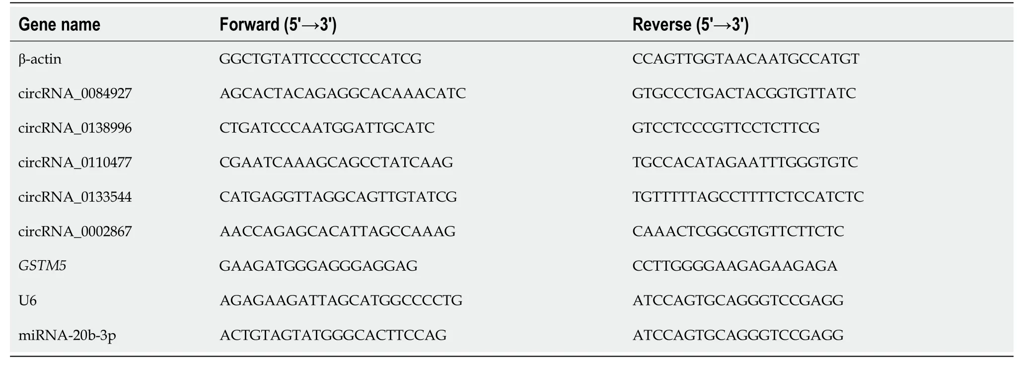
Table 1 Sequence of primers used in the study
Western blot analysis
Cells were collected and lysed.Protein concentration was determined using a protein assay kit (Beyotime, China).The protein was separated and transferred to a polyvinylidene fluoride membrane followed by incubation with 5% milk at room temperature for 1 h.The membrane was incubated at 4 °C overnight with the following antibodies: GSTM5 (GTX108776, Genetex, United States), PI3 kinase p85(PI3K, 4292S, CST, United States), phospho-PI3K (BS-3332R, Bioss, China), Akt (4685S,CST), phospho-Akt (4060S, CST), mTOR (2983S, CST), phospho-mTOR (5536S, CST),and β-actin (4970s, CST).Then, the secondary antibody was added and incubated at room temperature for 2 h, and protein expression was observed using a chemiluminescence gel imaging system (Tanon 5200, China).
Double luciferase reporter assay
HEK-293T cells were seeded in 24-well plates and transfected using FuGene HD transfection reagent (Promega, United States) according to previous studies[17,18].Briefly, HEK-293T cells were transfected using circ_0084927 wild type (circ_0084927-WT; Genomeditech, China) or circ_0084927 mutant (circ_0084927-Mut; Genomeditech)plasmid with or without miR-20b-3p mimic/inhibitor.After transfection for 22 h,luciferase activity was detected using the dual-luciferase reporter system (Promega).
Statistical analysis
Statistical analyses were conducted using SPSS 24.0 software.Data were assessed using Student’st-test or one-way ANOVA.Pearson correlation was used to analyze the correlation between circRNA and miRNA.Receiver operating characteristic (ROC)curve analysis was performed to evaluate the diagnostic value using MedCalc software.P< 0.05 was considered statistically significant.Dr.Ming Yang from the Good Clinical Practice Office, Longhua Hospital, Shanghai University of Traditional Chinese Medicine reviewed the statistical methods of this study before submission.
RESULTS
Circ_0084927 is markedly increased in CRC and associated with the pathological stage of CRC
Our previous study has proved that circRNAs play important roles in the transition of adenoma into CRC[16], but the function of circRNAs in advanced-stage CRC remains largely undetermined.According to previous circRNA sequencing data[16], five differentially expressed circRNAs were chosen and verified.The results showed that circ_0084927 expression was significantly increased in the CRC group compared with the normal group and was higher in advanced-stage CRC compared with early-stage CRC (Figure 1A and B).The other four circRNAs were not significantly different(Supplementary Figure 1).In addition, circ_0084927 expression in CRC cells (HCT116,HT29, and SW620) was also increased (Figure 1C).ROC curve analysis was used to evaluate the diagnostic performance of circ_0084927 in CRC.The area under the curve(AUC) of circ_0084927 was 0.806 (95%CI: 0.683 to 0.896) (P< 0.001).In addition, the AUC was 0.874 (95%CI: 0.738-0.956) in patients with advanced-stage CRC and 0.713(95%CI: 0.555-0.840) in those with early-stage CRC, indicating that circ_0084927 hadhigher diagnostic performance in advanced-stage CRC (P= 0.0153) (Figure 1D and E).

Figure 1 CircRNA_0084927 expression is markedly increased in colorectal cancer.A: Expression of circRNA_0084927 (circ_0084927) in colorectal cancer (CRC); B: Expression of circ_0084927 in advanced-stage and early-stage CRC; C: Expression of circ_0084927 in CRC cells; D: Receiver operating characteristic (ROC) curve analysis of circ_0084927; E: ROC analysis of circ_0084927 in advanced-stage and early-stage CRC.Data are presented as the mean ±SEM.aP < 0.05; bP < 0.01; cP < 0.001.Circ_0084927: CircRNA_0084927; Normal: Adjacent normal tissues; CRC: Colorectal cancer; ROC: Receiver operating characteristic; AUC: Area under the curve.
Knockdown of circ_0084927 inhibits HCT116 cell migration and invasion
To explore the function of circ_0084927 in CRC, circ_0084927 expression was inhibited in HCT116 cells using shRNA plasma (Figure 2A), and circ_0084927 knockdown markedly inhibited HCT116 cell proliferation at 48 h and 72 h (Figure 2B).In addition,circ_0084927 knockdown also significantly inhibited HCT116 cell migration and invasion (Figure 2C-G).These results demonstrated that circ_0084927 is an oncogene that promotes CRC function.

Figure 2 Knockdown of circRNA_0084927 inhibits the migration and invasion of HCT116 cells. A: Knockdown of circRNA_0084927(circ_0084927); B: Vability of HCT116 cells after circ_0084927 knockdown; C-G: Migration and invasion of HCT116 cells tested by wound healing assay (C and D)and transwell assay (E-G).Data are presented as the mean ± SEM.bP < 0.01; cP < 0.001.Circ_0084927: CircRNA_0084927; Sh-circ_0084927: Short hairpin circ_0084927 plasmid; Sh-NC: Short hairpin negative control plasmid.
Circ_0084927 acts as a sponge of miR-20b-3p in CRC
The biological function of circRNAs mainly involves acting as a sponge of miRNAs[19].In this study, circ_0084927 may act as a sponge of miR-20b-3p as demonstrated by bioinformatics analysis (Figure 3A).The miR-20b-3p mimic markedly reduced the luciferase activity of circ_0084927, whereas the miR-20b-3p inhibitor markedly increased the luciferase activity of circ_0084927, but had no effect on circ_0084927-Mut(Figure 3B and C).qPCR results also indicated that knockdown of circ_0084927 promoted the expression of miR-20b-3p (Figure 3D).We also tested the expression of miR-20b-3p in CRC tissue samples, and its expression was significantly reduced(Figure 3E).Spearman correlation coefficient analysis revealed that circ_0084927 was negatively correlated with miR-20b-3p (Figure 3F).All of the data suggested that circ_0084927 acts as a sponge of miR-20b-3p.

Figure 3 CircRNA_0084927 acts as a sponge of miRNA-20b-3p in colorectal cancer. A: CircRNA_0084927 (Circ_0084927) potentially acts as a sponge of miRNA-20b-3p (miR-20b-3p) in database; B: miR-20b-3p mimic could markedly reduce the luciferase activity of circ_0084927; C: miR-20b-3p inhibitor could markedly increase the luciferase activity of circ_0084927; D: circ_0084927 knockdown markedly increased the level of miR-20b-3p; E: Expression of miR-20b-3p in colorectal cancer (CRC); F: Correlation of circ_0084927 and miR-20b-3p.Data are presented as the mean ± SEM.aP < 0.05; bP < 0.01.Circ_0084927:CircRNA_0084927; MiR-20b-3p: MiRNA-20b-3p; NC: Negative control; CRC: Colorectal cancer; Normal: Adjacent normal tissues; Circ_0084927-WT:CircRNA_0084927 wild type; Circ_0084927-Mut: CircRNA_0084927 mutant; Sh-circ_0084927: Short hairpin circ_0084927 plasmid; Sh-NC: Short hairpin negative control plasmid.
The function of circ_0084927 in HCT116 cells with circ_0084927 knockdown is rescued by miR-20b-3p
To assess whether the function of circ_0084927 in HCT116 cells is mediated by miR-20b-3p, HCT116 cells with circ_0084927 knockdown was transfected with or without miR-20b-3p inhibitor (Figure 4A).The CCK-8 assay showed that circ_0084927 knockdown inhibited the proliferation of HCT116 cells, while the effect was rescued by the miR-20b-3p inhibitor (Figure 4B).Wound healing and transwell assays showed that circ_0084927 knockdown inhibited the migration and invasion of HCT116 cells,whereas miR-20b-3p inhibition rescued this effect (Figure 4C-G).

Figure 4 The function of circRNA_0084927 in HCT116 cells with circRNA_0084927 knockdown is rescued by miRNA-20b-3p. A: Expression of miRNA-20b-3p (miR-20b-3p); B: Viability of HCT116 cells detected after transfecting circRNA_0084927 (circ_0084927) or miR-20b-3p inhibitor; C-G: Migration and invasion of HCT116 cells tested by wound healing assay (C and D) and transwell assay (E-G) after transfecting sh-circ_0084927 plasmid or miR-20b-3p inhibitor.Data are presented as the mean ± SEM.aP < 0.05; bP < 0.01; cP < 0.001.Sh-circ_0084927: Short hairpin circ_0084927 plasmid; Sh-NC: Short hairpin negative control plasmid; miR-20b-3p: miRNA-20b-3p.
GSTM5 is a target of miR-20b-3p
According to bioinformatics analysis, we found that GSTM5 is a target of miR-20b-3p.We then assessed GSTM5 expression after overexpressing miR-20b-3p.The results showed thatGSTM5mRNA expression was significantly reduced after overexpressing miR-20b-3p (Figure 5A).GSTM5 protein level was consistent with its mRNA level(Figure 5B).Moreover, GSTM5 mRNA and protein expression was also markedly reduced after silencing circ_0084927 (Figure 5C and D).After transfecting the miR-20b-3p inhibitor into HCT116 cells with circ_0084927 knockdown, GSTM5 mRNA and protein expression was rescued (Figure 5E and F).The survival curve results using The Cancer Genome Atlas (TCGA) data also showed that patients with high GSTM5 levels had a poor survival, indicating that GSTM5 was negatively correlated with CRC patient survival (Figure 5G).

Figure 5 Glutathione S-transferase mu 5 is a target of miRNA-20b-3p. A and B: Expression of glutathione S-transferase mu 5 (GSTM5) after transfecting miRNA-20b-3p (miR-20b-3p) mimic; C and D: Expression of GSTM5 after transfecting circRNA_0084927 (circ_0084927); E and F: Expression of GSTM5 after transfecting sh-circ_0084927 plasmid and miR-20b-3p inhibitor; G: Overall survival by GSTM5 expression.Data are presented as the mean ± SEM.aP < 0.05, bP <0.01.NC mimic: Negative control mimic; GSTM5: Glutathione S-transferase mu 5; miR-20b-3p: miRNA-20b-3p; Sh-circ_0084927: Short hairpin circ_0084927 plasmid; Sh-NC: Short hairpin negative control plasmid.
The AKT-mTOR pathway is inactivated by circ_0084927 knockdown and rescued by miR-20b-3p
Further experiments found that circ_0084927 knockdown or miR-20b-3p overexpression both reduced the phosphorylation levels of AKT and mTOR (Figure 6A and B).In addition, AKT and mTOR phosphorylation levels were rescued after inhibiting miR-20b-3p in HCT116 cells with circ_0084927 knockdown (Figure 6C).These results showed that circ_0084927 regulated CRC migration and invasionviathe miR-20b-3p/GSTM5/AKT-mTOR pathway.

Figure 6 AKT-mTOR pathway is inactivated by circRNA_0084927 knockdown and rescued by miRNA-20b-3p. A: Expression of AKT-mTOR pathway molecules after transfecting miRNA-20b-3p (miR-20b-3p) mimic; B: Expression of AKT-mTOR pathway molecules after transfecting circRNA_0084927(circ_0084927); C: Expression of AKT-mTOR pathway molecules after transfecting sh-circ_0084927 plasmid and miR-20b-3p inhibitor.Data are presented as the mean ± SEM.aP < 0.05; bP < 0.01.Sh-circ_0084927: Short hairpin circ_0084927 plasmid; Sh-NC: Short hairpin negative control plasmid; NC mimic: Negative control mimic.
DISCUSSION
As a malignant tumor, the incidence of CRC has recently increased.Although the 5-year survival rate of CRC patients is 65%[2], it is very low in patients with advancedstage CRC.Therefore, it is urgent to develop techniques for the early diagnosis of CRC, which would improve patient survival.Moreover, new treatment strategies for advanced-stage CRC are also very important.Studies have indicated that circRNAs are very stable and not easily degraded due to loop properties[20] and have demonstrated the specificity of their expression in particular tissues and tumor developmental stages[20-23].Therefore, circRNAs, such as serum exosomal hsa-circ-0004771 and circ-PNN[24,25], could act as biomarkers for the diagnosis of CRC.In addition, circRNAs could promote the progression of CRC and act as therapeutic targets[6,8,26,27].Circ_0084927 originates from epithelial splicing regulatory protein 1(ESRP1), which is upregulated in CRC (Supplementary Figure 2) and associated with the pathogenesis of CRC[28].Studies have demonstrated that circ_0084927 facilitates cervical cancer advancement[29-31], but its function in CRC is unclear.In this study,we found that circ_0084927 expression was significantly increased in CRC tissues and cells, and was higher in advanced-stage CRC than in early-stage CRC.Further studies indicated that knockdown of circ_0084927 inhibited the migration and invasion of CRC cells, showing that circ_0084927 played an important role in CRC progression.
CircRNAs perform biological functions by acting as sponges of miRNAs[6,7].In cervical cancer, miRNAs related to circ_0084927 mainly include miR-142-3p, miR-634,and miR-1179[29-31].Our study found that circ_0084927 acts as a sponge of miR-20b-3p in CRC.In addition, miR-20b-3p activation reduced circ_0084927 level, whereas miR-20b-3p inhibition increased circ_0084927 level.The expression of circ_0084927 was not altered after the binding site with miR-20b-3p was mutated.In addition, miR-20b-3p expression in CRC patients was also reduced and was negatively correlated with circ_0084927.Studies have shown that miR-20b-3p is related to temozolomide resistance in glioblastoma[32] and diabetic retinopathy progression[33], but the function of miR-20b-3p in CRC has not been reported.In this study, we demonstrated that miR-20b-3p rescued the effect of circ_0084927 on CRC cells, indicating the role of miR-20b-3p in CRC.
Further studies found that GSTM5 is a target of miR-20b-3p.GSTM5 expression was significantly reduced after overexpressing miR-20b-3p or silencing circ_0084927.However, GSTM5 expression was rescued after silencing circ_0084927 and inhibiting miR-20b-3p.Studies have demonstrated that glutathione S-transferases are procarcinogenic in CRC[34].AS a glutathione S-transferase, GSTM5 is also involved in various tumors, including breast cancer[35,36], prostate cancer[36], ovarian carcinoma[37], and CRC[38,39].Although previous studies have demonstrated that glutathione metabolism is involved in the progression of tumors[40-42] and GSTM5 could be a prognostic and predictive marker in CRC[38,39], the regulatory mechanism is unknown.In this study, we demonstrated that circ_0084927 regulated the migration and invasion of CRC by the miR-20b-3p/GSTM5 axis and finally regulated the AKTmTOR pathway, which plays an important role in CRC[43-45].Therefore, our results revealed that circ_0084927 regulated the progression of CRCviathe miR-20b-3p/GSTM5/AKT-mTOR pathway.
CONCLUSION
In summary, we have demonstrated that circ_0084927 expression is significantly increased in CRC and is higher in advanced-stage CRC than in early-stage CRC.Mechanistically, circ_0084927 regulates the migration and invasion of CRC cellsviathe miR-20b-3p/GSTM5/AKT-mTOR pathway.This is the first study to demonstrate the role of circ_0084927 in CRC, and these findings provide a new perspective for targeted therapy of CRC with metastasis.
ARTICLE HIGHLIGHTS
Research background
Colorectal cancer (CRC) is the third most common cancer and the second most common cause of cancer-related death worldwide.Recent studies have shown that circular RNAs play important roles in regulating the progression of CRC.However,the biological role and underlying mechanism of circRNA_0084927 (circ_0084927) in CRC remain unclear.
Research motivation
Due to tumor metastasis and other complications, the 5-year survival rate of patients with advanced-stage CRC is very low.We hope to provide a new treatment strategy for CRC with metastasis.
Research objectives
The present study aimed to investigate the role and mechanism of circ_0084927 in regulating the progression of CRC.
Research methods
Clinical tissue samples and cells were collected, and the expression of circ_0084927 was detected by quantitative polymerase chain reaction (qPCR) The diagnostic performance of circ_0084927 was assessed by receiver operating characteristic curve analysis.The role of circ_0084927 in proliferation, migration, and invasion was determined using cell counting kit-8 assay, wound healing assay, and transwell assay,respectively.The regulatory relationship among circ_0084927, miRNA-20b-3p (miR-20b-3p), and glutathione S-transferase mu 5 (GSTM5) was identified using databases,luciferase reporter assays, qPCR, and Western blot analysis.AKT-mTOR signaling was also verified after circ_0084927 knockdown or miR-20b-3p mimic treatment.
Research results
The expression of circ_0084927 was significantly increased in CRC tissues and cells,and was increased in advanced-stage CRC compared with in early-stage CRC.The area under the curve (AUC) of circ_0084927 was 0.806 (95%CI: 0.683 to 0.896).In addition, the AUC was 0.874 (95%CI: 0.738-0.956) in patients with advanced-stage CRC and 0.713 (95%CI: 0.555-0.840) in those with early-stage CRC.Knockdown of circ_0084927 inhibited the migration and invasion of HCT116 cells.Moreover,circ_0084927 was found to act as a sponge of miR-20b-3p.MiR-20b-3p activation reduced the circ_0084927 level, whereas miR-20b-3p inhibition increased the circ_0084927 level.But the effect was not found after circ_0084927 mutation.In addition, miR-20b-3p expression in CRC patients was also reduced and negatively correlated with circ_0084927 expression.The function of circ_0084927 in HCT116 cells with circ_0084927 knockdown was rescued by miR-20b-3p.Moreover, GSTM5 expression was significantly decreased after overexpressing miR-20b-3p or inhibiting circ_0084927, but its expression was rescued when circ_0084927 and miR-20b-3p were both inhibited.Finally, AKT-mTOR signaling was markedly regulated by circ_0084927 and miR-20b-3p.
Research conclusions
The expression of circ_0084927 is significantly increased in CRC and higher in advanced-stage CRC than in early-stage CRC.Moreover, circ_0084927 potentially regulates CRC migration and invasionviathe miR-20b-3p/GSTM5/AKT/mTOR pathway.
Research perspectives
Circ_0084927 could promote CRC progression and provide a new treatment strategy for CRC with metastasis.
 World Journal of Gastroenterology2021年36期
- World Journal of Gastroenterology的其它文章
- Fluorescent cholangiography: An up-to-date overview twelve years after the first clinical application
- Histone methylation in pancreatic cancer and its clinical implications
- Hepatitis B virus infection and hepatoceIIuIar carcinoma in sub-Saharan Africa: ImpIications for eIimination of viraI hepatitis by 2030?
- Liver disease in the era of COVID-19: Is the worst yet to come?
- Treatment of hepatitis B virus infection in chiIdren and adoIescents
- Basic Study ExosomaI microRNA-588 from M2 poIarized macrophages contributes to cispIatin resistance of gastric cancer ceIIs