Mediastinitis for an infected lung’s teratoma: clinical and surgical challenges: a case report
Domenico Loizzi, Michele Piazzolla, Nicoletta Pia Ardò, Sara Tango, Roberto De Bellis, Francesco Lastaria, Francesca Cialdella, Giulia Pacella, Leonardo Fino, Rita Marasco, Francesca Sanguedolce, Francesco Sollitto
【Abstract】 Mediastinitis is a life-threatening condition caused by purulent effusion in the mediastinum. Rapid surgical treatment and proper clinical approach are the cornerstones for healing. This case report highlights an unusual cause of onset for this condition and describes how various approaches for this disease could be complementary. A 39-year-old man was referred to our department, with a history of recurrent pneumonia and upper left lung lobe’s opacity, from the intensive care unit (ICU) for the CT finding of mediastinitis. We performed a video-assisted left mini-thoracotomy and subxiphoid access to drain the purulent collection from mediastinum and pleural cavities. Then we started an unprecedented off-label multi-drug antibiotic treatment with ceftolozane/tazobactam plus fosfomycin and, after 15 days, we performed an upper left lobectomy. The histological finding was suggestive of the presence of a lung’s teratoma, which had caused the mediastinitis. The patient was dismissed and is, nowadays, in good health. Identifying mediastinitis is essential for his rapid and proper treatment, and the surgical approach is not always sufficiently effective. The present case report underlines that it is mandatory to remember that rapid surgical intervention, with the right timing, right clinical approach, and multidisciplinary approach, are critical factors for mediastinitis treatment.
【Key words】 Empyema; lung teratoma; mediastinitis; subxiphoid access; case report
Introduction
Mediastinitis is a life-threatening condition triggered by iatrogenic, traumatic, or infectious causes breaking mediastinum’s integrity. Timely surgical treatment and antibiotic therapy are the cornerstones of mediastinitis treatment.
Herein, we describe an uncommon case of mediastinitis caused by an infected lung teratoma that we believe is of interest for clinical practice given the innovative surgical approach and the aggressive antibiotic treatment adopted. We present the following case in accordance with the CARE reporting checklist (available at https://dx.doi.org/10.3877/cma.j.issn.2095-8773.2021.03.04).
Case presentation
A 39-year-old man with an 8-month history of recurrent pneumonia was admitted to the pneumology department for shortness of breath and cough. A contrast-enhanced chest CT scan demonstrated a mass in the lung with internal cavitation. Flexible bronchoscopy showed no significant alterations, and microbiological tests performed on bronchoalveolar lavage (BAL) were negative. Despite the antibiotic therapy, the patient’s clinical condition worsened with fever, S-T elevation, respiratory failure evidence, and sepsis then he was transferred to the intensive care unit (ICU).
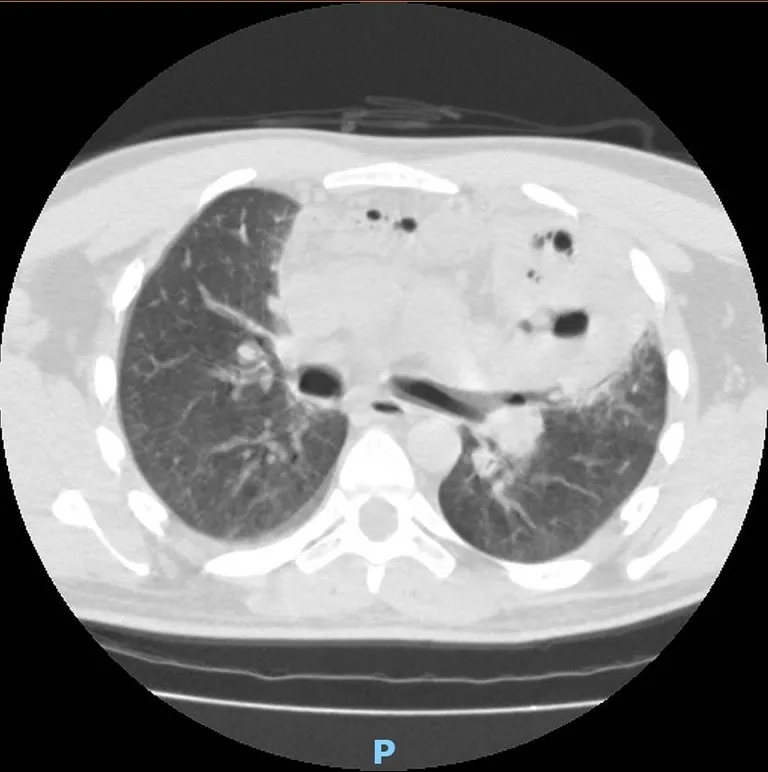
A second contrast-enhanced CT revealed air bubbles, fluid collection in the anterior mediastinum, and an excavated lesion close to the lingula (Figures 1,2).
The patient was then transferred to our thoracic surgery department with a diagnosis of mediastinitis, bilateral pleural empyema, and sepsis. At admission, blood tests showed: white blood cell (WBC) 21.0×10³/L, hemoglobin (HGB) 10.2 g/dL, hematocrit (HCT) 31%, international normalized ratio (INR) 1.36, D-dimer 16,547 ng/mL, aspartate aminotransferase (AST) 69 U/L, alanine transaminase (ALT) 87 U/L, gamma glutamyl transferase (GT) 111 UI/L, reactive C protein 123.90 mg/L, procalcitonin 1.72 ng/mL. The presence of phlegmon on the thorax and abdomen skin was detected.
The patient was submitted to a left video-assisted mini-thoracotomic and subxiphoid approach to the mediastinum and both pleural cavities. After the subxiphoid muscular layer’s incision, purulent exudate seeped out from the mediastinal collections, drained, and debrided before placing three chest tubes in the pleural spaces and in the mediastinum.
Based on the unsuccessful preoperatory treatment with broad-spectrum antibiotics (meropenem plus teicoplanin) and on the knowledge of the local epidemiology [recent clusters of P. aeruginosa multidrug-resistant (MDR)], the infectious disease specialist suggested a combination therapy with ceftolozane/tazobactam (“off-label” administration) (1) plus fosfomycin. Soon thereafter, the patient’s clinical conditions improved, allowing us to perform a definitive surgical treatment, a left upper lobectomy. The histological findings showed an infected mature ruptured dermoid cyst of the lung.
After 30 days of hospital stay, the patient was discharged and is now alive and in good health conditions (Figure 3). All procedures performed in studies involving human participants were in accordance with the ethical standards of the institutional and/or national research committee(s) and with the Helsinki Declaration (as revised in 2013). Written informed consent was obtained from the patient.

Figure 1 The first CT scan.

Figure 2 Same CT scan, mediastinal window.

Figure 3 Last chest X-ray.
Discussion
Kluge has recently described (2) the most common causes of acute mediastinitis, like esophageal, tracheal or bronchial rupture due to endoscopic procedures, intubation or traumatic lesions, the Boerhaave syndrome, and the descending necrotizing mediastinitis, which is often related to odontogenic, pharyngeal, cervical, or other head and neck infections (3).
To our knowledge, the mediastinal involvement in infection of a fistulized teratoma has not been previously described.
Intrapulmonary teratomas (IPTS), also called “dermoid cysts”, are sporadic tumors, often localized in the left upper lobe (4), whose rare complications include rupture (5). All intrathoracic teratomas produce proteolytic or digestive enzymes, leading to the rupture or the adjacent structures’ invasion, as previously widely described (6).
Common symptoms of pulmonary teratoma are fever, cough, chest pain, and superimposed lung abscess. A particular symptom is the trichoptysis (hair expectoration), considered pathognomonic for bronchial extension of an intrathoracic teratoma (7).
The crucial factors for the favorable outcome, in this case, were both the right timing and techniques for the surgical procedures and the choice of aggressive antibiotic therapy to achieve adequate infection control.
We performed a left mini-thoracotomic and videoassisted thoracoscopic surgery (VATS) subxiphoid access to the mediastinum and both pleural cavities, which has never been described in the literature to drain both pleural spaces in case of empyema and mediastinitis, but it has been proposed to drain the anterior mediastinum (3,8,9). Moreover, it must be considered that in the literature, only a few mediastinitis cases were treated with a video thoracoscopic approach, as this clinical condition usually affects different anatomical regions (10).
Another critical factor for the management of this patient was the close collaboration with the infectious disease specialist, as the particular combination therapy (ceftolozane/tazobactam and fosfomycin) designed to treat MDR pathogens (1) was successful and allowed the left upper lobectomy, thus eliminating the focus of infection.
In conclusion, this case shows that mediastinitis caused by an infected lung teratoma achieved a favorable evolution due to a tailored on-the-case surgical strategy and aggressive antibiotic treatment.
Acknowledgments
Funding: None
Footnote
Provenance and Peer Review: This article was commissioned by the editorial office, Chinese Journal of Thoracic Surgery for the “International Thoracic Surgery Column”. The article has undergone external peer review.
Reporting Checklist: The authors have completed the CARE reporting checklist. Available at https://dx.doi.org/10.3877/cma.j.issn.2095-8773.2021.03.04
Conflicts of Interest: All authors have completed the ICMJE uniform disclosure form (available at https://dx.doi.org/10.3877/cma.j.issn.2095-8773.2021.03.04). The “International Thoracic Surgery Column” was commissioned by the editorial office without any funding or sponsorship. FS serves as an unpaid editorial board member of Chinese Journal of Thoracic Surgery from May 2021 to April 2023. The authors have no other conflicts of interest to declare.
Ethical Statement: The authors are accountable for all aspects of the work in ensuring that questions related to the accuracy or integrity of any part of the work are appropriately investigated and resolved. All procedures performed in studies involving human participants were in accordance with the ethical standards of the institutional and/or national research committee(s) and with the Helsinki Declaration (as revised in 2013). Written informed consent was obtained from the patient.
Open Access Statement: This is an Open Access article distributed in accordance with the Creative Commons Attribution-NonCommercial-NoDerivs 4.0 International License (CC BY-NC-ND 4.0), which permits the noncommercial replication and distribution of the article with the strict proviso that no changes or edits are made and the original work is properly cited (including links to both the formal publication through the relevant DOI and the license). See: https://creativecommons.org/licenses/by-ncnd/4.0/.

