多导睡眠监测工程原理及在神经疾病中的应用
李依芃 方 圆 刘倩倩 陈文武
1.乔治华盛顿大学 工程与应用科学学院,华盛顿 20052;2.河南大学 第一附属医院,河南 开封 475001
0 Introduction
With faster pace of life and increased pressure,sleep disorders disturb more and more people.In particular,they seriously affect people's normal life and physical health.The sleep cycle refers to the biological rhythm of sleep that circulates in mammals (including humans) and birds.The International Academy of Sleep Medicine classifies normal sleep into four stages[1],and a structure cycle of sleep has two phases:non-rapid eye movement (NREM) and rapid eye movement(REM) sleep.NREM is variably synchronized with cortical electroencephalogram (EEG) and it is associated with lower muscle tone and mental activity.REM's EEG is out of this synchronization,and the body's muscles keep flabby,basically indicating a loss of muscular tone[2].Normally,humans go through a different phase in about 90 minutes[3].During the entire sleep cycle,NREM and REM occur alternately and in cycles[4].
Sleep plays an important role in human health.Generally,a reasonable sleep should be 7 to 9 hours per night.Adequate sleep can enhance immunity,cognitive effects,and memory[5].Deep sleep promotes the secretion of Growth Hormone,thus facilitates the growth and development of children.For adults,the secretion of growth hormones speeds up the metabolism of cells,motivates the synthesis of proteins,and expedites tissue repair[6].Research has shown that people who keep early hours are less stressed and with higher levels of mental health than those who stay up late[7].Elderly people tend to have poor quality of sleep and the prevalence of sleep disorders in this group is higher than the younger group[8],so those might be the reasons why COVID-19 is spreading among the elderly in the first place[9].Lack of sleep has been associated with multiple health problems[10],including increased burden on the heart and brain,absenteeism from work,and augmented risks of neurological diseases.Polysomnography can help doctors and patients better understand the causes of sleep disorders and then build healthy sleep cycles.This paper will introduce polysomnography and analyze its practical application in diseases,to provide a scientific basis for the follow-up treatments.
1 Engineering Principles of Polysomnography (PSG)
Sleep research is an important part of sleep science and electroencephalography (EEG).Behaviorism defines sleep as an unresponsive and reversible state of behavior that is divorced from the environment[3].Although electrocardiogram monitors (ECM) and respiratory recorders are usually used to monitor sleep,their effects are limited compared to PSG.PSG synchronously records of multiple physiological parameters during sleep and wakefulness.Therefore,the emergence of PSG has a promising future in promoting the study of sleep.
1.1 History of Polysomnography System
The application of polysomnography in clinical sleep medicine originated in the late 1950s[11],and it was first used in an overnight diagnostic sleep study in 1974.Previously,scientists thought sleep was a single physiological process,associated only with slow waves of synchronized electroencephalography (EEG).However,with the help of ancient PSG technology (Eye movement sensors),Kleitman and Aserinsky[12]in 1957 discovered that human sleep is punctuated by two different phases,NREM and REM.Subsequently,with the generations of PSG,the respiratory and cardiac sensors and leg leads are added to PSG.For the first time,scientists observed and described REM sleep.In this state,despite rapid eye movements,there are also various sensory impairments.For instance,the reduced skeletal muscle reflex activity and tension,and decreased but unstable vegetative nerve functions[13].
Besides,dream as a cognitive and behavioral level of subjective experience,is the metabolic product[14]that cerebral cortex neurons keep active during sleep state.The interpretation of dreams is a perennial topic.Neurons under multiple frequencies have formed brain rhythms (dream),and PSG plays an important role in the interpretation of dream generation and decoding dreams.Polysomnogram monitoring system found that there was a significant difference in electrical markers between NREM and REM states.In the REM stage,the metabolic process of the human body and the firing of neurons in the cerebral cortex are like those in the waking state,dreams are the typical characteristics of REM[15].Therefore,decoding the electrical activity of the brain under dream can be better applied to the study of human brain function and related behaviors.
1.2 Polysomnography Mechanism and Processing System
Polysomnography structure mainly comprises the host,display,sensor,acquisition box,amplifier,and other parts.Traditional PSG processing system merely includes electroencephalography(EEG),electrooculography (EOG),electromyography (EMG),electroencephalogram (EEG) and electromyography (EMG) to reflect sleep status and stage[16].For the needs of some patients requiring specific monitoring,such as the diagnosis of obstructive sleep apnea syndrome (OSAS),the latest PSG makes use of chest and abdomen motion sensors,respiratory rate,thermal airflow sensors,oxygen sensors,body position sensorset cetera[17]to record and determine the state of breathing.Some polysomnography also records snore to learn about snoring and the relationship between sleep apnea and its frequency spectrum[18].Others have postural sensors[19]that record changes in the patient's position during sleep to see how apnea is related to it.By recording and analyzing the above parameters,doctors can accurately analyze and diagnose neurological diseases,and then meet demands of different patients to the greatest extent[20].
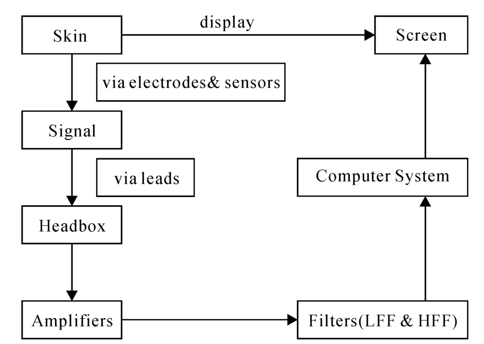
PSG has a complete process for the acquisition and processing system (Figure 1).First,PSG receives bioelectrical signals from each part of the body through electrodes and sensors attached to the skin of the patient.Then,A/D conversion is realized by pre-integrated amplifiers,transforming electrical signals to digital signals.Subsequently,the high-frequency filter (HFF) and low-frequency filter (LFF) are used to limit the signals which are passed by amplifiers to the frequency range that is needed.Finally,the computer system forms the images and displays them on screens.

Fig.1 Signal acquisition and processing system
1.3 Operation of Polysomnography in Clinic
A “10%~20% System” is used to determine electrodeposition for scalp EEG recording of patients,which is referred to as the International 10~20 system,including 19 head recording electrodes and two ear pole reference electrodes.First of all,doctors found the Median Sagittal section(the front line from the nose to the occipital tuberculum) and Coronal section (the front line between the anterior recesses of the ears) on the brain to determine the position of the Cz electrodes and take 10%~20% distance as the spacing between each electrode[21].Common lead combinations include ear reference lead,mean reference lead,and bipolar reference lead.In clinical practice,the left and right cerebral hemispheres were distinguished by odd and even numbers,respectively,and headphone reference guides are often used to mark the left and right earlobes as A1and A2.The distribution of the left side is successively marked as Fp1,F3…T3,and as Fp2,F4…T4on the right[21].Thus,the integrated lead pathways are formed.
2 Polysomnography in Neurological Diseases
It has long been known that REM sleep is associated with dreams,but it often associates with neurological diseases as well.In recent years,polysomnography has been widely used in clinical diagnosis and treatments.The PSG system can observe the EEG changes parallel to the severity of illness.The response of patients under external stimulation is also of great value for post-treatment evaluation.
Insomnia is the most common sleep disorder[22],and it is an important part and typical characteristic of the depressive symptom group.Sometimes,insomnia and depression are under cooccurrence.Some studies[23]have confirmed that there is a significant relationship between the degree of depression and sleep disturbance.The relationship between depression and insomnia are complex and bi-directional,as the depression and anxiety associated with sleep deprivation can in turn exacerbate insomnia[24].Traditionally,patients are asked to write a daily sleep diary[25],and doctors track this to understand the cause of their sleep disorders.However,such reports are difficult to ensure accuracy and objectivity,so they can have a big impact on evaluation results and subsequent treatment regimens.Therefore,polysomnography is necessary as an effective and objective measurement.
Introducing the poly-sleep chart confirmed the interference with sleep in depressed patients[26].It is not only a decrease of slow-wave sleep but also the rapid eye movements.Moreover,the time of REM sleep and the duration and dosage of drug treatments were significantly correlated.That is,almost all anti-depressant drugs suppress REM sleep.Therefore,PSG can help doctors and patients to analyze the sleep regulation mechanism,understand the sleep structure changes caused by depression,and pay more attention to the relationship between sleep and depression treatments.
In the study of epilepsy,PSG also plays a significant role.sleep and epilepsy interact with each other.Studies[27]have found that almost two-thirds of seizures occur during sleep.NREM sleep can be further divided into N1~N4.Of them,N1and N2are shallow sleep,N3and N4are slow-wave sleep(SWS) period.PSG found that seizures usually occurred in the N1or N2stages of NREM sleep[28].During this period,the brain wave tends to be slowed down and synchronized due to the decreased function of the brain stem reticular uplink activation system.Then,abnormal electrical activities in certain neurons appear and spread,as Interictal Epileptiform Discharges (IEDs)[29].However,the activation of such IEDs is not evenly distributed,so the location and identification of IEDs could be the key to diagnose seizures.
Some studies[30]have suggested that sleep structure affects epileptiform discharge and epileptic seizures.Epilepsy is circadian[31],so,circadian rhythm changes and sleep deprivation will all respond to the occurrence of epilepsy,but diverse types of epileptic seizures may have different circadian distributions.In these cases,PSG is a good way to observe epileptic patients.When the patients have high sleep quality,their limbs are generally at rest.When patients turn and move,the EMG sensor can detect and record these changes,and calculate the total sleep time and systematically sleep quality.Sensors can detect and record data from multiple directions,and EEG,EMG spectrum can reveal intuitively physiological signatures.Through the detected data,we can know the sleep state and quality,and then we can consult the doctor for intervention treatment accor dingly.Moreover,doctors not only can analyze the sleep architecture using PSG,synchronically showing the sleep status of patients and the incidence of seizure,but also possibly locate the epileptiform discharge (IED) during REM[32]and non-REM.
3 Conclusion &Limitations
Overall,polysomnography can provide a va riety of auxiliary analysis basis.For example,the PSG system can help doctors understand the actualsituation of insomnia,assess the degree of insomnia,and conduct a quantitative analysis of sleep.Also,it can find the cause of the neurological diseases and provide objective indicators for evaluating the efficacy of various drugs or nondrugs.Despite electroencephalogram (EEG),polysomnography (PSG) supervises physiological signals of multiple channels including electrocardiogram (ECG),electromyogram (EMG),eye movement (REM),thoracic and abdominal respiratory tension chart,nasal and oral ventilation volume,postural body movement,blood oxygen saturation,and cavernosal muscle volume.Besides,PSG can lead real-time monitoring,which is more conducive to the discovery of hidden problems.PSG system plays a qualitative and quantitative role in the diagnosis of diseases.It not only can diagnose diseases but also determine the severity of diseases,facilitating the designation of clinical treatment regimens and the quantitative evaluation of surgical or other treatment effects.In the treatment of sleep disorders,the presence of the PSG system can greatly improve control efficiency and quality.
However,despite the widespread applicability,PSG has some limitations.For doctors,it needs special training to use,record,and analyze the visual results of PSG[33].For patients,it is time-consuming and expensive,and they must go to a special laboratory or ward,which may lead to a “first-night phenomenon” and to a certain extent of error value in results.To overcome these difficulties,more portable monitors with high sensiti vity and low specificity are being developed,and the monitoring scope and results will get better through the joint efforts of the multi-monitor system.

