Lues Maligna in an Immune-Reconstituted Patient with Retroviral Infection: A Case Report
Louis Y.Tee*, Chee Hian Tan, Michelle M.F.Chan, Hei Man Wong, Shiu Ming PangYi Wei YeoDepartment of Dermatology, Singapore General Hospital, Singapore; National Skin Centre, Singapore; Department of Anatomical Pathology, Singapore General Hospital, Singapore; Department of Infectious Diseases, Singapore General Hospital,Singapore.
Introduction
Lues maligna is an uncommon, severe form of secondary syphilis.Most cases are linked to poorly controlled human immunodeficiency virus (HIV) infection with cluster of differentiation 4 lymphocyte (CD4) counts between 100 and 350,1and immune reconstitution inflammatory syndrome has been reported to increase the risk of lues maligna.2Here we describe an uncommon case of lues maligna complicated by syphilitic hepatitis in a patient with virologically suppressed and immune-reconstituted HIV infection during the brief period when he had stopped his medications, and review the literature about this rare and potentially fatal form of syphilis.
Case report
A 46-year-old man presented with a 1-month history of non-pruritic and painless nodules that started on the face,and subsequently spread to his upper limbs and trunk.Lesions were also noted in the nasal, oral, and groin regions.There was no fatigue, fever, chills, or loss of weight.Systemic review was otherwise unremarkable.
The patient has HIV infection since 2008, currently treated with tenofovir disoproxil fumarate,emtricitabine,and efavirenz in a once daily combination pill.Notably,he had stopped taking his medications for a month prior to the onset of the rash due to lethargy,which he attributed to his hectic work schedule.Prior to this, he had claimed complete compliance.There was no other new medication or supplement use.His last Treponema pallidum (T.pallidum) particle agglutination assay and Venereal Disease Research Laboratory (VDRL) test done in 2008 were negative for syphilis.The patient reports that he has a history of bisexual relationships and his last sexual contact was about 1 year ago,but was otherwise not forthcoming with his sexual history.
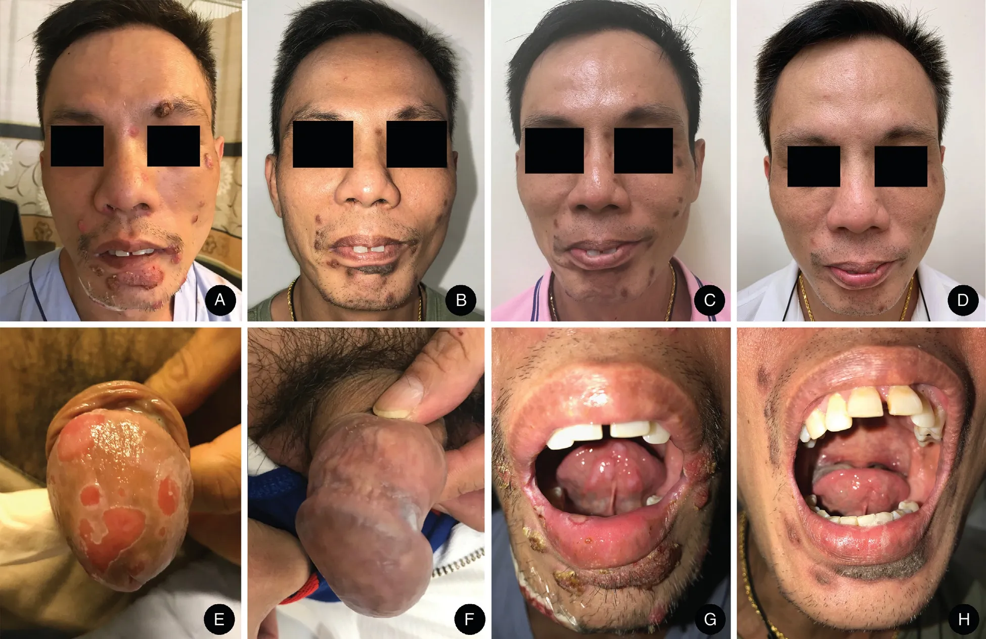
Physical examination was notable for multiple discrete,erythematous ulcerated nodules of varying sizes with overlying thick crust on the face (left periorbital area,cheeks, and the perioral area), left ear, trunk, and limbs(Fig.1A).In addition, there were mucosal lesions in bilateral nasal cavities and painless ulcers on the hard palate, under the tongue, and on the glans penis (Fig.1E and 1G).There was no cervical, axillary, or inguinal lymphadenopathy.Otherwise, he was systemically well.Blood investigations revealed a CD4 count of 663cells/mm3, and an undetectable HIV viral load.Herpes simplex virus polymerase chain reaction (PCR) from swabs of the oral and genital ulcers was negative.T.pallidum particle agglutination assay was positive,and he had a VDRL titer of 1:256.Incidentally,investigations for potential complications of syphilis revealed transaminitis with a serum alanine aminotransferase(ALT)of 206 and aspartate transaminase(AST)of 102.Subsequently,an enzyme immunoassay for hepatitis C antibodies was positive, leading to the discovery of a genotype 1A hepatitis C infection, with a viral load of 6.66 log.
Investigations for other infective diseases were unremarkable.Serum cultures for bacteria, fungal, and tuberculosis were unyielding, and PCRs for Epstein-Barr virus, cytomegalovirus, and herpes simplex virus were negative.Serum cryptococcus antigen and hepatitis A immunoglobulin M antibodies were negative.Hepatitis B serology was indicative of immunity to hepatitis B from prior vaccination: Hepatitis B surface antibodies were reactive,while hepatitis B surface antigen was nonreactive,and hepatitis B DNA levels were undetectable.

Figure 1.Clinical pictures of the patient with lues maligna.Multiple discrete,erythematous ulcerated nodules with thick crust present on the face,particularly around the periorbital and perioral regions(A).Facial nodules improved after administering intramuscular benzathine penicillin by 2 weeks (B), 4 weeks (C), and 3 months (D).Ulcers on the glans penis (E) and lips (G) resolved by 2 weeks after the initiation of treatment(F and H).
A skin punch biopsy was obtained from a representative nodule on his chin.The epidermis was hyperplastic with neutrophilic infiltration (Fig.2A and 2B).There were prominent perivascular infiltrates of plasma cells within the dermis (Fig.2C).Stains with Warthin-Starry, Ziehl-Neelsen,Fite acid fast stain,Periodic acid-Schiff(PAS),and Grocott-Gomori methenamine silver (GMS) stains were negative.Tissue cultures for bacteria, fungus, and tuberculosis were unyielding.
A diagnosis of lues maligna was made given the clinical presentation and investigation results.The patient received 2.4 million units of intramuscular (IM) benzathine penicillin weekly for three doses, with no development of Jarisch-Herxheimer reaction and subsequent resolution of skin lesions (Fig.1B-1D, 1F, and 1H).Three months after treatment, the VDRL titer decreased by fourfold to 1:64.
Moreover, the patient's lues maligna was likely complicated by syphilitic hepatitis.His transaminitis initially worsened to an ALT of 1,104 and AST of 1,012 when the third dose of IM benzathine penicillin was given.However,corresponding to the reduction in VDRL titer, both ALT and AST normalized 3 months after the third dose of benzathine penicillin.The resolution of the transaminitis with benzathine penicillin and the correlation with VDRL titer is characteristic of hepatitis due to syphilis rather than hepatitis C.Ultrasound of the abdomen and the international normalized ratio and bilirubin levels were normal,and the patient had no signs or symptoms of liver failure.
Discussion
Theincidenceof luesmaligna,anuncommon form of syphilis first described by Bazin in 1859 in patients with poor health and malnutrition, has risen since 1987 due to the HIV pandemic.3In fact, the frequency of lues maligna in HIVinfected patients is 60 times higher than in the general population.4Furthermore, 80% of HIV-infected patients with lues maligna have CD4 counts greater than 200 and there are case reports of lues maligna in HIV-infected patients with normal CD4 counts,suggesting that HIV infection itself may modulate the clinical presentation of syphilis.4The lesions usually appear after an incubation period of 4 weeks toayearfromtheprimaryinfection,andcommonlyaffectthe face and scalp.4The initial lesions are often papules that rapidly pustulate,and subsequently form ulcers with necrotic centers that may have a lamellated,brown-black crust.5The skin lesions may be preceded by a prodrome of arthralgia,malaise, and fever.6Additionally, patients may have signs and symptoms of syphilitic hepatitis,6ocular syphilis,7and accelerated neurosyphilis.8Indeed,lues maligna was historicallyfataldue tothesyphiliticinvolvement of multipleorgans when left untreated.5Therefore, prompt diagnosis of lues maligna is essential.

Figure 2.Hematoxylin-eosin stain of the skin biopsy specimen from the nodule on the chin.The imaging showed a hyperplastic epidermis(A)with mixed neutrophilic(B)and plasmacytic(C)infiltration.Original magnifications: A, × 40; and B and C, × 200.
In an immunocompromised host, the differential diagnoses for such presentation would include disseminated atypical infections, such as deep fungal infection or atypical mycobacterial infection, and cutaneous lymphoma.8The diagnosis is often confirmed with positive syphilis serological results and supported by skin histological findings.VDRL titers are generally very high in lues maligna,but notably the prozone effect may lead to a false negative VDRL result in HIV-infected patients.Therefore,alternative diagnostic methods such as the biopsy of skin lesions for histopathology with Warthin-Starry, Steiner,and immunohistochemical stains are often employed.Histopathology typically shows dermal plasma cell infiltrates with lymphocytes, histiocytes, and neutrophils,while spirochetes are occasionally detected with special stains and immunohistochemistry.6Notably, PCR to T.pallidum has recently emerged as a fast and reliable way to diagnose secondary syphilis,9but is not available in our laboratory.
The first-line treatment of malignant syphilis is penicillin.Appropriate preparations of penicillin include IM benzathine penicillin(2.4 mega units)weekly for 3 weeks,and procaine penicillin or intravenous methyl penicillin daily for 10-14 days.5For patients with drug allergies to penicillin, intravenous ceftriaxone for 10-14 days, and 750mg tetracycline given daily for 10 days have also been effective.6Patients may develop a Jarisch-Herxheimer reaction in the first 24hours,and constitutional symptoms usually resolve in a few days.In contrast, healing of skin lesions typically starts 2 weeks to 4 months after beginning treatment.Like ordinary syphilis,lues maligna shows good serologic response and excellent prognosis with treatment.In conclusion, malignant syphilis is an uncommon variant of secondary syphilis that should be included in the differential diagnosis of HIV-infected patients with ulcerated and necrotic lesions.This case adds to the body of knowledge about lues maligna in terms of sites of mucocutaneous involvement and the good response with penicillin.In addition,this case demonstrates that even in a HIV-positive patient with adequate CD4 counts, lues maligna should be considered,particularly when there is a history of interruption of anti-retroviral therapy.It also highlights the importance of obtaining an accurate drug history,including brief periods of nonadherence.Furthermore, this patient's transaminitis reminds us of the importance of being vigilant of syphilitic involvement of other organs, such as syphilitic hepatitis, ocular syphilis,cardiovascular syphilis, and neurosyphilis, which can be incapacitating and even fatal when left untreated.
- 国际皮肤性病学杂志的其它文章
- Targetoid Hemosiderotic Nevus
- Eccrine Spiradenoma of the Forehead: A Case Report
- Seborrheic Keratosis in a Young Woman:A Mimicker of Keratoacanthoma
- A Case of Condyloma Acuminatum on the Nipple Detected via Dermoscopy
- Azathioprine Hypersensitivity Syndrome in an Asian Woman
- Linagliptin-Associated Bullous Pemphigoid: The First Case in China

