Successful treatment of congenital palate perforation:A case report
Jin-Feng Zhang,Wen-Bin Zhang
Jin-Feng Zhang,Wen-Bin Zhang,Department of Oral and Craniomaxillofacial Surgery,Ninth People's Hospital,Shanghai Jiaotong University School of Medicine;National Clinical Research Center for Oral Diseases;Shanghai Key Laboratory of Stomatology,Shanghai 200011,China
Abstract
Key words: Congenital;Palate;Perforation;Craniomaxillofacial;Submucous cleft palate etiology;von Langenbeck palatoplasty
INTRODUCTION
Traumatic and postoperative palatal perforation is common,while congenital palatal perforation is rarely reported in the literature.The earliest mention of congenital palatal perforation was published by Roux[1].Then sporadic putative cases were reported[2-4].However,whether the perforation in these cases was congenital or acquired after birth was unmentioned[5,6].Also,whether the perforation was isolated or associated with submucous cleft palate (SMCP) was undocumented[7,8].There are only a few patients with palate perforation that was identified shortly after birth.Lynch reported a congenital cleft of the palate,which involved only the hard palate,with the alveolus and soft palate intact[9].Mehendaleet al[10]observed that one infant had a small hole at the junction of the hard and soft palate.Esheteet al[11]presented two cases of congenital palatal perforation associated with SMCP.Modified von Langenbeck palatoplasty was performed for the two patients.There was no recurrence of the fistula in the 2-year follow-up of one patient.However,speech evaluation revealed articulation and speech errors secondary to persistent mild hypernasality.Another patient was lost to follow-up.
Although there is not a consensus for congenital palatal perforation associated with SMCP,the goals of treatment are the same:Closure of the fistula,rearrangement of the palatal muscles,and lengthening of the short velum.The surgical treatment of the fistula is simple;however,satisfactory velopharyngeal competence is not easily achieved.In this paper,a case of congenital palatal perforation associated with SMCP is reported,and the treatment and outcomes are discussed.
CASE PRESENTATION
Chief complaints
A full-term neonate boy was referred to the oral and craniomaxillofacial surgery department with a chief complaint of nasal regurgitation of food and a hole in the palate at birth.
History of present illness
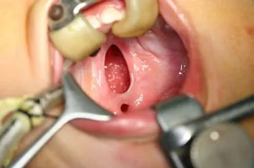
The patient was born with a 5 mm × 3 mm hole in the anterior part of the velum and a bifid uvula.And the hole was developed as the child grew.At the age of 10 mo,the patient was admitted to the hospital.
History of past illness
The patient was born after a healthy pregnancy without cleft lip or alveolar cleft.
History of family illness
There was no family history of cleft lip,cleft palate,or other craniofacial malformations,and the mother denied occupational or environmental chemical exposure.
Physical examination
Intraoral examination revealed a thin translucent central zone with a 5 mm × 3 mm hole in the anterior part of the velum and a bifid uvula.At the age of 10 mo,the patient was admitted to the hospital while the palate perforation had developed to 8 mm × 4 mm (Figure 1).
FINAL DIAGNOSIS
The patient was diagnosed with a congenital palatal perforation associated with SMCP.

Figure1 Congenital palatal perforation.
TREATMENT
This patient underwent modified von Langenbeck palatoplasty.The connective velum was excised,and the perforation was repaired,with radical dissection and reposition of the velar muscles for the reconstruction of the palatal muscular connection.Dissection showed that the levator muscles did not meet in the midline to form a sling.The soft palate closure was successful,and there was no fistula.
OUTCOMES AND FOLLOW-UP
Five days after the operation,the patient was discharged without any complications.The patient was followed for four years after surgery.His speech was satisfactory.
DISCUSSION
Most of the putative congenital palatal perforation cases are associated with SMCP.The clinical signs of a classic SMCP are well recognized and reported as a notch in the posterior edge of the bony palate,a thin translucent central zone with an absence of muscle union,and a bifid uvula.A submucous cleft may break open after birth,either spontaneously or (more probably) artificially,as has been described by Smariuset al[12].In SMCP patients with a significant bony defect or notch in the hard palate,the midline mucosa is thinner than that in the uvula.Thus,the hard posterior palate may be vulnerable to developing a perforation at a relatively early stage.Besides,the mucosa is tented across the edges of the bony defect in the hard palate.It is relatively immobile and liable to perforate in response to trauma,in contrast with the mucosa of the mobile velum[13].Therefore,it is very likely that in some of the patients,a thin central zone may have perforated within a few days of birth.It is also possible that minor trauma from swallowing or finger sucking may cause prenatal perforation.The prenatal or postnatal perforation hypothesis is plausible for many of the cases.However,the explanation does not account for some clinical findings in other reports.Lynch reported a defect extending from the incisive foramen to the junction of the bony and soft palate in a patient who was not thought to have an SMCP[9].So he postulated a malformation etiology.The duration and magnitude of lingual obstruction produced a cleft in the hard palate while the posterior soft palate closed by fusion.Jagannathan and Agarwal also reported cases of congenital palate fistula with a developed posterior palate[14,15].They believed that congenital perforation of the palate should be classified as a failure of the palatal differentiation of fetus,which is not caused by an accident or artificial factor.
In cases of isolated congenital palate perforation (not associated with SMCP),only simple repair of perforation was recommended.For those cases associated with SMCP,regardless of whether the perforations are congenital or acquired after birth,the aim of treatment for palatal perforation is the same.It includes the closure of the deformity,lengthening of the soft palate,and the reconstruction of the palatal muscular connection,especially of the uvula muscles,as in a complete cleft palate repair.These patients underwent closure of the fistula combined with a palatoplasty.More attention should be given to achieve adequate muscle approximation and excellent velopharyngeal competence.Closure of the fistula and satisfactory velopharyngeal competence were achieved in the present case.However,the longterm effect on maxilla growth should be followed.
CONCLUSION
Considering the anatomy and etiology,congenital palate perforation should be classified as isolated or associated with SMCP,and the treatment procedure should be altered accordingly.The case presented in this article is regarded as congenital palate perforation associated with SMCP.Therefore,reconstruction of the palatal muscular connection is fundamental.
 World Journal of Clinical Cases2020年1期
- World Journal of Clinical Cases的其它文章
- Role of oxysterol-binding protein-related proteins in malignant human tumours
- Oncogenic role of Tc17 cells in cervical cancer development
- Acute distal common bile duct angle is risk factor for postendoscopic retrograde cholangiopancreatography pancreatitis in beginner endoscopist
- Three-dimensional computed tomography mapping of posterior malleolar fractures
- Application of a modified surgical position in anterior approach for total cervical artificial disc replacement
- Potential role of the compound Eucommia bone tonic granules in patients with osteoarthritis and osteonecrosis:A retrospective study
