Application of ozonated water for treatment of gastro-thoracic fistula after comprehensive esophageal squamous cell carcinoma therapy:A case report
De-Di Wu,Ke-Nan Hao,Xiao-Jing Chen,Xin-Min Li,Xiao-Feng He
De-Di Wu,Ke-Nan Hao,Xiao-Jing Chen,Xin-Min Li,Xiao-Feng He,Division of Vascular and Interventional Radiology,Department of General Surgery,Nanfang Hospital,Southern Medical University,Guangzhou 510515,Guangdong Province,China
Abstract BACKGROUND Gastro-thoracic fistula is a serious complication after radical surgery for esophageal cancer,and a conservative approach or endoscopic intervention is commonly applied to treat most cases.CASE SUMMARY Here we describe the case of a patient with a gastro-thoracic fistula which could not be closed during gastroscopy after receiving postoperative radiotherapy,together with severe multiple drug-resistant bacterial infection and chest wall fistula.The abscess was drained and local irrigation applied with ozonated water,together with oral ozonated water,which achieved a good effect and highlighted a new way to cure fistula in such patients.CONCLUSION Patients with gastro-thoracic fistula that cannot be closed and severe infection can be treated by drainage and flushing with ozonated water.
Key Words:Esophageal squamous cell carcinoma;Ozonated water;Radiotherapy;Gastrothoracic fistula;Drug-resistant bacterial infection;Case report
INTRODUCTION
The incidence and mortality of esophageal cancer are high in China.Radical surgery is often the first-line treatment option for the majority of patients[1,2].One type of lifethreatening postoperative complication is gastro-thoracic fistula with an incidence of 14.1% worldwide[3,4],which may be associated with body mass index[5].Effective antiinfection treatment is the key to reducing mortality and improving patient quality of life.
Long-term clinical trials have proven that ozone can both effectively control infections caused by various bacteria,fungi,and viruses[6,7],and promote tissue repair[8].Here,we report a case of gastro-thoracic fistula after comprehensive treatment for esophageal cancer,complicated by severe thoracic infection,and successfully treated using ozone.
CASE PRESENTATION
Chief complaints
A 50-year-old woman developed an open wound in a surgical incision scar and was readmitted to our hospital on July 31,2019 because the wound in her left chest wall had reopened.
History of present illness
Following radical esophagectomy in June 2016 to treat esophageal squamous cell carcinoma,in May 2019,the patient developed an open wound in the surgical incision scar on her left chest wall(Figure 1).
History of past illness
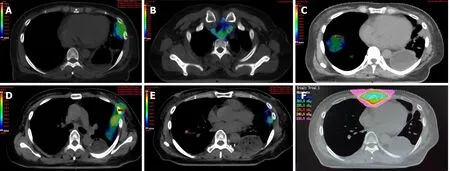
The patient was diagnosed with moderately differentiated esophageal squamous cell carcinoma in 2016.During the 3 years following surgery,the patient received repeated radiotherapy to metastatic tumor of the left lung(40 Gy/4 F),anastomotic site and lymph nodes(60 Gy/30 F),metastatic tumor of the right lung(50 Gy/5 F),metastatic tumors of both lungs(48 Gy/6 F),and sternal metastases(45 Gy/15 F)with time sequence(Figure 2).
Physical examination
When the patient was readmitted to our hospital on July 31,2019,she was pale and dehydrated on physical examination,and the wound in her left chest wall had reopened,was approximately 2 cm × 3 cm,and had malodorous purulent secretion emanating from it.
Laboratory examinations
Bacterial culture and drug sensitivity testing of the pleural drainage fluid on August 8,2019 revealed positivity for methicillin-resistantStaphylococcus aureusandCandida albicans.Regular bacterial culture of the pleural abscess drainage fluid indicated thatPseudomonas aeruginosa(August 29)andKlebsiella ozaena(September 30)were present.

Figure 1 Timeline.
Imaging examinations
When the patient presented in May 2019,whole body positron emission tomography(PET-CT)indicated the formation of a gastro-thoracic fistula,with lesions involving the lateral chest wall(Figure 3A and B)and active malignancies in the left lung,inferior segment sternotomy,and right lobe of the liver.A gastro-thoracic fistula of approximately 5 cm,located in the greater curvature of the stomach,was confirmed on endoscopy(Figure 3C and D).Poor drainage of the abscess by the drainage tube applied was detected on computed tomography(CT)examination in September 2019.
Personal and family history
The patient did not smoke or consume alcohol.Her mother and brother both died of esophageal cancer.
MULTIDISCIPLINARY EXPERT CONSULTATION
Rui-Jun Cai,Deputy Chief Physician,Department of Thoracic Surgery,Nanfang Hospital,Southern Medical University
Gastro-thoracic fistula was diagnosed clearly and the fistula is too large to perform reoperation or endoscopic treatment.
Hao Zhou,Deputy Chief Physician,Department of Infection Control,NanfangHospital,Southern Medical University
The images of PET-CT showed that the gastro-thoracic fistula was complicated with severe infection that was not controlled.
FINAL DIAGNOSIS
The final diagnosis was gastro-thoracic fistula following comprehensive treatment for squamous cell esophageal carcinoma.
TREATMENT
Following detection of the fistula in May 2019,her oral intake was restricted and she received gastrointestinal decompression therapy and nose-jejunum nutrition support at our hospital until the open wound was sealed.
On readmission on July 31,she received intravenous administration of imipenem and fosfomycin to treat the infection and a gastric tube was placed to irrigate the wound.She was also administered metronidazole,enteral nutrition support,and intravenous nutrition supplementation.The open wound at the site of her surgical scar was treated with local empyema and disinfection,and the dressing changed.A jejunal nutrition tube and gastric-thoracic fistula drainage tube were placed on August 7 and saline used to flush the fistula following surgery.Following detection of methicillinresistantStaphylococcus aureusandCandida albicanson August 8,sulfamethoxazole combined with fluconazole was administered intravenously to control the infection.Next,we began application of 15 µg/mL ozonated water to rinse the fistula once per day(500 mL each time)and administered 12 µg/mL ozonated water orally(500 mL per day)from August 10.Subsequently,following detection ofPseudomonas aeruginosa(August 29)andKlebsiella ozaena(September 30),intravenous antibiotics were adjusted to piperacillin sodium and tazobactam sodium injection(September 4),in addition to amikacin combined with ciprofloxacin(September 30),according to the results of drug sensitivity testing.
Leukocyte number and the proportion of neutrophils were found to have continued to increase on September 3(Figure 4A and B),indicating that the infection was unsatisfactorily controlled.On detection of poor abscess drainage,we applied closed thoracic drainage under the guidance of CT on September 10,resulting in drainage of thick,dark red liquid.
The patient then received 2 mo of adequate drainage and rinsing with ozonated water.

Figure 2 Cumulative isodose distribution.A:Radiotherapy for metastatic tumor of the left lung(40 Gy/4 F)in April 2017;B:Radiotherapy for anastomotic site and lymph nodes(60 Gy/30 F)from April to June 2017;C:Radiotherapy for metastatic tumor of the right lung(50 Gy/5 F)in June 2018;D and E:Radiotherapy for metastatic tumors of both lungs(48 Gy/6 F)in November 2018;F:Radiotherapy for sternal metastases(45 Gy/15 F)in February 2019.

Figure 3 Positron emission tomography-computed tomography and endoscopy examination images of a 50-year-old woman with gastrothoracic fistula after esophagectomy taken before treatment.A and B:Computed tomography(CT)and fused CT images showing the formation of a gastro-thoracic fistula,with lesions involving the lateral chest wall;C and D:Endoscopic images showing the purulent gastric contents and local necrosis.
OUTCOME AND FOLLOW-UP
The patient’s infection was satisfactorily controlled,leukocyte number and the proportion of neutrophils were subsequently decreased gradually after closed thoracic drainage(Figure 4A and B).Procalcitonin also declined,indicating that related symptoms,including severe bacterial infection and sepsis,were gradually improving(Figure 4C).C-reactive protein levels fluctuated,related to the changes in the underlying disease of the patient,including obstructive pneumonia(Figure 4D).

Figure 4 Infection indices of the patient during the course of treatment.A and B:Leukocyte number and neutrophil proportion continued to rise in early September because of obstruction of the drainage tube and decreased gradually after the position of the drainage tube was changed;C:Procalcitonin was also declining,indicating that related symptoms,including severe bacterial infection and sepsis,were gradually improving;D:C-reactive protein levels fluctuated,related to the changes in the underlying disease of the patient,including obstructive pneumonia.
CT examinations during treatment revealed that the wall of the gastro-thoracic fistula gradually thickened(Figure 5A-F).Three-dimensional stereograms of the fistula were reconstructed from CT images using Interactive Medical Image Control System 21 software(Materialise,Belgium).Fistula volumes were measured based on the sum of the voxel volume for the fistula,using ITK-SNAP 3.8.0(Yushkevich,University of Pennsylvania and Gerig,University of Utah),and were 131,144,155,162,98,and 96 mL on August 20,August 26,August 30,September 2,September 17,and October 8,respectively(Figure 5A’-F’).
The open wound on her left chest wall was sealed on September 20.She did not complain of chills,fever,or other types of discomfort.
DISCUSSION
First line treatments for patients with gastro-thoracic fistula generally comprise conservative approaches[9],including restriction of oral intake,infection control,thoracic drainage,and nutritional support.Patients with severe symptoms may also require endoscopic intervention,such as endoscopic clips,fibrin glue,sutures,stents,or a combination of two or more techniques[10,11];however,due to severe infection and poor physical condition,it is challenging to conduct endoscopic clipping or reoperation for these patients[12,13].Then,we can use the ozonated water,which has not been reported to be applied in esophageal carcinoma patients with fistula before.

Figure 5 Computed tomography images and 3D stereograms of the gastro-thoracic fistula(asterisk)during the course of treatment.A-F:Computed tomography examination of the patient on August 20,August 26,August 30,September 2,September 17,and October 8,2019,respectively,showing that the wall of the thoracic abscess gradually thickened after 2 mo of adequate drainage,together with ozonated water rinse;A’-F’:Three-dimensional stereograms confirming that the volume of the fistula cavity was reduced markedly in late September.
In the case presented here,the gastro-thoracic fistula was located in the greater curvature of the stomach with high tension,combined with brittle local tissue,as a consequence of repeated radiotherapy;hence,endoscopic intervention would have been technically challenging.Furthermore,other conventional conservative approaches,including antibiotic administration,oral intake,and parenteral nutrition,had minimal effect,due to the poor physical condition and severe infection of the patient,which left us with more effective anti-infection therapy and adequate drainage as the crucial options.During treatment of this patient,a thoracic drainage tube was placed in the lowest position in the purulent cavity,rather than through the original higher chest wall fistula,and together with daily flushing with ozonated water,the rupture of the chest wall gradually healed and the chest infection was satisfactorily controlled.
CONCLUSION
In conclusion,adequate local drainage and anti-infection treatment are likely the best choice to aid survival of patients with gastro-thoracic fistula which cannot be closed under endoscopy and is complicated with severe infection,as in our case.Combined with conventional conservative treatments,the application of local ozonated water irrigation to control infection can promote the healing of a chest wall fistula and improve patient quality of life.
ACKNOWLEDGEMENTS
Thanks to Dr.Wang YY for her valuable comments on the logic and writing of the paper,and to Dr.Sun JY and Dr.Cao CH for their assistance in checking and verifying the radiotherapy plan and radiation dose.
 World Journal of Clinical Cases2020年19期
- World Journal of Clinical Cases的其它文章
- Parathyroid adenoma combined with a rib tumor as the primary disease:A case report
- Displacement of peritoneal end of a shunt tube to pleural cavity:A case report
- Localized primary gastric amyloidosis:Three case reports
- Bochdalek hernia masquerading as severe acute pancreatitis during the third trimester of pregnancy:A case report
- Intravesically instilled gemcitabine-induced lung injury in a patient with invasive urothelial carcinoma:A case report
- Intraosseous venous malformation of the maxilla after enucleation of a hemophilic pseudotumor:A case report
