Parathyroid adenoma combined with a rib tumor as the primary disease:A case report
Lu Han,Xiao-Feng Zhu
Lu Han,College of Medicine,Jiamusi University,Jiamusi 154000,Heilongjiang Province,China
Xiao-Feng Zhu,Department of Thoracic Surgery,The First Affiliated Hospital of Jiamusi University,Jiamusi 154000,Heilongjiang Province,China
Abstract BACKGROUND Parathyroid adenoma is a benign parathyroid tumor,with serum parathyroid hormone and calcium ion concentrations as the typical basis for diagnosis.Its clinical manifestations are complex and changeable;thus it is easily missed or misdiagnosed.Approximately 85% of patients with parathyroid adenoma develop primary hyperparathyroidism,and abnormalities in bones,kidneys and other organs can occur.Brown tumors are rare.CASE SUMMARY We report a rare case of fibrocystic osteitis associated with a parathyroid adenoma,which was discovered by chance due to a rib tumor.Abnormally elevated serum parathyroid hormone and calcium ion were found before surgery.We suspected primary hyperparathyroidism,and color Doppler ultrasound suggested the presence of a thyroid mass.With informed consent by the patient and her family,we first removed the rib tumor,and one week later,resection of the parathyroid adenoma and thyroid mass was performed on both sides,and the patient recovered well after surgery.CONCLUSION In the case of parathyroid adenoma combined with brown tumor,the bone cyst will gradually decrease in size with time without treatment.If not,surgery should be performed as soon as possible.
Key Words:Parathyroid adenoma;Rib;Brown tumor;Primary hyperparathyroidism;Treatment;Operation;Case report
INTRODUCTION
Parathyroid adenoma is a benign parathyroid lesion,which is sporadic in the population.The incidence rate of parathyroid adenoma in the Asian population is lower than that in the Caucasian population,especially in middle-aged women.Parathyroid adenoma lacks typical clinical features in the early stage,and is easily neglected.It is worth noting that abnormal serological indicators often have important roles in the diagnosis of parathyroid adenoma,including significantly increased serum parathyroid hormone(PTH)and calcium,and enlarged parathyroid gland on auxiliary ultrasound,which often indicates abnormal parathyroid function.
Primary hyperparathyroidism(PHPT),often secondary to parathyroid adenoma,is often accompanied by structural and functional abnormalities of the corresponding target organs.At present,only a few cases of PHPT with pathological changes of bone tissue have been reported,and most of these changes are osteoporosis or fractures,and costal tumors are very rare.
We report a unique case of parathyroid adenoma which was found by chance after the diagnosis of a rib tumor.
CASE PRESENTATION
Chief complaints
The patient had a cough and sputum production for 20 d.Computed tomography(CT)identified a right rib tumor.
History of present illness
On August 17,2019,a 53-year-old woman attended the Respiratory Department of our hospital for treatment due to a cough and sputum production for 20 d,occasionally yellow sticky sputum.Self-static anti-inflammatory,expectorant and other symptomatic drugs were administered(specific drug and dosage are unknown).Two days previously,CT examination of the chest in our hospital suggested a mass of the right rib,bone destruction and expansive growth.The right lower lobe of the lung showed a scattered area in the form of a cord and an increase in light lamellar density.The patient was admitted for thoracic surgery.Since the onset,the patient has had a normal diet and no significant weight loss.On preoperative examination,it was found that the patient's serum PTH was 527.60 pg/mL(reference range:15-65 pg/mL)and serum calcium ion was 3.67 mmol/L(reference range:2.2-2.7 mmol/L).Abnormal parathyroid gland function was suspected.We then invited the general surgeon to consult.HPHT was suspected,and after consultation with the family,it was decided to transfer the patient to general surgery for further treatment after thoracic surgery.
After undergoing rib tumor resection,anti-inflammatory,expectorant and other drugs were administered.The patient was in a stable condition and was transferred to general surgery for further treatment.
Physical examination
The superficial lymph nodes in the entire body were not enlarged,the neck was soft,the jugular veins were not filled or dilated,the trachea was centered,and there was no obvious enlargement of bilateral thyroid.In the left lying position,with the muscles relaxed,the inner edge of the shoulder blade on the right thorax could touch the local ridge of the sixth rib.The thoracic cage was normal with no intercostal space widening.The lungs were percussively unvoiced,breathing unvoiced,no dry and wet rales,and no wheeze.
Laboratory examinations
PTH was 527.60 pg/mL(reference range:15-65 pg/mL),serum calcium ion was 3.67 mmol/L(reference range:2.2-2.7 mmol/L),alkaline phosphatase was 455 U/L(reference range:50-135 U/L),uric acid was 440.3 μmol/L(reference range:155-357 μmol/L),serum phosphorus was 0.70 mmol/L(reference range:0.85-1.51 mmol/L),triglyceride was 4.32 mmol/L(reference range:Cholesterol:5.75 mmol/L(reference range:2.3-5.18 mmol/L),and no obvious abnormalities in other indicators were found.
Imaging examinations
CT scan of the chest revealed right rib bone destruction with expansive growth,visible separation,and tracheobronchial patency.The shadow in the lung hilum was not large.In the right lower lung,there were strip-like,increased flaky density shadows,fuzzy edges,and no obvious thickening of bilateral pleura(Figure 1).Thyroid color Doppler ultrasound showed a solid mixed tumor of the right thyroid leaf cyst(TIRADS:Class 3)(Figure 2).
FINAL DIAGNOSIS
The diagnosis of parathyroid adenoma with brown tumor was considered,but the nature of the tumor was unclear.
TREATMENT
The patient was admitted for thoracic surgery on August 19,2019.The tumor was removed on August 22.A right posterolateral chest wall incision was selected,and it was found that the irregular mass in the posterior costal part of the right 6th rib was approximately 10 cm long,with an uneven internal density,invasion of the parietal pleura,abundant nourishing blood vessels and bleeding.Complete resection of the damaged rib tissue was performed,followed by postoperative chest CT(Figure 3),and pathology showed an aneurysmal bone cyst(Figure 4).The patient underwent parathyroidectomy in the general surgery department on August 30.The right thyroid mass was removed during the operation.Intraoperative pathology indicated a right thyroid follicular tumor(1.5 cm in diameter),with abundant cells and cystic changes,and infiltration of the capsule is not excluded in the cell.Another tumor 1.5 cm × 1.0 cm in size was removed during surgery.The pathological results showed that the tumor was a parathyroid adenoma(Figure 5).
OUTCOME AND FOLLOW-UP
On September 2,2019,serum PTH(28.71 pg/mL)and calcium(2.43 mmol/L)were in the normal range.The patient was discharged on September 6,2019,with regular follow-up.
DISCUSSION
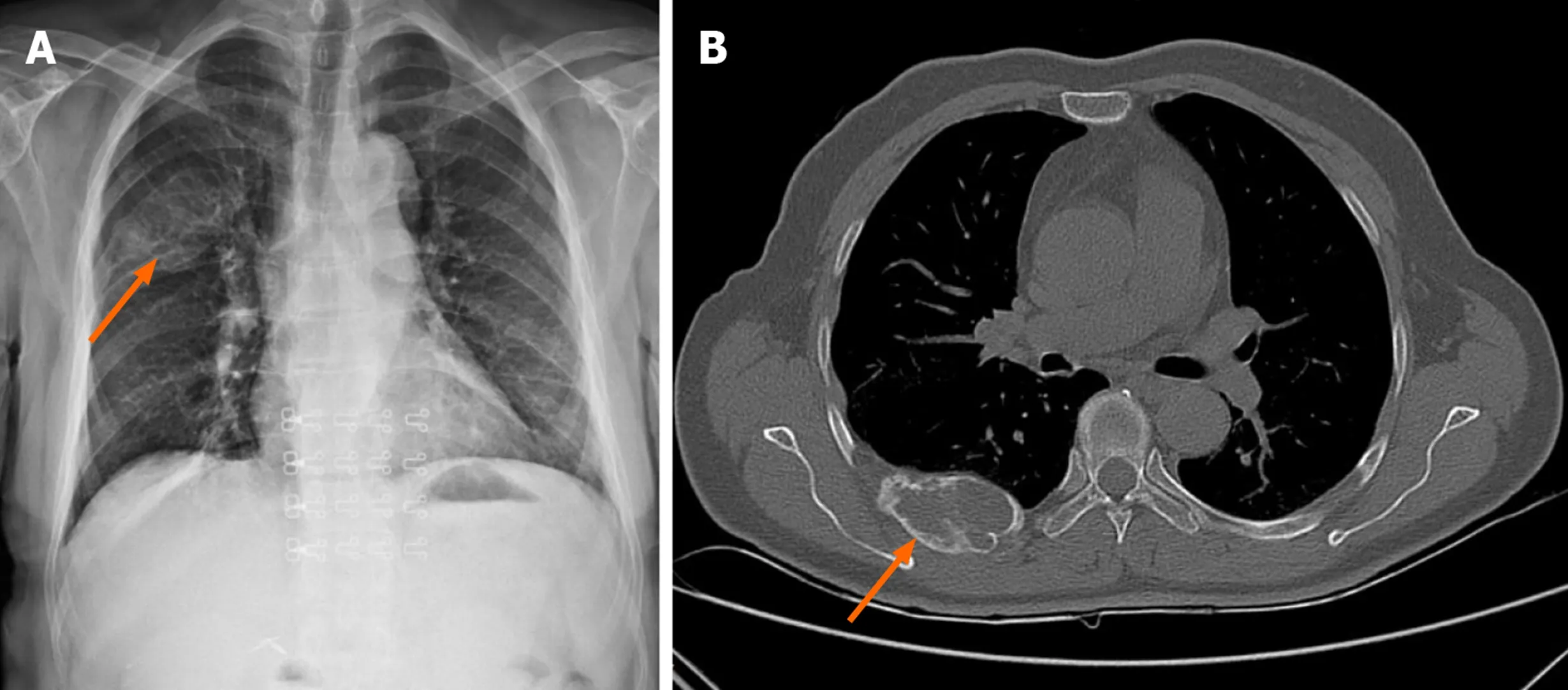
Figure 1 Chest X-ray and computed tomography before surgery.
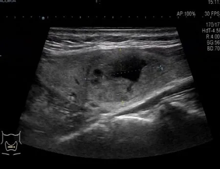
Figure 2 Ultrasound image of the thyroid.
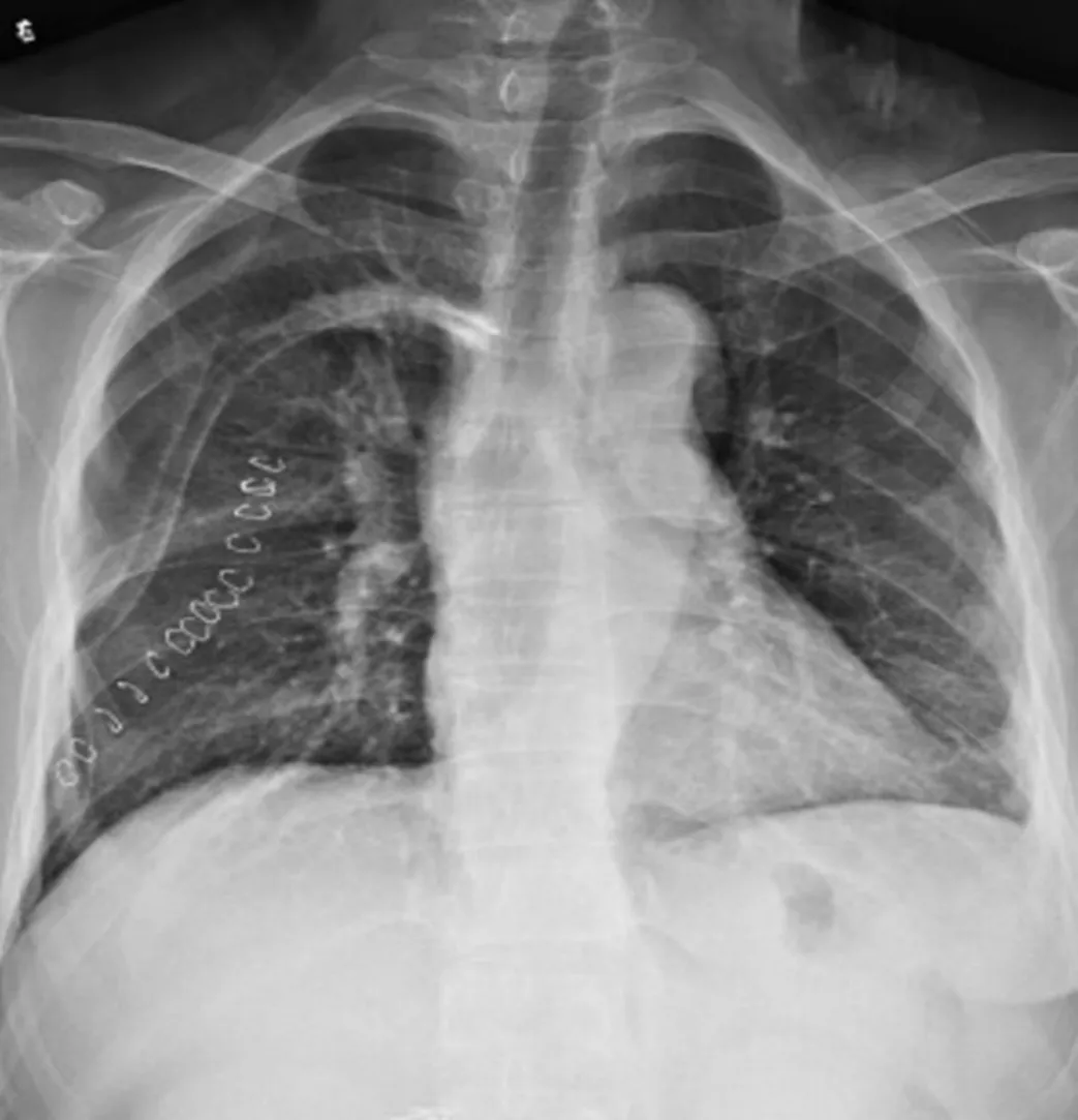
Figure 3 Chest X-ray after surgery.
Parathyroid adenoma is a common benign parathyroid tumor.Eighty-five percent of patients can have PHPT[1,2].The symptoms involve multiple systems,including hypercalcemia and urinary tract abnormalities.Stones caused by calcium salt deposition can also manifest as osteoporosis,pathological fractures and other symptoms.Most patients have symptoms at the time of consultation.A few patients lack typical clinical manifestations,and often have kidney,bone and other diseases[3].According to the different manifestations of affected organs,PHPT can be divided into bone disease-based types,where patients may have bone pain,are prone to fracture,and fibrocystic osteitis is a typical skeletal lesion of PHPT combined with bone cysts,brown tumors,osteoporosis and fragile fractures,as shown by conventional bone radiography[4].The primary target organ of PHPT is the kidney,which can develop into kidney stones[5].It can also manifest as urinary calculi and osteolytic bone changes,as well as other systemic lesions[6,7].
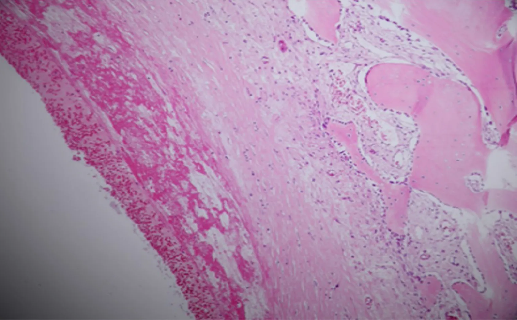
Figure 4 Pathological results of the rib tumor.A solid section of the cut surface,bleeding and bloody fluid are seen in the cyst cavity,fibrous tissue hyperplasia around the cystic cavity,reactive hyperplasia of bone-like tissue and trabecular bone in the deep cystic cavity,solid part of the area containing hemosiderin deposition,and multinucleated giant cell reaction.
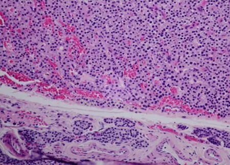
Figure 5 Pathological results of the parathyroid adenoma.Left lower parathyroid gland combined with HE morphology and immunohistochemistry results diagnosis:parathyroid adenoma.Immunohistochemical results:CgA part(+),PTH(+),Ki67(marker of proliferation Ki-67)approximately 1%(+),TG(-),and TTF-1(thyroid transcription factor 1)(-).
In the present case,the patient presented with a rib tumor,and CT was suggestive of giant cell tumor of bone,and osteolytic changes on admission.Preoperative examination revealed a significant increase in serum calcium and serum PTH.We considered this to be parathyroid gland dysfunction.With the consent of the patient and her family,we first performed rib tumor resection,and the pathology revealed a cyst.The patient was in good physical condition after surgery.Parathyroidectomy was subsequently performed in the general surgery department.Postoperative serum calcium and serum PTH levels decreased.We consider that the rib tumor may have been fibrocystic osteitis secondary to parathyroid adenoma.
Fibrocystic osteitis,also known as brown tumor,mostly manifests in the middle and late stages of hyperparathyroidism,and is a metabolic bone disease caused by calcium and phosphorus abnormalities[8].Fibrocystic osteitis occurs in the clavicle,long bones,and pelvis[9].The imaging manifestations of osteolytic bone destruction include clear boundaries and it often causes bone expansion,and other subperiosteal bone resorption.It is not a real tumor,but abnormally increased PTH in the body stimulates osteoclast activity,which leads to osteolysis and destruction[10].
At present,there is no clear treatment plan for parathyroid adenoma complicated with fibrocystic osteitis,and there is no consistent guideline recommendation whether to treat fibrocystic osteitis surgically.Parathyroidectomy was performed in this patient with PHPT and fibrocystic osteitis.Postoperative blood calcium returned to normal levels and bone changes were reversed.In such patients,parathyroidectomy is the first treatment choice for fibrocystic osteitis.Postoperative PTH is reduced,bone destruction is controlled,fracture risk is reduced,and bone density gradually increased in the later stage,thereby completely inhibiting fibrocystic changes.During the development of osteitis,Vander Walde found that irrespective of age,and blood calcium levels,patients with surgically removed parathyroid glands have a lower risk of fractures than those without surgery;thus,surgical treatment of fibrocystic osteitis can be eliminated[11].Of course,different patients have different outcomes due to the severity of the disease,whether they have fractures,and their vitamin status.When pathological fractures occur,the reversion time of bone lesions is relatively long,which may be related to the slow recovery of cortical bone density.However,for obvious local bone and joint pain,pathological fracture or high fracture risk,and recurrence after reversal,orthopedic surgery can be performed to relieve the symptoms of local pain and fix the fracture,but only if the diagnosis is clear,in order to reduce secondary damage to patients caused by overtreatment.
Agarwal G followed the clinical recovery,bone turnover,bone mineral density,and biochemical indicators on bone radiology of 51 patients with PHPT and fibrocystic cysts[12].Qualitative destruction recovered,and bone tumors persisted in a few patients,requiring further surgical intervention.Wang performed a large rib brown tumor resection in patients who had been treated with zoledronic acid for 17 mo with elevated serum calcium and PTH.One patient reported hip pain and abnormal pelvic X-rays after surgery.A parathyroid adenoma was subsequently found.Pain relief was achieved after adenoma resection.It took at least 2 years for fibrotic osteitis to recover[13].Khalil reported a patient with brown tumor lesions in the right pelvis,and surgery was considered due to a persistent increase in focal pain.Pain was relieved postoperatively.When there is a high risk of pathological fractures or patients have persistent focal bone pain,surgery should be considered at the appropriate site[14].Satpathy reported a case of parathyroid adenoma with mandibular fibrocystic osteitis.After parathyroidectomy,the patient's bone lesions were significantly reduced during the 6 mo of regular follow-up,while complete bone reconstruction took longer.The author believed that the removal of parathyroid adenomas and achieving normal blood calcium levels are options for treating parathyroid glands with fibrocystic osteitis.If primary bone disease does not shrink significantly after 6 mo of treatment,surgical curettage should be considered[15].
As the incidence of fibrocystic osteitis is extremely low,and the primary manifestations in parathyroid glands are not fully understood,doctors are often confused by the association with bones.Surgical removal of bone tumors does not include a comprehensive preoperative examination.Although the pain caused by the patient's skeletal disease can be relieved temporarily,after a period of time,the bone disease will recur and brown tumors in other areas can occur.Therefore,an appropriate preoperative examination is necessary to diagnose parathyroid adenoma with fibrocystic osteitis and plan reasonable treatment.Giant cell tumors of bone and brown tumors have osteolytic lesions of cortical bone,which often confuse doctors and can be misdiagnosed.Therefore,it is necessary to combine the patient's other clinical manifestations,medical history,and reasonable examination methods for the diagnosis of brown tumors,such as preoperative color Doppler ultrasound.With regard to the treatment of parathyroid adenoma with fibrocystic osteitis,previous studies have reported that skeletal lesions will gradually be controlled and bone cysts will be reversed in the majority of patients.However,the reconstruction of intact bone takes a long time,and long-term skeletal lesions cannot be completely controlled,which may cause pathological fractures,and although there are certain risks,surgical intervention is still required.Some brown tumors can occur on the face.If they cannot be treated within a short time,they will also affect the patient's mental status.Therefore,surgical removal can be performed at the patient's request,and the disease should be detected early to establish a correct diagnosis.The patient's symptoms should be comprehensively evaluated,not only to avoid misdiagnosis,but also to reduce trauma.
CONCLUSION
Parathyroid adenoma with brown tumor is relatively rare,a comprehensive preoperative physical examination and evaluation of the patient's condition are conducive to a more accurate diagnosis and treatment.At present,in patients with a brown tumor,most are treated with parathyroid adenoma resection,and after a period of time,the brown tumor may gradually disappear.However,if the brown tumor does not improve and the patient's physical condition is affected,surgical treatment with regular follow-up is necessary.
 World Journal of Clinical Cases2020年19期
- World Journal of Clinical Cases的其它文章
- Displacement of peritoneal end of a shunt tube to pleural cavity:A case report
- Localized primary gastric amyloidosis:Three case reports
- Bochdalek hernia masquerading as severe acute pancreatitis during the third trimester of pregnancy:A case report
- Intravesically instilled gemcitabine-induced lung injury in a patient with invasive urothelial carcinoma:A case report
- Intraosseous venous malformation of the maxilla after enucleation of a hemophilic pseudotumor:A case report
- Solitary hepatic lymphangioma mimicking liver malignancy:A case report and literature review
