Eight-year follow-up of locally advanced lymphoepithelioma-like carcinoma at upper urinary tract:A case report
Che H Yang,Wei C Weng,Yi S Lin,Li H Huang,Chin H Lu,Chao Y Hsu,Yen C Ou,Min C Tung
Che H Yang,Wei C Weng,Yi S Lin,Li H Huang,Chin H Lu,Chao Y Hsu,Yen C Ou,Min C Tung,Department of Surgery,Division of Urology,Tungs’ Taichung MetroHarbor Hospital,Taichung City 435403,Taiwan
Abstract BACKGROUND Urinary tract lymphoepithelioma-like carcinoma is rarely seen.Although it is termed after lymphoepithelioma at the nasopharynx,it behaves more like high grade urothelial carcinoma by immunohistochemical features.Most published literatures focused on its rarity but few discussed results of long-term follow-ups.As no available guidelines are applicable,we postulated that principles should be similar to that of urothelial carcinoma at urinary tract.As of now,this work features the longest follow-up of this cancer at the upper urinary tract.CASE SUMMARY A 63-year-old female had a chief complaint of intermittent left flank pain for 2 mo,along with accompanying symptoms including vomiting and body weight loss,about 7 kg over 2 mo.Laboratory data showed normocytic anemia,mildly poor renal function,and hyperparathyroidism.Urine analysis showed mild hematuria.Computed tomography showed a 4.2-cm-width irregular mass over left renal pelvic and enlarged lymph node at the left renal hilum.Whole-body bone scan was negative of active bone lesions.Biopsy from ureteroscopy showed urothelial carcinoma.Specimen from laparoscopic nephroureterectomy with bladder cuff resection showed lymphoepithelioma-like carcinoma with muscular invasion(pT3).She took adjuvant chemotherapies of 2 cycles and full courses of radiation therapy.No recurrence was observed with designed investigative programs.CONCLUSION Locally advanced urinary tract lymphoepithelioma-like carcinoma could benefit from nephroureterectomy and bladder cuff excision in terms of recurrence-free survival.
Key Words:Urologic neoplasms pathology;Kidney pelvis;Tomography X-ray computed;Carcinoma mortality;Kidney neoplasms mortality;Case report
INTRODUCTION
Urinary tract lymphoepithelioma-like carcinoma(LELC)is termed after lymphoepithelioma(LE)at the nasopharynx but is actually a variant of urothelial carcinoma(UC).LELC is infrequently,2% occurrence,identified at both upper and lower urinary tract.Malignancy is constituent of epithelial parts and inflammatory cells,such as myeloid cells or lymphoid cells under hematoxylin and eosin stain,making chemotherapy feasible.Immunohistochemical analysis differentiates urinary tract LELC from LE at the nasopharynx.No hybridization to Epstein–Barr virus,which is thought to be essential in cell expression and maintenance in LE of the nasopharynx,could be found in urinary tract LELC[1].On the other hand,other characteristics such as presence of p53 make urinary tract LELC more resembling to high grade UC.In our previous published literature[2],we deliberated our postulation on urinary tract LELC,applying the same therapeutic principle as UC.From our experiences[2],nephroureterectomy with bladder cuff excision without adjuvant therapies on urinary tract LELC showed no recurrence within one year after surgery.However,most published literatures tend to have more discussion on the lower and few on the upper urinary tract.Among case studies of upper urinary tract LELC,few had discussions on longterm follow-ups.Furthermore,compared to stages T1 and T2 upper urinary tract LELC,stage T3 is proved to be worse in terms of overall survival[3].To date,this documentation has the longest investigations on the locally advanced upper urinary tract LELC.
CASE PRESENTATION
Chief complaints
On July 2012,a 63-year-old female came to the urology office with symptoms of intermittent left flank pain for 2 mo.
History of present illness
The pain was described to be localized at left fank.Neither precipitating factors nor exaggerating factors were mentioned.It occurred randomly without any special time or occasions,and can be suppressed by painkillers.Accompanying symptoms include vomiting and body weight loss,about 7 kg over 2 mo.She denied having visible red color urine,neither fever.Appetite was not changed.Neither specific travel histories nor cluster histories were mentioned.
History of past illness
She had uterine prolapse diagnosed 2 years before this episode,but did not have any interventions about it.
Physical examination
Her temperature was 35.9 °C,and heart rate was 74 bpm.Respiratory rate was 17 breaths per minute,and blood pressure was 127/81 mmHg.Oxygen saturation in room air was 98%.She ranked the pain subjectively 4 out of 10 on visual analog scale pain score.No knocking pain was examined at costovertebral angle,neither the tenderness on abdomen.
Laboratory examinations
Normocytic anemia was seen on complete blood cell(White blood cell:4 500/UL,Red blood cell:3370000/UL,Hemoglobulin:10.4 g/DL,Mean corpuscular volume:93.5 FL).Mildly insufficiency was seen on renal functions tests(Creatinine:1.4 mg/DL,Blood urea nitrogen:16.2 mg/DL).Elevated Intact parathyrin(EIA/LIA)was 245 pg/ML.Free T4(EIA/LIA)was 1.070 ng/DL,and TSH(EIA/LIA)was 1.5 UIU/ML.Tumor markers of carcino-embryonic antigen,CA-199,and alpha-fetoprotein were all within normal limits.Urine examination showed hematuria(red blood cell 5-10/HPF)and pyuria(white blood cell:10-20/HPF).Urine cytology was gained,but no atypical cells were examined.
Imaging examinations
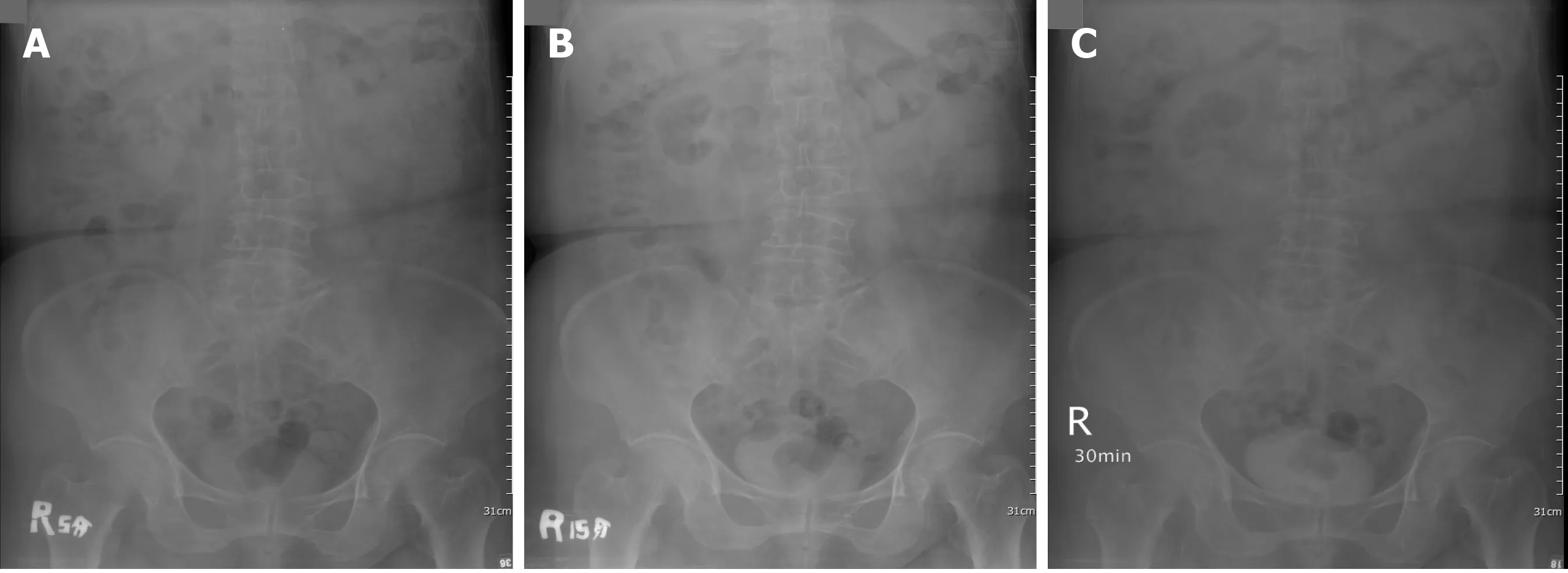
Left side hydronephrosis was evident on ultrasound.Intravenous pyelography showed a non-enhanced left kidney(Figure 1)and then computed tomography urogram was arranged(Figure 2)for comprehensive studies,which showed an enlarged lymph node close to the left renal pedicle and one irregularly ill-defined 4.2-cm width mass at the left renal pelvic.
Further diagnostic work-up
Ureteroscopy was arranged and biopsy was done to this 4.2-cm mass at left renal pelvis.Pathology from the biopsy was proved to be high-grade(grade III)UC,which is invasive to lamina propria.
FINAL DIAGNOSIS
The final diagnosis of this case is upper urinary tract UC with clinical stage of T3N1Mx at left renal pelvis.
TREATMENT
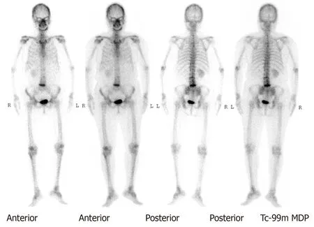
A whole-body bone scan showed a negative finding(Figure 3).After securing informed consent,the patient underwent laparoscopic left nephroureterectomy with bladder cuff excision.
A tumor size of 3 cm × 2.5 cm× 2.5 cm from left nephroureterectomy with bladder cuff resection was examined as undifferentiated LELC(Figure 4),high grade(undifferentiated type,grade IV),and invasive into muscle at renal pelvis and peripelvic fat(pT3).Surgical margin,Gerota’s fascia,ureter,and resected bladder cuff were all free of tumor.No lymph node was identified from the specimen.Tissues from the other non-tumor kidney displayed chronic pyelonephritis with focal global sclerosis.
OUTCOME AND FOLLOW-UP

Figure 1 Intravenous pyelography.No enhancement of urinary tract at 5 min(A),15 min(B)and 30 min(C)after administration of the contrast.

Figure 2 Computed tomography urogram.One 4.2 cm irregular mass was seen occupying the left renal pelvis on computed tomography urogram(A)(B);One enlarged lymph node was observed at the left renal pedicle(C).

Figure 3 No observed active lesions were demonstrated on whole-body bone scan.
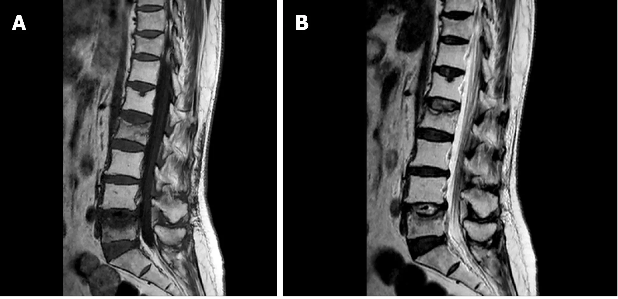
She received two cycles of gemcitabine(1000 mg/m2)and cisplatin(35 mg/m2)chemotherapy after the operation but stopped due to unbearable adverse effects,which was assessed to be grade I nausea and vomiting on World Health Organization criteria.Palliative radiation therapy(RT)was targeted to the enlarged lymph node at the left renal pedicle which had no proven pathologies.A total dosage of 5580 cGy was divided into 31 doses.However,2 mo later,she suffered from low back pain and osteoporotic compression fractures were shown on X-ray and magnetic resonance imaging(Figure 5)of the L-spine(L2 and L5),which was a suspicious pathological change secondary to LELC.A second whole-body bone scan revealed no new onset changes compared to the preoperative one.The orthopedist performed palliative resection of the bone lesions and fixation with cages for symptomatic treatment.Fortunately,the specimen taken from bones showed null of metastatic cancer.Active follow-ups with urine cytology and cystoscopy every three months were planned.Computed tomography scan was done every six months postoperatively in the succeeding five years.After five years,biannual cystoscopy and annual computed tomography scan were done.She has no evidence of tumor recurrence postoperatively for 8 years till now.In conclusion,this pathological T3N1Mx mixed LELC with two cycles’ adjuvant chemotherapy and a full dose of palliative RT can lead to a recurrence-free survival up to 8 years after left nephroureterectomy with bladder cuff excision.

Figure 4 Hematoxylin and eosin stain.A:Generally,with hematoxylin and eosin stain the tumor was seen with invasion to muscular layer;B and C:Under augmentation(400 ×),the tumor was examined with epithelial cells and lymphoid cells.

Figure 5 Magnetic resonance imaging.T1-weight(A)and T2-weight(B)image revealed destructive L2 and L5 spine.Related involvement of nearby spinal cord was also noted.
DISCUSSION
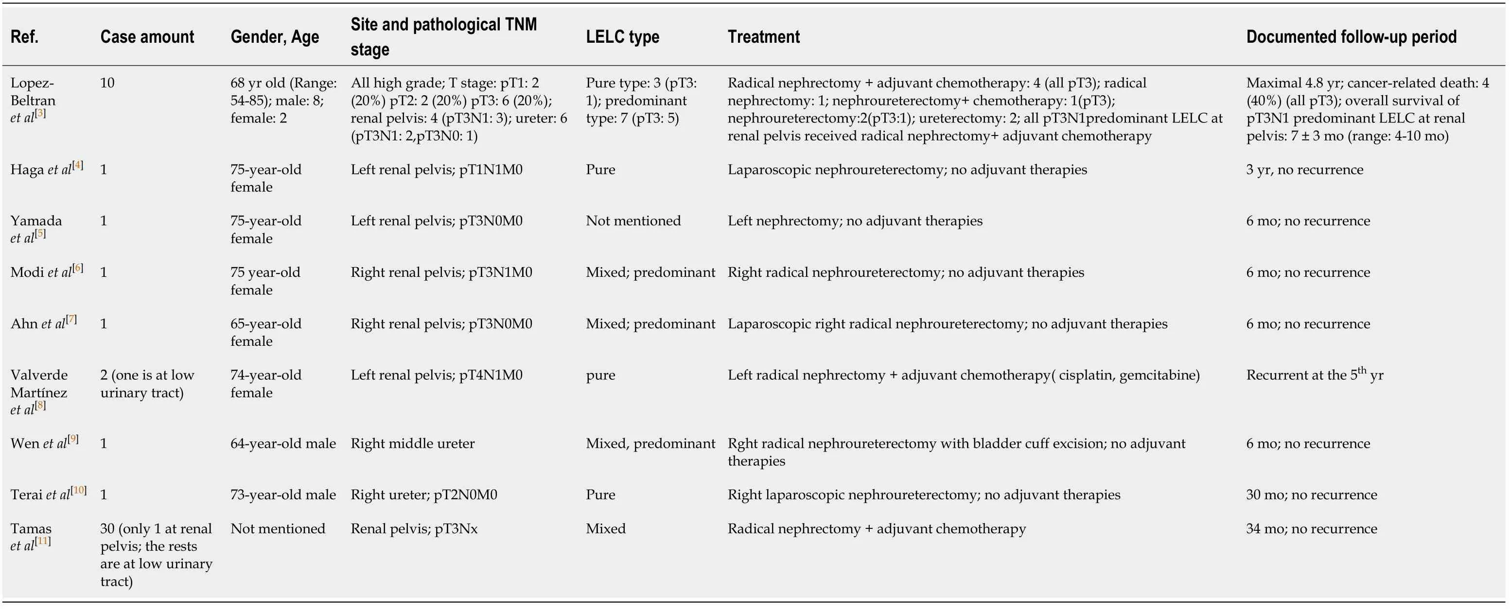
Generally speaking,urinary tract LELC can be categorized into pure(100% LELC)or mixed type(predominant:>50% LELC),and pure type is reported to have more favorable prognosis.The case,described in this article,comprises of some of the worst scenarios of LELC at the upper urinary tract,with muscular invasion,lymph node involvement,and undifferentiated mixed type histology.Lopez-Beltranet al[3]issued the largest series of follow-ups on upper urinary tract LELC[3]with a maximal 4.8-year span.Their data suggests that the overall survival rate is comparable to conventional UC at the upper urinary tract(about 60% survival rate and 40% cancer-related death)across all stages.However,in their statistics,all cancer-related deaths are at pT3 stage.They also find that prognosis will be influenced by T stage,and T3 stage(time before cancer-related death:15 ± 16.2 mo;median:8.5 mo;range:4-39 mo)will significantly be worse than T1 and T2.The other related reports about LELC at the upper urinary tract in the past 20 years are listed in Table 1[3-11].
Among most publications,very few are documented with follow-ups long enough to represent meaningful survival analysis[3,4,8,10,11].In article by Lopez-Beltranet al[3],radical nephrectomy with adjuvant chemotherapy is performed to all pT3N1 predominant LELC,which is similar to our case,at renal pelvis but they all die from cancer within 7 ± 3 mo.Judging from our case,literature from Lopez-Beltranet al[3]and case reports from Valverdeet al[8]and Tamaset al[11],patients can benefit more from the operative method of nephroureterectomy than nephrectomy in terms of recurrence-free survival and overall survival.Adjuvant chemotherapy and palliative RT,by comparing our case with T1 from Hagaet al[4]and T2 from Teraiet al[10],might provide beneficial credits to prolong the recurrence-free survival for those with unfavorable advanced stage.However,there are still some examples with radical nephrectomy and adjuvant chemotherapy[8]that demonstrated patients could live without recurrence to a maximal 5-year interval.This phenomenon of prognostic variations might imply that some other factors will further affect recurrence-free survival.

Table 1 Published literatures of upper urinary tract lymphoepit helioma-like carcinoma
CONCLUSION
From our experiences and published literature,operative methods still possess definite roles,and locally advanced LELC at the upper urinary tract could benefit from nephroureterectomy and bladder cuff excision over nephrectomy in terms of recurrence-free survival and overall survival.Adjuvant chemotherapy and palliative RT might have positive roles for locally advanced LELC at upper urinary tract to extend the recurrence-free survival parallel to those with T1 and T2 ones,but a largescale analysis is necessary to further prove the hypothesis.
ACKNOWLEDGEMENTS
We thank Chung Hsiao Chin(Pathologist,Tungs’ Taichung MetroHarbor Hospital,Taiwan)for helping re-stain the pathological specimen.
 World Journal of Clinical Cases2020年19期
- World Journal of Clinical Cases的其它文章
- Parathyroid adenoma combined with a rib tumor as the primary disease:A case report
- Displacement of peritoneal end of a shunt tube to pleural cavity:A case report
- Localized primary gastric amyloidosis:Three case reports
- Bochdalek hernia masquerading as severe acute pancreatitis during the third trimester of pregnancy:A case report
- Intravesically instilled gemcitabine-induced lung injury in a patient with invasive urothelial carcinoma:A case report
- Intraosseous venous malformation of the maxilla after enucleation of a hemophilic pseudotumor:A case report
