
Antibacterial activity of bacillomycin D-like compounds isolated from Bacillus amyloliquefaciens HAB-2 against Burkholderia pseudomallei
2020-04-02 07:34:20MamyJayneNellyRajaoferaXunKangPengFeiJinXinChenChenChuLiLiYinLinLiuQingHuiSunNanZhangChuiZheChenNaHeQianFengXiaWeiGuoMiao
Mamy Jayne Nelly Rajaofera, Xun Kang, Peng-Fei Jin, Xin Chen, Chen-Chu Li, Li Yin, Lin Liu, Qing-Hui Sun, Nan Zhang, Chui-Zhe Chen, Na He, Qian-Feng Xia✉, Wei-Guo Miao
1Key Laboratory of Tropical Translational Medicine of Ministry of Education and School of Tropical Medicine and Laboratory Medicine, Hainan Medical University, Haikou, Hainan, China
2Key Laboratory of Green Prevention and Control of Tropical Plant Diseases and Pests (Hainan University), Ministry of Education, Haikou 570228, Hainan, China
ABSTRACT Objective: To investigate the inhibitory effect on Burkholderia pseudomallei (B. pseudomallei) strain HNBP001 of a bacillomycin D-like cyclic lipopeptide compound named bacillomycin DC isolated from Bacillus amyloliquefaciens HAB-2.Methods: The antibacterial effect of bacillomycin DC on B. pseudomallei was determined using the disk diffusion method. The minimum inhibitory concentrations were evaluated by microdilution assay. In addition, transmission electron microscopy was performed and quantitative real-time polymerase chain reaction assay was carried out to determine the expression of MexB, OprD2, and qnrS genes. Results: Bacillomycin DC produced an inhibition zone against B. pseudomallei with minimum inhibitory concentration values of 12.5 μg/mL 24 h after treatment and 50 μg/mL at 48 and 72 h. Transmission electron microscopy showed that bacillomycin DC resulted in roughening cell surface and cell membrane damage. Quantitative real-time polymerase chain reaction analysis showed low expression of MexB, OprD2 and qnrS genes. Conclusions: Bacillomycin DC inhibits the growth of B. pseudomallei and can be a new candidate for antimicrobial agents of B. pseudomallei.
KEYWORDS: Bacillomycin DC; Bioactive compound; Burkholderia pseudomallei
1. Introduction
Burkholderia pseudomallei (B. pseudomallei) is a Gram-negative environmental bacterium that is a causal agent of melioidosis, a major community-acquired infection in endemic areas such as Southeast Asia and northern Australia. The disease has a wide spectrum of clinical manifestations, ranging from acute pneumonia or septicaemia to chronic abscesses[1,2]. Relapsing melioidosis is a common disease, causing a high mortality rate. The overall mortality rate in individuals with melioidosis varies by country. In Thailand, the mortality rate is around 40.0%, while approximately 10.0% in Northeast Australia[3,4]. However, the mortality rate of septicaemic melioidosis can be up to 90%, even with appropriate antibiotic therapy[2,4,5]. A recent report estimates the incidence of melioidosis disease was 165 000 cases per year[6]. Despite all this, there is no licensed vaccine to protect against melioidosis[5].
Treatment of melioidosis often involves antibiotics therapies with prolonged course[7]. Among the used antibiotics, ceftazidime and cephalosporins displayed great effect, but the toxicity and side effects of these antibiotics could not be neglected. The thirdgeneration cephalosporins were reported to result in some adverse reactions in clinical application[8]. Furthermore, the abuse of antibiotics also leads to an increase in antibacterial resistance. It was reported that B. pseudomallei is resistant to a wide range of antimicrobial agents including cephalosporins, trimethoprimsulfamethoxazole, and amoxicillin–clavulanic acid[9-14]. Therefore, it is considerable to search for a newer and effective antimicrobial agent.
Many pieces of literature have reported the potential use of the secondary metabolites produced by the natural organisms exhibiting a selective spectrum of inhibition. Among them, antimicrobial peptides including lipopeptide compounds have been especially useful to control the growth of different pathogenic organisms[15-17]. They induce a defense system against invading pathogenic bacteria and are effective therapeutically, causing the rapid killing of the target organisms, including the antibiotic-resistant microorganisms[18]. Thus, screening of these antimicrobial peptides has attracted the attention of many researchers due to their potential applications as therapeutics against biological warfare agents. In the previous study, we have reported that a bacillomycin D-like cyclic lipopeptide compound, known as bacillomycin DC which was isolated from Bacillus amyloliquefaciens (B. amyloliquefaciens) strain HAB-2, had strong antifungal activity against Colletotrichum gloeosporioides[19]. Reports on the antibacterial effect of the natural product against pathogenic bacteria are rare. Boottanun et al. have reported that the secondary metabolites from B. amyloliquefaciens strains N2-4 and N3-8 were able to kill B. pseudomallei, but the antibacterial component was not identified[20]. Therefore, in this study, the effect of bioactive compound bacillomycin DC against the causative agent of melioidosis, B. pseudomallei was explored.
2. Materials and methods
2.1. Materials
Bacillomycin DC, a cyclic lipopeptide compound, was isolated from B. amyloliquefaciens HAB-2[19] and stored in our laboratory at 4 ℃. Bacillomycin DC was dissolved in 2% (v/v) dimethyl sulfoxide to produce stock solutions of 10 mg/mL. Ceftazidime dissolved with distilled water was used as a positive control.
2.2. Test organism
All experiments were carried out under biosafety laboratory level 2 (BSL-2) containment using standard operating procedures. B. pseudomallei HNBP001 which was a clinical strain was previously isolated from the blood of a melioidosis patient with pneumonia at the First Affiliated Hospital of Hainan Medical University and used as the test organism[21]. As previously reported, the strain was sensitive to trimethoprim-sulfamethoxazole, ceftazidime, meropenem and imipenem[22]. The strain was grown in Luria Bertani broth and incubated at 37 ℃ for 18 h before the concentration adjusted to a 0.5 McFarland turbidity standard. Thereafter, the adjusted bacterial cultures were diluted to approximately (A600of 0.8) 3×10⁸colony forming unit (CFU)/mL for the antibacterial activity assay.
2.3. Antibacterial assay of bacillomycin DC against B. pseudomallei
The capacity of bacillomycin DC to inhibit the growth of B. pseudomallei was determined using the disc diffusion method[20]. Briefly, the test organism was mixed with the Luria Bertani agar medium pre-cooled to 45 ℃ and then poured immediately into sterile Petri dishes. Filter paper disk (6 mm in diameter) was impregnated with 10 mL of bacillomycin DC (50 μg/mL), dried and placed on the surface of the plates which were previously seeded with the test organism, and then incubated in the dark at 37 ℃. After 48 hours of incubation, the antibacterial activity was determined. The same concentration of ceftazidime was used as a control. The experiment was carried with three replicates.
2.4. Determination of minimum inhibitory concentration (MIC)
For antimicrobial assay, the MIC was also studied using a microtiter plate dilution assay. Bacillomycin DC and ceftazidime were first diluted into 2 000 to 0 μg/mL. About 200 μL of sterile Luria Bertani broth was poured in each well of a 96-well microtiter plate (Greiner, Nurtingen, Germany) and 50 mL of test organism in different rows was then added into plates. Subsequently, 50 mL of different dilutions of bacillomycin DC was added to each well. Each dilution series included control wells containing bacteria without the test organism. The plates were incubated at 37 ℃ incubators for 24, 48 and 72 h and the inhibition of bacterial growth was determined by measuring the absorbance at 664 nm. The lowest concentration that inhibited the growth of test organisms that did not show any increase in absorbance was considered as MIC. The experiment was replicated three times.
2.5. Transmission electron microscopy (TEM)
TEM analysis was used to determine the effect of bacillomycin DC on the structure of B. pseudomallei. One milliliter of the bacteria suspension was added with the bioactive compound and then incubated at 37 ℃ at 180 rpm. After 24 hours of incubation, bacterial cells were treated with 50 μg/mL bacillomycin DC and centrifuged. The filtrate was harvested after centrifugation, washed twice with sterile distilled water and then fixed with 2.5% (v/v) glutaraldehyde in 0.1 M phosphate buffer (PBS, pH 7.4) at 4 ℃ for 2 h. The fixed cells were washed three times for 10 min with 100 mM phosphate of PBS followed by post fixing with osmium tetroxide for 3 h at room temperature. Samples were dehydrated by a graded series (70%, 80%, 90%, and 100%) of ethanol solutions and then coated with gold and analyzed on a HitachiS-3000N scanning electron microscope (Hitachi, Japan). The sections were double-stained with saturated uranyl acetate and lead citrate. The grids were examined with a TEM (Hitachi H-600, Japan)[23]. The plate which was not inoculated with the bacterium was used as a negative control and ceftazidime was used as a positive control.
2.6. Quantitative real-time polymerase chain reaction (qRTPCR) analysis
B. pseudomallei was inoculated with 50 μg/mL of bacillomycin DC and grown overnight at 37 ℃. Total RNA was isolated after 24, 48 and 72 hours of culture following the manufacturer’s instructions (Beijing, China Co., LTD). Complementary DNA (cDNA) was synthesized with the Fast Quant RT SuperMix Kit (Tiangen). Three drug-assistant genes were selected for expression analysis (Table 1). SYBR Green Master Mix (Agilent Technologies, Santa Clara, CA, USA) was used for qRT-PCR. B. pseudomallei identification gene (P1/P2) was used as an internal reference. The threshold (Ct) value for each gene was normalized against the Ctfor EF1α. Relative expression levels were analyzed using the delta-delta-Ctmethod[28]. The following protocol was used for amplification: 94 ℃ for 5 min followed by 35 cycles at 94 ℃ for 30 s, 58 ℃ for 40 s and 72 ℃ for 60 s. Ceftazidime subjected to the same treatment was used as a positive control. The experiment was performed in three replicates.

Table 1. Primers used in this study.
2.7. Statistical analysis
All analyses were carried out in triplicate and the data were expressed as mean ± standard deviations (SD). One-way ANOVA and Dunnet’s test were performed to analyse the differences between test and control groups (P < 0.05). Statistical analysis was performed using SPSS version 16.0 software (SPSS Inc., Chicago, IL, USA).
2.8. Ethical statement and informed consent
The present study was approved by the Ethics Committee of Hainan Medical University (Haikou, China; CEEA2018-213) and patient provided written informed consent.
3. Results
3.1. Bacillomycin DC exhibited antibacterial activity against B. pseudomallei
At 24 hours post-inoculation, 50 μg/mL of bacillomycin DC showed inhibitory effect on B. pseudomallei with a diameter of inhibition zone (17.00 ± 0.21) mm. In addition, the results showed that the MICs of bacillomycin DC were 12.5 μg/mL at 24 h, and 50 μg/mL at 48 and 72 h. For ceftazidime, it was 25 μg/mL at 24, 48 and 72 hours post-treatment.
3.2. Bacillomycin DC caused changes in the morphology of B. pseudomallei

Figure 1. Transmission electron microphotographs of Burkhoderia pseudomallei: (A, B) untreated bacteria, (C, D) bacteria treated with bacillomycin DC, (E, F) bacteria treated with ceftazidime. Arrows indicate cell membrane damage. Magnification: 25× (A, C, E) and 120× (B, D, F).
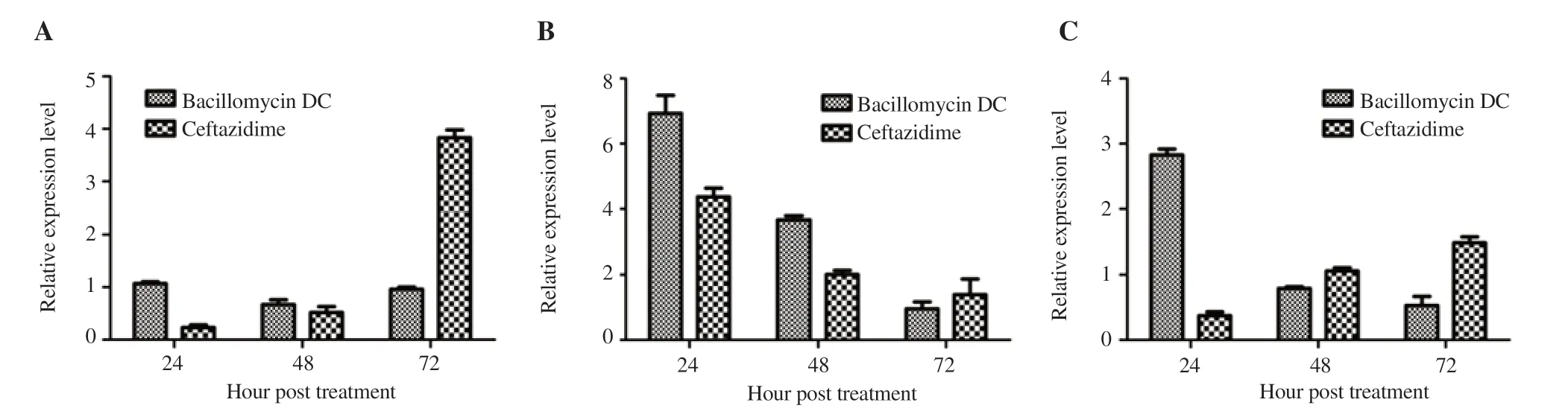
Figure 2. Relative expression level of drug resistance genes. A: MexB, B: OprD2, C: qnrS. Total RNA was isolated after bacillomycin DC and ceftazidime treatment, and then subjected to qRT-PCR analysis. Values were presented as mean ± SD from three independent replicates.
In order to gain insight into the bactericidal mechanism of the bacillomycin DC on B. pseudomallei, TEM was performed. Compared to the untreated bacteria, B. pseudomallei cells preincubated with the compound for 24 h displayed significant changes in cell morphology (Figure 1), including cell damage with roughening cell surface (Figure 1C and D). The antibacterial mechanism of ceftazidime was found different, demonstrating cell elongation with distortion on B. pseudomallei (Figure 1E and F).
3.3. Gene expression involved in the antibacterial activity of bacillomycine DC
The effect of bacillomycin DC on the expression profile of the drug resistance gene in B. pseudomallei by qRT PCR analysis is shown in Figure 2. The expression of MexB gene showed similar results both in bacillomycin DC and ceftazidime treatment. No significant difference was found between the two treatments in 24 h and 48 h post-treatment. At 72 h post-treatment, bacillomycin DC treatment lowered the gene expression by 3.99 fold compared with ceftazidime treatment (Figure 2A). The OprD2 gene decreased gradually in a time-dependent manner. Compared with the ceftazidime treatment, the expression of OprD2 gene was found 1.59 fold higher in bacillomycin DC treatment at 24 h post-treatment (Figure 2B). The expression of qnrS after bacillomycin DC treatment was high at 24 h and then decreased gradually in a time-dependent manner. Conversely, its expression was increased by ceftazidime treatment. The expression of qnrS after bacillomycin DC treatment reached 7.64 fold higher than that of ceftazidime treatment at 24 h posttreatment (Figure 2C).
4. Discussion
B. pseudomallei has become a major public health concern since it poses a significant threat to human and animal health. This organism is widely known for its resistance to many classes of antimicrobial agents[9-14], which is one of the major factors preventing the treatment of melioidosis; an infectious disease caused by B. pseudomallei. Therefore, it is of considerable interest to develop antimicrobial agent which can potentially decrease the emergence of drug resistance. In this context, the discovery of new approaches or novel therapeutic alternatives that can replace or complement the existing antibiotics is urgently required.
The origin and nature of certain drugs used against specific diseases make a sound difference in prognosis and therapy. The efficacy and safety of natural and synthetic drugs have been investigated and compared in different researches. The natural sources derived from drugs have been found comparatively safe and preferred against synthetic drugs. Different natural products have been reported to possess antimicrobial activity on different pathogenic organisms[17,18,29]. There is an interest in lipopeptide compounds for their potential applications as therapeutics against different pathogenic microorganisms[15-17]. We found that bacillomycin DC, a cyclic lipopeptide compound isolated from B. amyloliquefaciens HAB-2, showed an effective antimicrobial activity on B. pseudomallei. The result showed that bacillomycin DC caused a destructive effect on B. pseudomallei by destroying the bacterial structure thus damaging bacterial cells.
The MIC determination is important in the selection of an appropriate and effective concentration of antimicrobial substances. At 24 h post-treatment, the bioactive compound had lower MIC compared to that of ceftazidime. The MIC value of this new compound proves itself as a kind of fast-acting drug. In the present study, its inhibitory effect at 24 h was faster than ceftazidime, suggesting this compound is effective in the initial stage of infection. Changes in the expression of related genes usually occur soon after drug treatments. In the present study, the effect of bacillomycin DC on the expression of drug resistance gene was determined by real-time PCR analysis. It is reported that efflux pumps contribute crucially to antibiotic resistance in Gram-negative bacteria pathogens. Earlier studies have shown that if the expression of efflux pumps increases, the antibiotic output will enhance, resulting in bacterial resistance to antimicrobial agents[30-32]. The expression of multidrug efflux pumps is described as important resistance mechanisms of B. pseudomallei[33]. In this study, MexB gene had a low expression after bacillomycin DC treatment.
The channel formed by the outer membrane protein is considered to be the small specific channel of carbapenem antibiotics, a small molecule that can quickly enter the bacteria[34,35]. OprD2 is a specific channel protein for a certain drug that is well known to have great relevance to clinically reasonable and effective use of antibiotics. It is the only pore protein in Pseudomonas aeruginosa known to allow antibiotics to pass smoothly. Research has reported that a decrease or deficiency in OMP OprD2 expression was found to affect the permeability of the outer membrane protein that blocks the drug to reach the target site, thus making the bacteria resistant. It is considered to be a specific channel for the rapid entry of small molecule of carbapenem antibiotics into bacteria that can form a specific binding site between bacteria and carbapenem drugs. Moreover, the deficiency of OprD2 was reported to be the main factor for the resistance of Pseudomonas aeruginosa to carbapenems[24,36]. In the present study, we found that OprD2 gene was highly expressed in bacillomycin DC treatment, reaching 1.59 fold higher compared to ceftazidime treatmen after 24 h. Similar to the previous report, the bioactive compound leads to the high expression of this outer membrane protein, and it is able to enter into the cells through the channel protein and then kill them[37].
The qnrS gene consists of one amino acid, such as glycine, and two pentapeptide repeats. Combined with bacterial DNA gyrase or topoisomerase quinolone on a specific target, the gene can reduce the sensitivity of bacteria to the antibacterial drug of quinolones[38-40]. In this study, qnrS gene was also found highly expressed after bacillomycin DC treatment. The expression level was higher at 24 h post-treatment and then declined with the exposure time, which can be explained by the mass death or serious injury, resulting in the bacteria unable to express this gene to protect its DNA helicase and topoisomerase[41]. qnrS gene was found reversely expressed in ceftazidime treatment. This might be that the drug had difficulty in entering the cell[42].
The results described in the current study provide an insight into a possible protective activity of bacillomycin DC against B. pseudomallei. The mechanism of antibacterial activity of this compound against B. pseudomallei is worthy of further investigation.
Conflict of interest statement
No potential conflict of interest was reported by the authors.
Acknowledgments
We thank Dr. Nasr Ullah Khan for the critical improvement of this manuscript.
Funding
This study was supported in part by the Key Research and Developement Program of Hainan Province (ZDYF2018240), National Natural Science Foundation of China (31660033, 81560002 and 81960002), National Key R&D Program of China (No. 2018YFD0201105) and National Science and Technology Major Project (No. 2018ZX10101003-001-009). The funder had no role in data collection and interpretation, or the decision to submit the work for publication.
Authors’ contributions
WGM and QFX conceived and designed the experiments. XK, XC and CCL performed the experiments. QHS, NZ, and CZC analyzed the data. PFJ, LY, LL, and NH contributed reagents/materials/analysis tools. RMJN wrote the paper.
 Asian Pacific Journal of Tropical Biomedicine2020年4期
- Asian Pacific Journal of Tropical Biomedicine的其它文章
- Information for Authors Asian Pacific Journal of Tropical Biomedcine
- Identification and investigation of Calodium hepaticum in rodents and insectivores from Wuhan section of the Yangtze River in China
- Anti-Acinetobacter baumannii activity of Rumex crispus L. and Rumex sanguineus L. extracts
- Free and liposome form of gallic acid improves calvarial bone wound healing in Wistar rats
- A novel polyherbal formulation containing thymoquinone attenuates carbon tetrachloride-induced hepatorenal injury in a rat model