Involvement of insulin receptor substrates in cognitive impairment and Alzheimer's disease
Daisuke Tanokashira, Wataru Fukuokaya, Akiko Taguchi,
1 Department of Integrative Aging Neuroscience, National Center for Geriatrics and Gerontology, Obu, Aichi, Japan
2 Division of Neurology, Endocrinology, and Metabolism, Faculty of Medicine University of Miyazaki, Miyazaki, Japan
Funding: This work was supported by a MEXTGrant-in-Aid for Scientific Research on Innovative Areas (brain environment) (JP24111536;to AT), JSPS KAKENHI (JP24650201, JP26282026, JP17K19951, JP17H02188; to AT), and grants from the Mitsubishi Foundation (to AT)and NOVARTIS Foundation Japan for the Promotion of Science (to AT).
Abstract Type 2 diabetes-associated with impaired insulin/insulin-like growth factor-1 (IGF1) signaling (IIS)-is a risk factor for cognitive impairment and dementia including Alzheimer's disease (AD). The insulin receptor substrate (IRS) proteins are major components of IIS, which transmit upstream signals via the insulin receptor and/or IGF1 receptor to multiple intracellular signaling pathways, including AKT/protein kinase B and extracellular-signal-regulated kinase cascades. Of the four IRS proteins in mammals, IRS1 and IRS2 play key roles in regulating growth and survival, metabolism, and aging. Meanwhile, the roles of IRS1 and IRS2 in the central nervous system with respect to cognitive abilities remain to be clarified. In contrast to IRS2 in peripheral tissues, inactivation of neural IRS2 exerts beneficial effects, resulting in the reduction of amyloid β accumulation and premature mortality in AD mouse models. On the other hand, the increased phosphorylation of IRS1 at several serine sites is observed in the brains from patients with AD and animal models of AD or cognitive impairment induced by type 2 diabetes. However, these serine sites are also activated in a mouse model of type 2 diabetes, in which the diabetes drug metformin improves memory impairment. Because IRS1 and IRS2 signaling pathways are regulated through complex mechanisms including positive and negative feedback loops, whether the elevated phosphorylation of IRS1 at specific serine sites found in AD brains is a primary response to cognitive dysfunction remains unknown. Here, we examine the associations between IRS1/IRS2-mediated signaling in the central nervous system and cognitive decline.
Key Words: type 2 diabetes; insulin/insulin-like growth factor-1; insulin receptor substrate; Alzheimer's disease;aging; serine phosphorylation; metformin; neuroprotective effects; high-fat-diet
Insulin Receptor Substrates-Mediated Signaling Pathways
The binding of insulin and insulin-like growth factor-1(IGF1) to the insulin receptor (IR), IGF1 receptor (IGF1R),or the hybrid between these receptors (IR/IGF1R) promotes tyrosine kinase activities of these receptors, subsequently inducing tyrosine phosphorylation of cellular substrates including IRS1-4 (Schlessinger, 2000; Taguchi and White,2008). Studies in genetically engineered mice have shown that the biological effects of insulin/IGF1 signaling (IIS) on glucose or lipid metabolism are mediated via insulin receptor substrate (IRS)1 and IRS2 (White, 2003) whereas the tyrosine kinase activation of IR/IGF1R stimulates the phosphorylation of other scaffold proteins such as SH2B, GABs,DOCKs, and CEACAM1. The IRS proteins are composed of an NH2-terminal pleckstrin homology and phosphotyrosine-binding domains, followed by a tail of tyrosine and serine/threonine (Ser/Thr) phosphorylation sites (Yenush et al., 1996). The morphological change of tyrosine-phosphorylated IRS proteins by IR/IGF1R increases the flexibility of binding to Src homology 2 domain proteins including phosphatidylinositol-3 kinase and Src homology 2 domain-containing protein tyrosine phosphatase-2 (Hanke and Mann,2009). Phosphatidylinositol 3,4,5-trisphosphate, the downstream mediator of phosphatidylinositol-3 kinase, recruits the Ser/Thr kinase phosphoinositide-dependent kinase 1 to the plasma membrane, where AKT and atypical protein kinase C isoforms (aPKCs ι/λ and ζ) are activated (Franke et al., 1997; Pearce et al., 2010). AKT activation also requires the mammalian target of rapamycin complex 2-dependent phosphorylation at Ser473 (Sarbassov et al., 2005; Hancer et al., 2014). The biological effects of IIS are regulated through alteration in IRS protein functions by Ser/Thr phosphorylation (Hancer et al., 2014). Studies of knockout mice of IRS1 and/or IRS2 in insulin-target tissues-liver, muscles, pancreas, and brain-have revealed tissue-specific roles of IRS1 and IRS2 (Morino et al., 2008; Copps et al., 2010); however,the molecular mechanisms underlying the functions of IRS1 and/or IRS2 in memory abilities still remain unclear.
Neural Insulin Receptor Substrate 2:Beneficial Effects of Insulin Receptor Substrate 2 Inactivation in Mouse Models of Alzheimer's Disease
Systemic heterozygous inactivation of IGF1R (IGF1R+/-) or neuronal deletion of IGF1R (nIGF1R-/-) improves survival in the Tg2576 mouse model of AD that harbors the Swedish mutation in the amyloid precursor protein while reducing behavioral impairment and amyloid β accumulation (Cohen et al., 2009; Freude et al., 2009). Similarly, deletion of one copy of neuronal IGF1R partly rescues premature mortality without decreasing amyloid β deposition in Tg2576 mice(Stohr et al., 2013). By contrast, neuron-specific ablation of IR fails to rescue premature death of Tg2576 mice while it reduces amyloid β accumulation (Freude et al., 2009; Stohr et al., 2013) (Table 1). Reduced IRS2 signaling throughout the body or in the brain prolongs life span (Taguchi et al.,2007); moreover, systemic reduction of IRS2 (IRS2-/-) improves cognitive function and reduces amyloid β deposition and premature mortality in Tg2576 mice with normal blood glucose levels (Freude et al., 2009; Killick et al., 2009). It is noteworthy that the expression and the activity of downstream signaling components of the IGF1R-IRS2 pathway such as AKT and glycogen synthase kinase 3β are unchanged when neuronal IGF1R deletion rescues the neurological phenotype in Tg2576 mice (Freude et al., 2009; Stohr et al., 2013). Thus, animal studies have demonstrated that a reduction in intracellular signaling mediated by IGF1R-IRS2 signaling but not the IR cascade in the central nervous system (CNS) exerts neuroprotective effects in Alzheimer's disease (AD) animal models.
Neural Insulin Receptor Substrate 1: Altered Serine Phosphorylation of Neural Insulin Receptor Substrate 1in Cognitive Decline
Preclinical studies
The increased phosphorylati on of IRS1 at human(h)Ser312/mouse(m)Ser307, a positive regulatory site essential for normal insulin signaling (Copps et al., 2010), and hSer636/mSer632, a negative regulatory site on the tyrosine phosphorylation of IRS1 (Hancer et al., 2014), is observed in the hippocampus and the temporal cortex of cynomolgus monkeys when injected amyloid β oligomers (Bomfim et al.,2012). Similarly, APP/PS1 (a chimeric mouse/human amyloid precursor protein and a mutant human presenilin 1)transgenic (Tg) mice, a mouse model of AD, have elevated phosphorylation of IRS1 at hSer312/mSer307 and hSer636/mSer632 residues (Bomfim et al., 2012) or hSer636/mSer632 alone (Lourenco et al., 2013) in the hippocampus. In addition, 3xTg-AD (APPSwe, tauP301L, and a PSEN1M146L knock-in/PSEN1-KI) mice, which is another AD mouse model, display increased phosphorylation of hSer312/mSer307 (Barone et al., 2016) or hSer616/mSer612 (Ma et al., 2009), suggesting that the sites may have a similar function to hSer636/mSer632 on IRS1 in the hippocampus. On the other hand, high-fat-diet (40% energy from fat)-induced type 2 diabetes mice that exhibit cognitive impairment also display elevated phosphorylation of IRS1 at the hSer312/mSer307 and hSer341/mSer336 sites in the hippocampus(Liang et al., 2015; Kothari et al., 2017). However, highfat-diet (60% energy from fat)-induced cognitive deficit in mice is accompanied by the activated phosphorylation of IRS1 at hSer1101/mSer1097 known as a potential target of mammallian Target Of Rapamycin signaling on IRS1 in the hippocampus (Liang et al., 2015; Kothari et al., 2017). Additionally, histological analysis of human tau-overexpressing Tg mice, a mouse model producing robust tau pathology similar to human AD and tauopathies, has shown that phosphorylated IRS1 on hSer616/mSer612 is co-localized in tangle-bearing neurons in these mice (Yarchoan et al.,2014). Together, serine phosphorylation of neural IRS1 may be involved in cognitive decline, whereas the various serine phosphorylation statuses of IRS1 appear to be dependent upon conditions, such as age of exposure, types of disease model, or severity of disease.
Clinical studies
Analyses of postmortem AD brain tissue demonstrated increased phosphorylation levels of IRS1 at hSer312/mSer307 and hSer616/mSer612, the sites also phosphorylated in the mouse models for AD described above, whereas the protein levels of total IRS1 and IRS2 are diminished (Moloney et al., 2010). In the AD patient brain, the protein level of IGF1R is robustly increased, whereas the IR protein levels are comparable between control and AD patients. Similarly,another study reported that the phosphorylation levels of hSer312/mSer307, hSer616/mSer612, hSer636/mSer632,and hSer639/mSer635 on IRS1 are significantly elevated in the postmortem AD brain compared with non-AD controls regardless of the presence or absence of diabetes (Talbot et al., 2012). Furthermore, a recent study of postmortem brains of patients with cognitive decline including AD, tauopathy,a-synucleinopathy, and TAR DNA-binding protein 43 kDa proteinopathy has shown that the phosphorylation levels of IRS1 at hSer312/mSer307 and hSer616/mSer612 are prominently elevated in both the AD and the tauopathy groups(Yarchoan et al., 2014). Consistent with preclinical study using human tau-overexpressing Tg mice, pIRS1hSer616/mSer612 is co-expressed with the disease-causing lesion proteins in both groups (Yarchoan et al., 2014). Studies of postmortem brains of AD or tauopathy demonstrate the correlation between cognitive decline and serine phosphorylation of neural IRS1. However, it remains unknown whether the phosphorylation of specific serine residues of neural IRS1 is the cause or an effect of the disease.

Table 1 The phenotypes of double mutants by crossing AD model mice with mice lacking IRS2, IGF1R, or IR
Hippocampal Insulin Receptor Substrate 1:Repurposing Metformin for Memory Deficit and Insulin Receptor Substrate 1 in the Hippocampus
Metformin, a biguanide antidiabetic medication, is the firstline therapy for patients with type 2 diabetes (Bailey and Turner, 1996). Metformin lowers blood glucose levels by decreasing basal hepatic glucose output and increasing glucose uptake by skeletal muscle through activation of the AMP-activated protein kinase (AMPK), an effector of metformin(Kahn et al., 2005; Buse et al., 2016).
Accumulating clinical evidence shows that metformin treatment decreases cognitive impairment and the risk of dementia in patients with type 2 diabetes compared with non-treated patients with type 2 diabetes, suggesting a beneficial effect of metformin against cognitive deficit (Hsu et al.,2011; Imfeld et al., 2012; Ng et al., 2014; Buse et al., 2016).
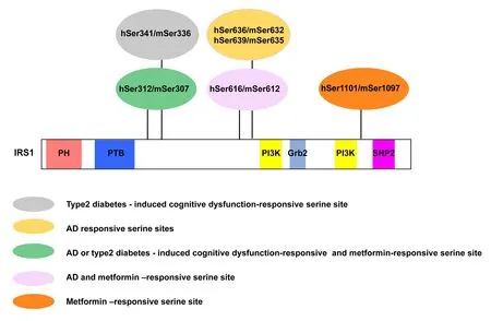
However, the mechanism underlying the beneficial effect of metformin on cognitive function remains to be elucidated.Preclinical studies also reported that metformin treatment improves cognitive deficits in animal models of cognitive impairment (Mousavi et al., 2015; Zhou et al., 2016). Additionally, intraperitoneal (i.p.) administration of metformin for 1 or 14 days increases the phosphorylation level of AMPK in the hippocampus while enhancing hippocampal neurogenesis and spatial memory formation in adult wildtype mice (Wang et al., 2012). Consistent with previous studies describing that metformin stimulates aPKC ζ/λ activity in cell culture (Wang et al., 2012), chronic metformin administration in drinking water increases the phosphorylation levels of both AMPK and aPKC ζ/λ in the hippocampus of middle-aged high-fat-diet (60% energy from fat)-type 2 diabetic mice when it improves hippocampal neurogenesis and spatial memory in these mice without lowering blood glucose levels (Tanokashira et al., 2018). At this time,chronic oral metformin treatment also increases the phosphorylation of hSer312/mSer307 and Ser616/mSer612 on IRS1 in the hippocampus of middle-aged high-fat-diet (60%energy from fat)-type2 diabetic mice and further promotes the phosphorylation of IRS1 at hSer1101/mSer1097 (Tanokashira et al., 2018) (Figure 1). These results suggest that metformin-stimulated serine phosphorylation of IRS1 in the hippocampus is involved in the mechanism underlying the beneficial effect of metformin on cognitive function via interactions with AMPK/aPKC ζ signaling.
Conclusions

Figure 1 AD or cognitive impairment-related serine phosphorylation sites of IRS1 in the brain are activated by metformin.Human(h)Ser341/mouse(m)Ser336 (gray), type 2 diabetes-induced cognitive dysfunction-responsive serine site; hSer636/mSer632 and hSer639/mSer635(yellow), AD-responsive serine sites; hSer 312/mSer307 (green),AD or type 2 diabetes-induced cognitive dysfunction-responsive and metformin-responsive serine site; hSer616/mSer612 (pink), AD and metformin-responsive serine site; hSer1101/mSer1097 (orange),metformin-responsive serine site.PH: Pleckstrin homology domain;PTB: phosphotyrosine-binding domain; PI3K: region containing multiple phosphoinositide 3-kinase binding motifs; Grb2:Grb-2 binding site; SH2 domain containing protein tyrosine phosphatase (SHP-2): SHP-2 binding site; AD: Alzheimer's disease; IRS:insulin receptor substrate.
Deficiency of IRS2 or IGF1R in the neurons and reduced IGF1R (partial deletion) in Tg2576 mice improve AD-like phenotypes; however reduced IRS2 also leads the mice to recover motor performance and extend their life span in the mouse model of Huntington's disease (Sadagurski et al.,2011). These findings indicate that IGF1R-IRS2 mediated IIS in the brain negatively regulates higher brain functions.Although IGF1R appears to be a primary upstream factor of IRS2 in the CNS, the function of IGF1R-IRS2-mediated IIS in cognitive abilities and the mechanism underlying the neuroprotective effect of reduced IGF1R-IRS2 signaling remain unknown. Given that IGF-1 is synthesized in the brain (Daftary and Gore, 2005; Wrigley et al., 2017) and can immediately promote the activation of intracellular signaling through tyrosine kinase activities of IGF1R and/or the IGF1R/IR hybrid in the CNS, it is unclear whether insulin in the CNS is dominantly involved in pathogenesis of neurodegenerative disease such as AD, because it is believed that a majority of insulin in the brain is secreted by pancreatic β-cells. Furthermore, ligands for receptor tyrosine kinase in the brain such as brain-derived neurotrophic factor, nerve growth factor, and neurotrophin-3 (Kruttgen et al., 2003;Lawn et al., 2015) affect IRS2-mediated intracellular signaling (Miranda et al., 2001; Russo et al., 2007; Lao-Peregrin et al., 2017). Thus, studies over the past two decades suggest that insulin-independent pathways might dominantly activate intracellular signaling mediated by IRS2 in the CNS,whereas intranasal insulin rescues memory deficits (Mao et al., 2016; Guo et al., 2017).
The multiple Ser/Thr sites on IRS1 positively and negatively modulate intracellular signaling in a context-dependent manner through feedback loops. Although Ser phosphorylation residues on IRS1 were suggested as markers of a detrimental consequence on cognitive function, some of these sites on IRS1 are phosphorylated and linked to beneficial effects of metformin treatment that improves cognitive dysfunction. There are discrepancies between the increased phosphorylation of these Ser sites on IRS1 and their outcomes; however the activation of these Ser sites on IRS1 in the CNS may play an important role in cognitive function through regulating IIS similarly as in the peripheral tissues including the liver and muscles (Morino et al., 2008; Copps et al., 2010; Hancer et al., 2014). Understanding the links between Ser phosphorylation of IRS1 in the brain and cognitive functions remains challenging because little has been reported on the function of IRS1 in the CNS. Although IRS1 and IRS2 share overlapping downstream signaling, the functions and the regulatory mechanisms through the interaction between IRS1- and IRS2-mediated pathways in the CNS are largely unknown.
Further studies are needed to clarify the role of IRS1 and IRS2 and the integrated signaling networks via IRS1/2 in the brain, in particular for their roles in modulating memory functions. Elucidating these pathways might provide a new therapeutic opportunity to prevent cognitive impairment and dementia including AD.
Author contributions:Creation of conceptual structure and definition of intellectual content: AT; table and figure design and preparation: DT and WF; table and figure modification: AT; manuscript editing and reviewing: DT. All authors approved the final submitted version.
Conflicts of interest:None declared.
Financial support:This work was supported by a MEXTGrant-in-Aid for Scientific Research on Innovative Areas (brain environment)(JP24111536; to AT), JSPS KAKENHI (JP24650201, JP26282026,JP17K19951, JP17H02188; to AT), and grants from the Mitsubishi Foundation (to AT) and NOVARTIS Foundation Japan for the Promotion of Science (to AT).
Copyright license agreement:The Copyright License Agreement has been signed by all authors before publication.
Plagiarism check: Checked twice by iThenticate.
Peer review:Externally peer reviewed.
Open access statement:This is an open access journal, and articles are distributed under the terms of the Creative Commons Attribution-Non-Commercial-ShareAlike 4.0 License, which allows others to remix,tweak, and build upon the work non-commercially, as long as appropriate credit is given and the new creations are licensed under the identical terms.
Open peer reviewer:George D. Vavougios, Athens Naval Hospital,Greece.
Additional file:Open peer review report 1.
- 中国神经再生研究(英文版)的其它文章
- Improvement of ataxia in a patient with cerebellar infarction by recovery of injured cortico-ponto-cerebellar tract and dentato-rubro-thalamic tract: a diffusion tensor tractography study
- Tandem pore TWIK-related potassium channels and neuroprotection
- Dendritic shrinkage after injury: a cellular killer or a necessity for axonal regeneration?
- Regenerative biomarkers for Duchenne muscular dystrophy
- Exploring the efficacy of natural products in alleviating Alzheimer's disease
- Role of macrophages in peripheral nerve injury and repair

