Galectin-3 prospects as a therapeutic agent for multiple sclerosis
Galectin-3 (Gal-3) in oligodendrocyte (OLG) differentiation:OLGs are the cells in charge of myelination in the central nervous system (CNS), allowing rapid conduction of the neural action potential and giving trophic support to axons. OLGs undergo a series of changes throughout their life cycle: first, upon neural stem cell commitment to the OLG lineage, cells referred to as OLG precursor cells (OPC) present a bipolar morphology,have proliferative and migratory capacity and express molecular markers like platelet-derived growth factor receptor alpha and neural/glial antigen 2; next, in an intermediate stage called pre-OLG, OLGs are more ramified and express CNPase, Olig1 and O4, among others; finally, cells develop into myelin forming cells which express molecular markers like myelin basic protein (MBP), adenomatous polyposis polyposis and proteolipid protein (Franklin et al., 2017). Worth pointing out, the actin cytoskeleton plays an important role in OLG maturation, as it evolves from pro-polymerization to pro-depolymerization dynamics, which allows axon ensheathing. These mechanisms are controlled in part by the relationship between MBP and actin disassembly proteins such as cofilin-1 and gelsolin. The latter are normally sequestered and inactivated by phosphatidylinositol 4,5-bisphosphate present in the plasma membrane. When MBP is expressed in mature OLG, it competes with gelsolin and cofilin-1 for phosphatidylinositol 4,5-bisphosphate binding,displacing and hence activating them, to trigger the disassembly of actin filaments (Zuchero et al., 2018).
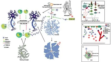
Gal-3, a β-galactoside binding lectin, has a myriad of functions depending on cell type and context. It is widely described in immune responses presenting both pro- and anti-inflammatory properties. Our group has thoroughly researched the role of this protein in OLG biology, first observing that LGALS3-/-mice had severe myelin defects, including fewer myelinated axons, decreased g-ratio and loosely wrapped myelin. Moreover,OLG treatment with media conditioned by microglia from wild type mice promoted OLG differentiation. However, this effect was impaired when OLG were cultured in media conditioned by LGALS3-/-microglia, suggesting that the expression of Gal-3 by microglia is necessary for OLG maturation. In addition, using neurosphere cultures, our group has also shown that Gal-3 promotes cellular commitment to the oligodendroglial lineage.Furthermore, these studies demonstrated that extracellular Gal-3 derived from microglia exerted a dose-dependent pro-differentiating effect in close relationship with the glycoconjugates present at the time of action, mainly in OPC (Figure 1) (Pasquini et al., 2011). Interestingly, extracellular Gal-3 accelerated OLG maturation in vitro by modulating Akt, Erk 1/2 and β-catenin signaling pathways and cytoskeleton dynamics (Thomas and Pasquini, 2019). Briefly, extracellular Gal-3 activated Akt,possibly through the mTORC1 pathway, in tight relationship with Erk 1/2 inhibition, leading to increased MBP expression(Figure 1i). These pathways were also critical for the accelerated actin cytoskeleton dynamics observed with Gal-3 treatment(Figure 1ii). In summary, our results indicate that Gal-3 plays a key role in both OLG differentiation and myelination.
Gal-3 in animal models of multiple sclerosis (MS):Autoimmune diseases or genetic defects, like MS or leukodystrophies,respectively, can cause dysfunction and consequent loss of OLG. In the course of MS, the inflammatory response generates OLG lesions which can in turn be followed by a repair response consisting in OPC activation, in which cells return to the cell cycle to proliferate and migrate, and recruitment to the damaged area. Once recruited, OPC may proliferate and differentiate to form new myelin sheaths for axon remyelination and thereby restore saltatory conduction. However, this process often fails and thus leads to axonal damage and neuronal death,and eventual progressive disability (Franklin et al., 2017). The most frequent type of MS, called relapsing remitting MS, consists in recurrent presentations of clinical signs followed by partial or total recovery. After 10 or 15 years of illness, symptoms become progressive and lead to continuous clinical deterioration, a phase called secondary progressive MS (SPMS). Nevertheless, in some patients, MS is relentless from the beginning,which constitutes the primary progressive MS (PPMS) form.In this scenario, several efforts are being made all around the world to improve remyelination by approaching the migration,proliferation and differentiation of OLG in the site of injury.
Research in this field relies on MS experimental demyelination models mediated by immunity, virus or toxins. Even if these models fail to replicate the full complexity and heterogeneity of MS features, they have allowed the development of various treatments. Experimental autoimmune encephalomyelitis is induced by immunization of mice with different myelin antigens such as myelin oligodendrocyte glycoprotein, MBP,proteolipid protein and myelin-associated glycoprotein. Despite experimental autoimmune encephalomyelitis widespread use, numerous beneficial effects obtained in this model have not been replicated in MS treatment. This model applied in LGALS3-/-mice displayed a decrease in CNS macrophage infiltration and in disease severity (Thomas and Pasquini, 2018),suggesting a key role for Gal-3 in promoting inflammation by leukocyte recruitment (Figure 1). In contrast, further studies in this model showed a Gal-3-induced neuroprotective role through cell debris removal, axon regeneration and remyelination (Itabashi et al., 2018).
Several authors hypothesize that environmental factors such as viral infections are involved in MS and may actually trigger the disease, which led to the development of virus-induced MS models like Theiler's Murine Encephalomyelitis Virus (TMEV)infection. Subventricular zone (SVZ) cell proliferation may decrease as a consequence of MS-induced inflammation and thus hinder recovery. Gal-3 expression increases in active human MS lesions (Stancic et al., 2011), periventricular regions in human MS and after murine TMEV infection, whereas Gal-3 loss restores SVZ proliferation in the TMEV model through a reduction in the number of immune cells (James et al., 2016).
Our group has studied Gal-3 involvement in the demyelination/remyelination process using the cuprizone (CPZ) model(Figure 1), in which 8-week-old LGAL3-/-and wild type mice were fed a diet containing 0.2% CPZ w/w for 6 weeks to evaluate demyelination, followed by two more weeks on a CPZfree diet to assess remyelination. CPZ administration produces massive demyelination in the CNS through pathogenic T cell-independent mechanisms, the corpus callosum being particularly affected. CPZ administration allows the investigation of the remyelination process independently of peripheral immune system contribution, as the model keeps the blood-brain barrier (BBB) intact. Both in 8-week-old LGALS3-/-and wild type mice, CPZ administration induces demyelination up to the fifth week of treatment. However, OPC response to demyelination in LGALS3-/-exhibits reduced branching, which indicates reduced OPC differentiation and is in line with our previous results showing Gal-3 participation in OLG differentiation.Most interestingly, wild type mice show spontaneous remyelination during the fifth week of CPZ treatment even if the toxic diet is kept during six weeks. In contrast, LGALS3-/-mice suffer continuous demyelination up to the sixth week with a sharp astroglial response. Gal-3 is upregulated in microglia in CPZ-induced demyelination and kept absent in astroglial cells. In addition, only wild type mice show ED1 (CD68) expression and TREM-2b upregulation during CPZ-induced demyelination,whereas LGALS3-/-mice display a large number of microglial cells which express activated caspase-3. Phagocytosis of myelin debris by microglia in CPZ demyelination requires phagocytic receptor TREM-2b expression and is essential to the onset of remyelination through oligodendroglial differentiation (Thomas and Pasquini, 2018). Strikingly, myelin phagocytosis relies on CR3/MAC-1 and SRAI/II, in turn regulated by Gal-3-dependent activation of PI3K; therefore, myelin phagocytosis by LGALS3-/-microglia is usually deficient (Thomas and Pasquini,2018). In agreement with the TMEV model described above(James et al., 2016), Gal-3 regulates SVZ progenitor response to CPZ-induced demyelination, asits loss is associated with increased SVZ progenitor migration to demyelinated regions(Hillis et al., 2016).
Taken together, the results described so far demonstrate that Gal-3 acts as a regulator of the microglial response to promote remyelination (Thomas and Pasquini, 2018).
Subsequent studies by our group further showed an increase in metalloproteinase-3 expression and a decrease in CD45+, TNFα+and TREM-2b+cells during remyelination only in wild type mice, parameters which remained unaltered in LGALS3-/-mice during demyelination and remyelination. Ultrastructural studies following remyelination revealed defective myelin sheaths and collapsed axons in the corpus callosum of LGALS3-/-mice but no relevant myelin disruption in wild type mice. These results add up to the knowledge of the mechanisms underlying Gal-3 impact on remyelination, as they show that the tuning of microglial cells involves the modulation of metalloproteinase activity (Thomas and Pasquini, 2018).
Gal-3 in MS:The role of Gal-3 in patients diagnosed with MS has been scarcely documented so far. It was recently shown that OPC treated with cerebrospinal fluid (CSF) obtained from patients with PPMS presented a significantly more ramified morphology than control CSF accompanied by a pro-differentiating transcriptome, as evidenced by lower platelet-derived growth factor receptor α and LINGO1 mRNA levels and higher MAG mRNA. However, this transcriptome was different than that of normal OPC. Of note, this report showed the upregulation of the LGALS3 gene only in OPC treated with CSF from PPMS patients (Figure 1), establishing a link between the upregulation of LGALS3 and the increase in OPC ramification. These findings received support from similar studies in post mortem human brain tissue from patients with PPMS (Haines et al.,2015).
It should be pointed out that autoimmune diseases such as MS present an aberrant glycosylation pattern. For instance, IgG from MS patients CSF exhibits a greater component of N-acetylglucosamine but lesser residues of sialic acid and galactose, the natural ligand of Gal-3 (Wuhrer et al., 2015). Given that proteins in general have a smaller amount of galactose residues in the course of MS, Gal-3 may not be able to exert its pro-differentiating action on OLG, either through higher secretion rates by microglia or OLG themselves.
Furthermore, sera from patients with SPMS present auto-antibodies against Gal-3, which could be in part responsible for the progressive damage to the BBB perceived in patients with MS (Nishihara et al., 2017). As part of the same study, the authors also determined that Gal-3 bound to the membrane of brain microvascular endothelial human cells (BMEC) was a target for the auto-antibodies present in sera from patients with SPMS but not in sera from healthy or other CNS illnesses patients. These authors also inhibited the expression of Gal-3 in these cells, which triggered an increase in the expression of intracellular adhesion molecule 1 (ICAM-1) and p-NFκB p65,both of them described as responsible for the leak of leukocytes to the CNS. Taken together, these results suggest that Gal-3 positively influences OLG differentiation in human brain tissue with MS and drives a downregulation of ICAM-1 in BMEC,thereby mediating a protective effect and highlighting its potential therapeutic in MS (Figure 1iii).
In summary, the findings obtained in different MS animal models such as experimental autoimmune encephalomyelitis, CPZ and TMEV support the notion that Gal-3 is secreted by microglia, although the exact role of this secretion has not been fully elucidated yet and seems controversial (Hillis et al.,2016; Thomas and Pasquini, 2019). For instance, it is known that Gal-3 may be secreted through a non-classical pathway and/or by exosomes. Our studies have demonstrated that Gal-3 derived from microglia exerts a pro-differentiating effect on OLG during CPZ-induced demyelination, favoring the onset of remyelination (Pasquini et al., 2011; Thomas and Pasquini,2018). Gal-3 modulates microglia toward a phagocytic and anti-inflammatory M2 phenotype, which increases the removal of myelin debris interfering with oligodendroglial differentiation (Thomas and Pasquini, 2018; Itabashi et al., 2018). Some studies, however, indicate that Gal-3 exacerbates the disease, a discrepancy probably explained by the use of different demyelinating models which simulate distinct MS features (James et al., 2016; Thomas and Pasquini, 2018). Our in vitro studies have elucidated that Gal-3 pro-differentiating effect from a mechanistic point of view, demonstrating that Gal-3 drives early OLG process outgrowth and branching through enhanced actin assembly and a decrease in Erk 1/2 activation, and also regulates OLG maturation by inducing Akt activation and an increase in MBP expression, promoting gelsolin release and actin cytoskeleton disassembly. Therefore, Gal-3 expressed by microglial cells could favor the onset of remyelination through the induction of M2 cell polarization and/or by a direct effect on OLG differentiation. Furthermore, recent evidence has strongly supported Gal-3 involvement in MS disease. Diminished expression of Gal-3 in BMEC increases ICAM-1 and leukocyte infiltration to the CNS (Nishihara et al., 2017), although Gal-3 expressed by these cells binds to autoantibodies from SPMS patients, which leads to BBB damage. In addition, Gal-3 upregulation in OPC treated with PPMS CSF culture promotes OPC ramification(Haines et al., 2015). However, and possibly due to an aberrant glycosylation pattern in MS (Wuhrer et al., 2015), Gal-3 cannot bind properly to glycoconjugates to exert its pro-differentiation activity and lead to fully mature OLG. Taken together, these results support Gal-3 as a novel extracellular mediator of glial crosstalk to promote accelerated OLG maturation and to favor remyelination. Future studies will be necessary to evaluate possible pathways for non-invasive delivery of Gal-3 to the CNS and strategies to specifically target oligodendroglial precursors.These studies may have major significance in the development of future therapies for a variety of demyelinating diseases and especially for MS.
This work was supported by grants from Agencia Nacional de Promoción Científica y Tecnológica (Argentina, PICT 2014-3116) and Universidad de Buenos Aires (20920160100683BA, to LAP).
Laura Thomas, Laura Andrea Pasquini*
Department of Biological Chemistry, School of Pharmacy and Biochemistry, Institute of Chemistry Biological
Physicochemistry (IQUIFIB), University of Buenos Aires and
National Research Council (CONICET), Buenos Aires, Argentina
*Correspondence to: Laura Andrea Pasquini, PhD,
laupasq@yahoo.com.

Figure 1 Gal-3 influencing OLG differentiation and (re)myelination.Spectrum analysis adopted ICBM 152 and imagetwork in depression patients, and blue represents brain regions with decreasing node degree of Gal-3 expressed by microglial cells during remyelination, favors an M2 microglial phenotype and, therefore, enhances myelin phagocytosis through phagocytic receptors and consequently, OLG differentiation. In OLG precursor cells (OPC), (i) extracellular Gal-3 activates Akt, possibly through the mTORC1 pathway, in tight relationship with Erk 1/2 inhibition, leading to increased MBP expression. (ii) These pathways are also critical for the accelerated actin cytoskeleton dynamics observed with Gal-3 treatment. Also, microglia-released Gal-3 induces OLG fate in neural stem cells. (iii) Gal-3 downregulation in brain microvascular endothelial human cells, decreases intracellular adhesion molecule 1 to impede leukocyte infiltration. Furthermore, Gal-3 has proven to be a target for antibodies in primary progressive multiple sclerosis (PPMS) cerebrospinal fluid (CSF) and in OPC, treatment with PPMS CSF, increases Gal-3 expression and branching (Thomas and Pasquini, 2018). OLG: Oligodendrocyte; Gal-3: galectin-3; BBB: blood-brain barrier; MS: multiple sclerosis; IGFR-1: insulin-like growth 1-receptor; ICAM-1: intercellular adhesion molecule 1.
orcid: 0000-0003-4292-3463 (Laura Andrea Pasquini)
Received: December 5, 2018
Accepted: January 25, 2019
doi: 10.4103/1673-5374.253521
Copyright license agreement:The Copyright License Agreement has been signed by both authors before publication.
Plagiarism check: Checked twice by iThenticate.
Peer review: Externally peer reviewed.
Open access statement:This is an open access journal, and articles are distributed under the terms of the Creative Commons Attribution-NonCommercial-ShareAlike 4.0 License, which allows others to remix, tweak, and build upon the work non-commercially, as long as appropriate credit is given and the new creations are licensed under the identical terms.
- 中国神经再生研究(英文版)的其它文章
- Improvement of ataxia in a patient with cerebellar infarction by recovery of injured cortico-ponto-cerebellar tract and dentato-rubro-thalamic tract: a diffusion tensor tractography study
- Tandem pore TWIK-related potassium channels and neuroprotection
- Dendritic shrinkage after injury: a cellular killer or a necessity for axonal regeneration?
- Regenerative biomarkers for Duchenne muscular dystrophy
- Exploring the efficacy of natural products in alleviating Alzheimer's disease
- Role of macrophages in peripheral nerve injury and repair

