应激颗粒与病毒的相互制约
黄羽,胡斯奇,郭斐
应激颗粒与病毒的相互制约
黄羽,胡斯奇,郭斐
中国医学科学院/北京协和医学院病原生物学研究所,国家卫生健康委员会病原系统生物学重点实验室,北京 100730
哺乳动物细胞受到热休克、氧化应激、营养缺乏或者病毒感染等环境压力时,能够迅速启动细胞的压力应答机制,终止细胞内的蛋白翻译,形成应激颗粒(stress granules, SGs)。SGs作为胞浆中翻译起始复合物的聚集产物,在细胞的基因表达和稳态中发挥着重要的作用,与细胞凋亡以及核功能具有密切联系。尤其是当病毒感染细胞时,SGs的形成可以使细胞内病毒蛋白翻译水平大大降低,从而抑制入侵病毒的复制。然而,病毒在长期进化过程中也衍生出了对抗细胞压力应答的相应机制,如与SGs关键组分相互作用,甚至切割等方式。本文对SGs的组成及诱发机制,特别是多种病毒诱导eIF2α磷酸化促成SGs组装的机制,以及病毒进化过程中形成的应对措施等方面进行了综述,旨在进一步阐释病毒感染与应激颗粒形成之间的相互影响和调控,为人们深入理解人体先天性免疫防御提供参考。
应激颗粒;翻译阻滞;病毒;先天性免疫
当哺乳动物细胞遇到压力环境(如热刺激、电离辐射、低氧、内质网压力和病毒感染等)时,细胞内会发生一系列的应激反应,其中细胞质中会形成一种可逆性动态结构,被称为应激颗粒(stress granules, SGs)。SGs是一种核糖核蛋白(ribonucleoprotein, RNP)聚集物,包括翻译起始需要的mRNAs、40S核糖体亚基、翻译起始因子以及一些RNA结合蛋白[1]。病毒感染宿主细胞后在其体内复制,对于宿主而言,无疑是一种强烈的刺激,可促进SGs的聚集;而SGs的形成又能够干扰病毒在宿主细胞内的复制,这是因为所有病毒的复制都无一例外的需要宿主翻译系统的帮助和参与。因此,病毒感染与SGs的形成两者间的相互作用关系对于先天性免疫的调节存在重要意义。本文通过对病毒感染诱导SGs的形成的机制及其相互作用作以总结,以期为人们深入理解先天性免疫调节在宿主抗病毒免疫系统中的作用提供参考。
1 SGs的形成及调节机制
SGs最早是在植物细胞中被发现,当植物细胞受到热刺激后,会出现包含RNA结合蛋白的TIA-1、TIAR以及poly (A)-RNA的细胞质内颗粒物质,被称作为SGs。在20世纪末期,研究发现在真核细胞内,当翻译起始因子elF2α磷酸化后,会导致胞质内出现TIA-1/TIAR阳性的SGs的出现,将未翻译的mRNA隔离,并且这种变化是可逆的[2]。
在真核细胞内,SGs形成的明显特征为mRNA翻译起始的暂时停滞。所形成的SGs主要包含以下组分:(1)靶向mRNA的翻译起始因子(elF4G、elF3、PABPC1、p-elF2α和elF5a)[3,4];(2)调控翻译和保护mRNA稳定性的mRNA结合蛋白(TIA-1、TIAR、HuR/ ELAVL1、FMRP和Pum1)[3,5];(3) mRNA代谢有关的蛋白质(G3BP1、G3BP2、p54/rck/DDX6、PMR1、SMN、Staufen1、DHX36、Caprin1、ZBP1、HDAC6和ADAR)[6~11];(4)信号传递蛋白(mTOR、RACK1和TRAF2)[12];(5)干扰素诱导基因的表达产物(interferon- stimulated gene, ISG),如PKR、ADAR1、RNA-sensing RIG-I-like receptors (RIG-I、MDA5和LGP2)、RNase L和OAS;(6)对SGs的形成有调控作用的蛋白(APOBEC3G、Ago2、BRF1、DDX3、FAST和TTP等)[13,14]。
目前研究表明,引发真核细胞SGs生成的机制主要有3种,分别为:elF2α的磷酸化、mTOR的失活以及干扰elF4F复合物的生成。这其中研究最多的是elF2α的可逆性磷酸化作用。磷酸化状态下的elF2α可与elF2B稳定结合,这种结合作用使elF2B不能催化GDP再生成GTP,也就阻碍了翻译起始过程中eIF2–GTP–Met–tRNAMet三元复合物的形成,诱导SGs的形成[2]。在哺乳动物细胞中,目前共发现了4种能够使elF2α磷酸化的激酶,分别可被不同种类的刺激反应激活。HRI (heme-regulated inhibitor)可被氧化损伤和热刺激激活[15,16];GCN2 (general control non-derepressible 2)的激活可由于氨基酸的缺乏[17]和紫外辐射损伤[18]所引起;PKR可被特殊的dsRNA所激活[19],如病毒RNA复制中间产物等;PERK (PKR-like endoplasmic reticulum (ER) kinase)可被内质网刺激作用激活[20]。另外,由于eIF2–GTP– Met–tRNAMet三元复合物的缺乏,使翻译起始区域不能被识别,也影响了核糖体大亚基的招募,结果造成了mRNP的累积和聚集[2]。这也是病毒感染引发细胞内SGs聚集的主要因素。
哺乳动物mTORC1是一种调控细胞代谢、营养分配和促进生长的激酶[21]。mTORC可对许多底物蛋白产生磷酸化作用,包括elF4E-binding proteins (4EBPs)。亚硒酸钠(sodium selenite, Se)和雷帕霉素(rapamycin)等药物和饥饿诱导等可明显抑制mTORC的活性。去磷酸化状态下的4EBPs可与elF4E结合,阻止elF4E与elF4G的相互作用[22],也抑制了翻译的起始,从而导致SGs的聚集。另外,pateamine A和hippuristanol等化合物可抑制RNA解旋酶elF4E的活性,使其不能与elF4G相互作用,干扰了elF4F 复合物的形成阻碍了翻译的起始,也是诱发SGs的重要机制。
压力刺激引起的翻译抑制可快速下调大量帽子结构依赖的翻译起始的蛋白质合成,不过,也有一些mRNA可被选择性翻译增强。例如热休克蛋白可被热刺激所诱导,并且其mRNA可在高水平的磷酸化elF2α存在下被翻译[23,24]。当elF2α磷酸化导致eIF2–GTP–Met–tRNAMet三元复合物的再生延迟时,位于uORF下游区的ATF4 (activating transcription factor 4)则被翻译合成。ATF4又可激活GADD34 (growth arrest and DNA damage-inducible protein 34),与蛋白磷酸酶1 (protein phosphatase 1, PP1)一起,催化elF2α去磷酸化,进而缓解翻译的抑制作用[25]。
在实验过程中,研究人员通常需要诱导哺乳动物细胞产生SGs的聚集,进而探究外界条件对其影响。比较常见的亚砷酸钠就是通过HRI磷酸化elF2α,进而诱导SGs的生成[15];另外,pateamine A和hippuristanol作为elF4A解旋酶的干扰物质,在48S复合物形成后,使mRNA二级结构不能打开,影 响核糖体小亚基通过mRNA 5¢UTR,进而阻滞翻 译[26,27]。过氧化氢以及亚硒酸钠可通过抑制mTOR使4EBP去磷酸化诱导非经典SGs,但不导致elF2α的磷酸化[28,29]。这种非经典的SGs较经典SGs相比缺少elF3等分子,其形成机制目前还有待研究。此外,TIA-1/TIAR和G3BP1等SG标志蛋白的过表达也可导致SGs的产生。
许多SGs的组成蛋白可通过翻译后修饰来调节SGs的形成,主要包括泛素化[30]、多聚ADP核糖基化(poly (ADP)-ribosylation)[31]、O连接的N乙酰氨基葡糖基化(-linked-acetylglucosamination)[32]、精氨酸脱甲基作用[33]、类泛素化修饰[34]、磷酸化及去磷酸化的修饰作用[8]。结果就导致了一些酶通过这些修饰作用来调节SGs的形成,并被招募至SGs,成为了其组成成分。还有一些酶在免疫信号途径中起了至关重要的作用如RIG-I、MDA5和LGP2等解旋酶,被招募至SGs,也印证了SGs作为调节抗病毒反应的平台,介导下游信号传递存在的生物学意义[35]。
SGs也是相分离(phase separation)的一种存在形式。相分离概念提出主要针对于一些细胞内存在的无膜结构,他们多数由蛋白质或RNA蛋白复合物聚集物的方式相对独立的存在于细胞中,主要包括信号复合物(signaling complexes),突触后聚集物,P小体(P bodies),细胞核内斑点,应激颗粒等[36]。在细胞承受外界刺激条件下所形成的SGs为无膜聚集结构,其中包括mRNAs和与之结合的多种翻译起始因子,以及RNA结合蛋白。细胞通过这种相对的隔离方式,将这些重要的翻译原件储存起来,待到刺激因素解除,细胞从这种应激状态恢复后,被SGs所隔离的这些翻译起始因子及RNA可恢复工作,从而帮助细胞渡过难关[37]。另外SGs可在空间上将某些毒性蛋白和致病成分与细胞内环境相对分离,随后通过p62介导的自噬溶酶体途径将其降解,来维持细胞内环境的稳定[38,39]。
2 病毒与SGs的相互作用
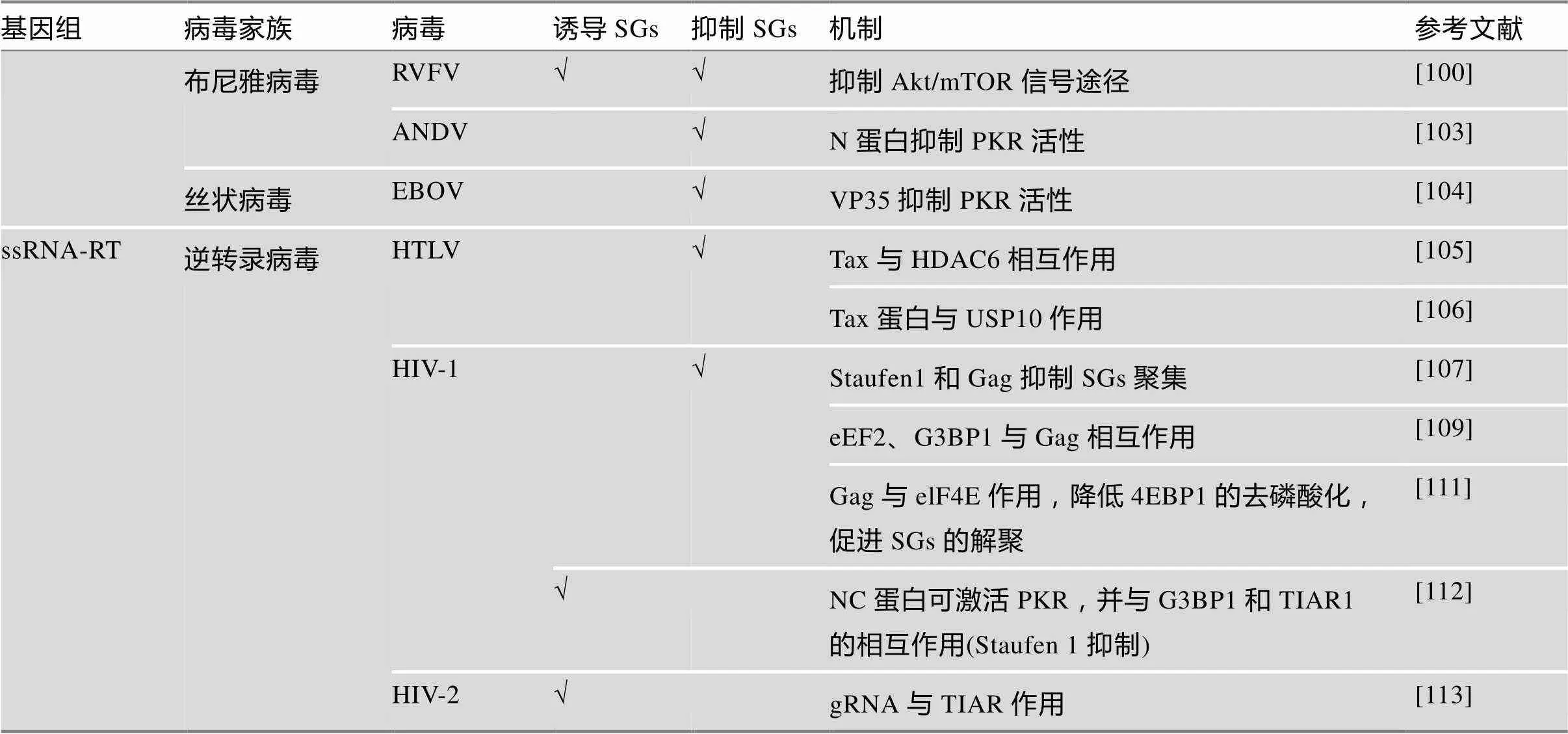
从最开始有关SGs的形成可抑制病毒复制的报道至今,10余年的时间里[40],研究人员将病毒感染与SGs形成及其相互作用的关系的研究,已经遍及多种病毒:从简单的正向单链RNA病毒,到较复杂的逆转录病毒。许多病毒在复制的不同阶段抑制SGs的形成。很多时候病毒抑制SGs的生成,目的在于适应病毒自身的复制反应,这也提示SGs形成所导致的蛋白质翻译的抑制作用,拥有一定的抗病毒功能。另外一些报道称,SGs形成为病毒的模式识别分子提供一个平台,募集天然免疫中的重要分子[14],进而为下游的抗病毒反应传递信号。病毒的感染作为一种强烈的外源刺激,可诱导哺乳动物细胞产生SGs。下文通过不同病毒家族讨论了其与SGs的相互作用关系(表1)。
2.1 双链DNA病毒
许多疱疹病毒科()及痘病毒科()都属于具有包膜的dsDNA病毒。单纯性疱疹病毒(herpes simplex virus type 1, HSV1)可通过自身的vhs (virionhostshutoff)蛋白[41]、US11[42]、ICP34.5[43]和gB (glycoprotein B )[44]影响elF2激酶的激活作用,从而影响宿主蛋白质的合成。当HSV-1感染时,宿主细胞TIA-1、TIAR和TTP水平上调,但不形成明显的SGs,主要原因为病毒RNA茎环结构可捕获TIA-1/TIAR来协助自身的复制[45];vhs缺失的HSV-1感染细胞后,则可通过激活PKR引起SGs的聚集[46,47]。Finnen及其同事报道,感染HSV-2可下调亚砷酸盐诱导的SGs,而对于pateamine A诱导的SGs则没有作用[48]。这种抑制亚砷酸盐诱导的SGs作用也依赖于vhs,感染vhs缺失的HSV-2突变体则可在感染后期诱导SGs的形成[49]。

表1 病毒与应激颗粒的相互作用

续表1
痘苗病毒(vaccinia virus, VV),是痘病毒科()家族的一员,其在宿主细胞复制过程中在胞质内形成大颗粒,被称作病毒DNA工厂,其中招募了许多SG蛋白质,如G3BP1、Caprin1、eIF4E、PABP (poly (A)-binding protein)以及eIF4G[50,51]。VV将这些蛋白利用在自身复制的不同阶段,如G3BP1/ Caprin1复合物可增强VV的转录[50];病毒翻译的起始依赖eIF4E/eIF4G/PABP;另外,病毒蛋白I3可与elF4G相互作用,从而招募病毒ssDNA[52],这都说明了SG的组分对于VV的转录和翻译都有协助作用。然而,TIA-1并没有出现在这个病毒DNA工厂中[53];当利用缺失E3L的突变体VV感染宿主细胞时,则可激活PKR,导致形成含有TIA-1、eIF3b、G3BP1和USP10的大颗粒,由于其具有抑制病毒复制的功能,所以称之为抗病毒颗粒(antiviral stress granules, avSGs)[54]。
2.2 双链RNA病毒
呼吸孤病毒科()为包含9~12条dsRNA基因组的无包膜病毒。典型的代表为轮状病毒(rotavirus),其感染宿主细胞同样可引起宿主蛋白质合成的抑制。由于大量病毒dsRNA在宿主细胞内生成,激活了PKR引起elF2α的磷酸化,但由于SGs的核心成分PABP从胞质向细胞核发生了转移,胞质内SG聚集受阻[55,56]。相反,哺乳动物正呼肠孤病毒(mammalian orthoreovirus, MRV)可在感染早期(脱壳后和mRNA转录之间)能够诱导SG的形成,这种SG的形成依赖于elF2α的磷酸化,对病毒的复制也有促进作用[57]。而到了MRV感染的晚期,尽管elF2α的磷酸化水平还很高,但SG的形成水平则有所降低[58]。还有研究报道SGs可招募病毒非结构蛋白μNS与G3BP1产生相互作用,进而干扰SGs的聚集形成,在G3BP1敲除的细胞中,MRV的复制效率明显提升[59]。
2.3 单股正链RNA病毒
小RNA病毒科()的所有成员都是无包膜病毒颗粒。骨髓灰质炎病毒(poliovirus, PV)蛋白酶2A可在感染早期诱导SGs生成[60],而到了感染晚期,PV 3C蛋白酶切割G3BP1[61],则可能导致了此类SG的解聚。但在PV感染的晚期,还有一种细胞质颗粒稳定存在,其组成不是典型的SGs,包含病毒RNA和TIA-1,但不包括elF4G和PABP[62]。脑心肌炎病毒(encephalomyocarditis virus, EMCV)、柯萨奇病毒(coxsackievirus B3, CVB3)也通过切割G3BP1来干扰SGs的形成[63,64]。小鼠脑脊髓炎病毒(Theiler’s murine encephalomyelitis virus, TMEV)则通过表达leader (L)蛋白与G3BP1产生稳定的相互作用,来干扰SG的合成[65]。门戈病毒(mengovirus) L蛋白的锌指结构域突变后,则导致了PKR依赖的G3BP1聚集,说明G3BP1-Caprin1-PKR复合物在门戈病毒感染相关的抗病毒免疫反应中有重要的作用[66]。手足口病病毒(foot-and-mouth disease virus, FMDV)则通过L蛋白来切割G3BP1和G3BP2抑制SGs的生成[67];而EV71可通过蛋白酶2A来切割elF4GI来诱导非经典的SGs生成,却抑制了经典的SGs的生成[68]。
黄病毒科()是一类包膜病毒,西尼罗河病毒(West Nile virus, WNV)为此类病毒家族中首次报道的具有抑制SG聚集的成员,其病毒基因组3¢茎环结构可与TIA-1和TIAR相互作用,抑制SG的形成[69],而嵌和WNV W956IC则通过激活PKR,在感染早期引起大量的SGs的形成[70]。不过,在WSN感染后期,可通过激活mTOR来解除对翻译的抑制,有利于病毒的复制[71]。另外,Xia等[72]报道登革病毒(dengue virus, DENV)感染的A549细胞可诱导非TIA-1依赖的G3BP1的聚集。蛋白组学分析显示,G3BP1、G3BP2、Caprin1和USP10与DENV 2 sfRNA (subgenomic flavivirus RNAs, sfRNA)相互作用,并且G3BP1、G3BP2和Caprin1通过调控ISG mRNA的转录,有利于DENV的复制[73,74]。森林脑炎病毒(tick-borne encephalitis virus,TBEV)感染宿主细胞后将TIA-1和TIAR招募至其复制的位点,形成包含G3BP1、elF3和elF4B的SGs[75]。另一方面,日本脑炎病毒(Japanese encephalitis virus, JEV)核心蛋白可通过与Caprin 1相互作用,从而隔离G3BP1和USP10,导致SG形成受阻[76]。
与上述黄病毒的作用方式略有不同,HCV是通过控制elF2α的磷酸化来实现其对于SGs形成的调控。Garaigorta等[77]的研究表明HCV感染宿主细胞可引起PKR依赖的SGs聚集,且TIA-1,TIAR和G3BP1对于HCV的复制周期具有重要调控作用。另外,Ruggieri等[78]研究表明,HCV感染宿主细胞,早期可以迅速诱导SGs的产生,在感染的后期又出现SGs解聚的现象,这一系列的变化依赖于elF2α磷酸化水平的变化。HCV感染前期的SGs聚集是由于细胞内病毒dsRNA激活了PKR,促进了elF2α的磷酸化水平;而SGs的解聚则是由于elF2α的去磷酸化,这种变化是通过蛋白磷酸酶1 (PP1)和GADD34的作用[77]。还有报道称,SGs的另一种成分DDX3可与HCV 3¢UTR和IKKα相结合,并激活IKKα,诱导LD产生基因表达[79],从而达到抗病毒的作用。这些发现解释了在HCV感染过程中SGs产生水平波动的现象。寨卡病毒(Zika virus, ZIKV)作为近几年的新发病毒广受关注,ZIKV感染宿主细胞,可引起elF2α磷酸化水平的增高,整体翻译水平的抑制,但并没有明显SGs聚集。病毒蛋白NS3和NS4A与细胞整体的翻译抑制有关,而病毒capsid protein、NS3/NS2B-3和NS4A干扰SG的聚集,而G3BP1、TIAR、Caprin-1与ZIKV在宿主细胞内的复制有关,且G3BP1与病毒基因组RNA和Capsid相互作用。这些表明ZIKV利用多种病毒组分来抑制SG的形成,从而促进自身的复制[80]。另外一篇报道显示,ZIKV可通过elF2α的去磷酸化来抑制亚砷酸钠引起的SGs聚集;而对于亚硒酸钠和PatA诱导的SGs没有抑制作用[81]。
冠状病毒家族是一类有包膜的病毒。已有报道显示,鼠肝炎冠状病毒(mouse hepatitis coronavirus, MHV)和猪传染性胃肠炎病毒(transmissible gastroenteritis virus, TGEV)引起TIA-1/TIAR的聚集和elF2α的磷酸化[82,83]。MHV在感染早期SGs的聚集,而TGEV则是感染的晚期引起SG聚集。在TGEV感染时,多聚嘧啶区结合蛋白(polypyrimidine tract-binding protein, PTB)被重新分配于细胞质中,与病毒的基因组RNA和亚基因组RNA共同聚集于包含有TIA-1/TIAR的聚集体中。中东呼吸综合征(Middle East respiratory syndrome, MERS)病毒(MERS-CoV)可通过结合dsRNA,抑制PKR介导的elF2α的磷酸化来抑制SGs的形成,但缺乏4a和4b的MERS-CoV (MERS-CoV-Δp4)则可诱导SGs的生成[84]。
甲病毒属的基孔肯雅病毒(Chikungunya virus, CHIKV)非结构蛋白nsP3可同nsP1、dsRNA一起稳定定位于包含有G3BP1/2的SGs内,并靠近核膜和核孔蛋白Nup98,限制了病毒的复制[85]。
2.4 单股负链RNA病毒
正粘病毒科()家族为一类包含反向ssRNA基因组的包膜病毒。流感病毒A (influenza A virus, IAV)通过表达非结构蛋白NS1来抑制PKR的活性,进而抑制SGs的聚集,如感染NS1缺失或突变的IAV,则可诱导形成SGs[86]。NS1介导的SGs的抑制作用,依赖于NS1于RNA相关蛋白55 (RNA associated protein 55, RAP55)的相互作用[87]。除了NS1、IAV的NP和PA-X都能通过非elF2α磷酸化依赖的方式来抵抗SGs的聚集[88]。此外,当感染IAV后,DDX3可与NP相互作用,并调节IFN的产生及SGs的聚集;当感染宿主的IAV缺失NS1,DDX3还能够与产生的SGs共定位[89];DDX6也可结合病毒RNA,促进RIG-I介导的干扰素应答反应,起到抗病毒作用[90]。
水泡性口炎病毒(vesicular stomatitis virus, VSV)属于弹状病毒科(),感染宿主细胞可诱导elF2α磷酸化,并促进SG样颗粒形成聚集。这种SG样颗粒包含TIA-1、TIAR、PCBP2、病毒复制蛋白和RNA,但缺少elF3和elF4A[91]。
副黏病毒科()家族由一类包膜病毒组成。呼吸道合胞体病毒(Respiratory syncytial virus, RSV)的感染可以引起PKR介导的SGs聚集,而这种变化又能够促进RSV的复制[92]。RSV感染人上皮细胞可产生一种细胞质包涵体(cytoplasmic inclusion bodies, IBs)其中包含多种病毒蛋白,RSV利用这种结构来调节病毒的复制[93]。这种IBs中包含了SGs的组成成分HuR、MDA5和MAVS,病毒通过这种方式,抑制了IFN的产生[94]。另有报道称,RSV的感染可利用形成IBs隔离磷酸化的p38和O-linked N-acetylglucosamine transferase (OGT),从而抑制了MAPK-activated protein kinase 2 (MK2)途径,也就抑制亚砷酸钠诱导的SGs聚集[95]。麻疹病毒(Measles virus, MeV)可通过病毒蛋白C和V来影响宿主的先天性免疫和适应性免疫[96]。并且,当蛋白C缺失的MeV感染宿主细胞时,可诱导PKR依赖的SGs聚集,而野生型的MeV则不能。但是,野生型MeV感染ADAR1敲除细胞则可诱导SGs的产生[13]。仙台病毒(sendai virus, SeV)感染可以引起轻微的SGs的聚集,而病毒3¢末端反向基因组RNA的转录产物,可与TIAR相互作用,而下调SGs的生成[97]。另外,还有研究报道仙台病毒C蛋白在影响SGs聚集和IFN长生上有重要作用,当用缺失C蛋白的SeV感染宿主细胞,会产生包含RIG-I和病毒RNA片段的SG样结构[98]。
布尼亚病毒家族由许多基因组为(-)ssRNA的包膜病毒组成。这类病毒的特点是需要一种“帽子”结构来协助起始mRNA合成,这个“帽子”是从宿主mRNA上获取来的。首先,病毒N蛋白与“帽子”结合,由RNA依赖的RNA聚合酶(RNA-dependent RNA polymerase, RdRp)的核酸内切酶domain将宿主mRNA“帽子”切下,作为引物来进行病毒mRNA的合成[99]。裂谷热病毒(rift valley fever virus, RVFV)可下调Akt/mTOR信号途径,从而增加4EBP1/2蛋白的活性来抑制翻译过程,可引起短暂的SGs聚集,而这种SGs的聚集也可能为病毒获取“帽子”结构提供便利[100]。另外,布雅病毒家族病毒蛋白也像其他病毒一样,进化出了下调PKR活性和抑制IFN产生的机制。Hantaviruses和Phleboviruses中S片段的非结构蛋白可抑制IFN反应[101,102];汉坦病毒(Andes Hantavirus, ANDV)的衣壳N蛋白可抑制PKR的二聚化,并影响其激活[103]。
另外,丝状病毒科()家族的埃博拉病毒(Ebola viruse, EBOV)是一种包膜病毒,当其感染宿主,SGs成分Staufen1可与病毒基因组RNA的3'和5'外显子区域结合,并与结构蛋白NP、VP30和VP35等产生结合作用,从而促进病毒基因组RNA的合成[104]。
2.5 逆转录病毒
逆转录病毒都是含有正向ssRNA的包膜病毒,通过逆转录合成cDNA并整合到宿主基因组DNA上。人T细胞白血病病毒(human T-cell leukemia virus, HTLV-1)是一种致癌的反转录病毒,可通过病毒蛋白Tax与组蛋白脱乙酰酶6 (histone deacetylase 6, HDAC6)的相互作用抑制SGs的形成,HDAC6是SGs的组成成分[105]。另有报道称,Tax可与USP10相互作用来抑制SGs的生成[106]。另外,人免疫缺陷病毒1 (human immunodeficiency virus type 1, HIV-1)感染宿主细胞可通过形成包含Staufen1的HIV-1依赖的核糖核蛋白(staufen1-containing HIV-1-dependent ribonucleoproteins, SHRNP )来明显的抑制SGs的形成[107]。核定位信号(nuclear localization signal, NLS)突变的Sam68能够诱导应激颗粒的形成,在HIV-1感染过程中,Sam68 突变体特异的与HIV- 1nef mRNA发生相互作用,将nef mRNA募集到应激颗粒中,抑制了nef蛋白表达,最终导致HIV-1复制受阻[108]。并且,HIV-1 Gag可通过非elF2α磷酸化依赖的方式来抑制SGs的聚集。HIV-1 capsid的N末端可通过与eEF2的相互作用来行使抵抗SGs形成的作用,若eEF2缺失,不仅SGs的形成受到抑制,HIV-1的在宿主细胞内的产生及感染性也会受到影响;HIV-1 Gag也可与G3BP1相互作用,来干扰SGs的生成[109],且G3BP1可与巨噬细胞内HIV-1未剪切的mRNA (gRNA)形成相互作用,来抑制病毒的复制[110]。HIV-1 Gag对于不同类型刺激产生的SGs的作用机理是有差异的。亚硒酸钠(Se)诱导宿主细胞可4EBP1介导产生的翻译抑制,形成非经典的SGs[28]。Cinti及其同事有报道显示,当HIV-1感染Se诱导的宿主细胞时,HIV-1 Gag可通过与elF4E相互作用,来降低4EBP1的去磷酸化,进而促进SGs的解聚[111]。另有最新报道表示,HIV-1 NC (nucleocapsid)蛋白过表达可引起细胞内mRNA聚集进而激活PKR,并与G3BP1和TIAR1的相互作用来诱导含有elF3的一型经典的SGs,并不会被前文所述的Capsid蛋白以及Gag全长蛋白所解聚;而宿主因子Staufen 1可与NC产生相互作用,来调节PKR的反应,抑制NC诱导的SGs的产生[112]。与HIV-1相反,HIV-2则通过gRNA招募TIAR来诱导SGs的生成[113]。
综上所述,多种病毒在感染早期,可引起PKR的激活,进而形成elF2α的磷酸化作用,如MRV、PV、HCV和MHV等;另外,RVFV也可通过下调Akt/mTOR通路,来引起SGs的快速聚集。而在病毒与宿主间千万年的作用中,也进化出了多种措施来调节这种翻译抑制的限制作用,其中一些可与SGs核心成分相互作用,比较有代表性的包括HSV-1和WNV的RNA茎环结构可与TIA-1相互做用;MRV、IIKV和HIV-1 Gag等多种病毒可结合G3BP1;VV与elF4G的相互结合;EBOV、HIV-1等与Staufen1的相互作用等都可引起SGs的解聚或抑制其聚集;另外,MERS和ANDV等还可通过抑制PKR二聚化的方式来影响SGs的聚集;PV和TMEV等则进化出了切割G3BP1的酶活性来阻止SGs对病毒复制的限制作用。通过对多种病毒的研究,研究人员也发现,当对病毒进行适当改造后,如vhs缺失的HSV-1,E3L缺失的VV等,则恢复了诱导SGs生成的特性,这些研究也为人们日后应对病毒感染提供了更多的提示和研究靶点。
3 应激颗粒与抗病毒先天性免疫
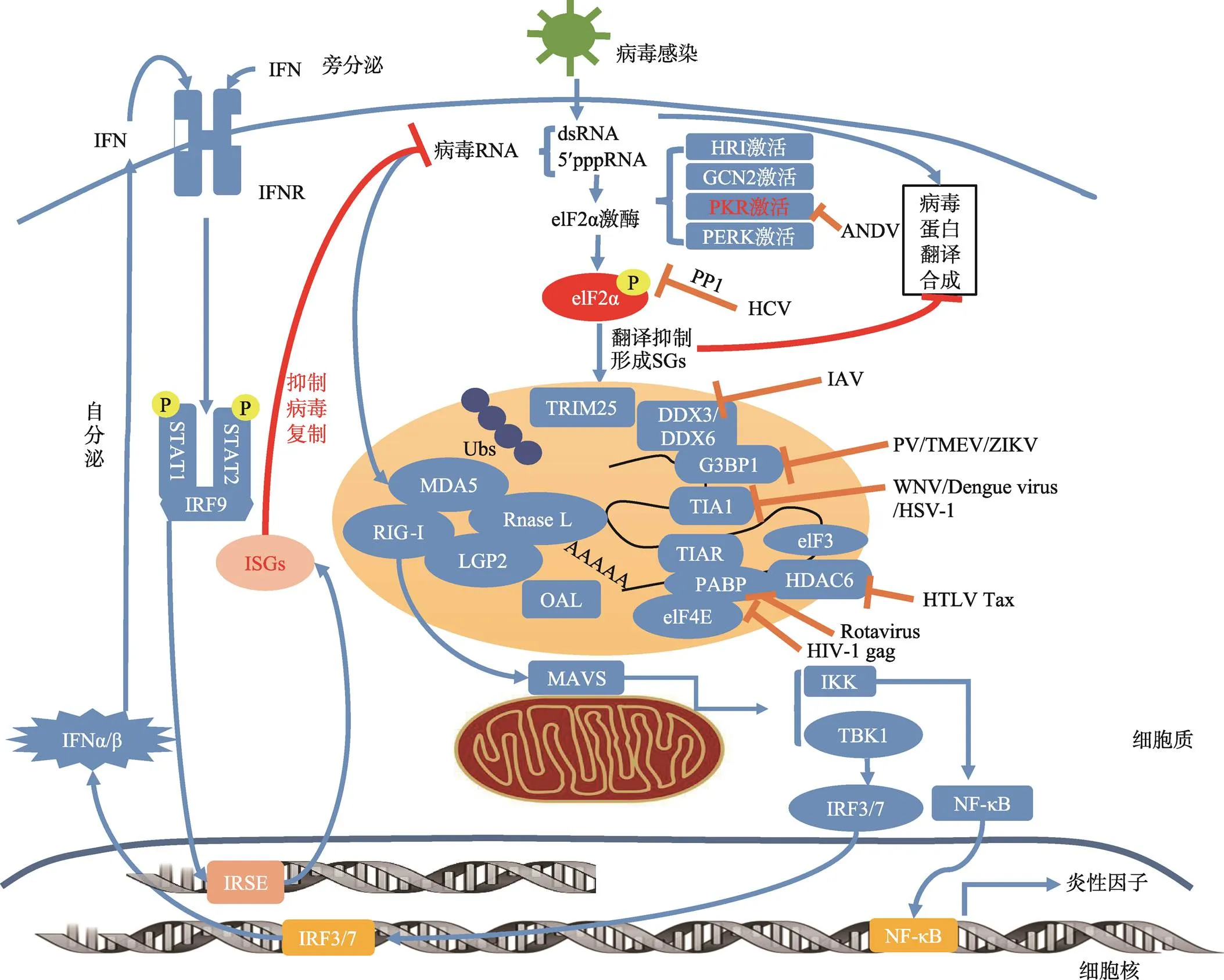
许多研究都指出,病毒感染引起的SGs的形成与抗病毒先天性免疫存在着千丝万缕的联系。宿主抗压力应答的过程中,或多或少都会有先天性免疫相关蛋白成分的参与,有些蛋白成分还起到至关重要的作用。病毒感染诱导SGs与天然免疫概况如图1所示。
PKR是一个典型的干扰素应答蛋白分子,它能够识别病毒的dsRNA及5'ppp RNA,感知细胞内病毒核酸的存在情况,介导下游的干扰素反应。与此同时,PKR的活化诱导了eIF2α的磷酸化,阻滞了细胞内的翻译进程,引起应激颗粒的产生。此外,PKR在细菌和双链RNA病毒感染时,还可以激活炎性小体和巨噬细胞的应答作用,引起细胞因子IL-1b和HMGB1的释放[114]。在先天性免疫的转录应答中,PKR也发挥着重要作用[115]。
胞质内的dsRNA及5′ppp-RNA还可被模式识别分子MDA5及RIG-I所识别,这些都是SGs的组成成分[116]。病毒RNA经MDA5,RIG-I或LGP2识别后,将信号传递至线粒体外膜上的MAVS (mitochondrial antiviral signaling protein),而后将信号传递至IKK和TBK-1,导致转录因子NF-kB和IRF3的激活,从而导致抗病毒的IFN及其他炎症因子的转录,而后激活ISGs表达,行使抗病毒免疫的功能。这表明,病毒诱导的SGs在细胞内提供了一个模式识别分子的平台,调控着下游信号的传导[117]。
IFN的诱导表达在先天性免疫应答中有着举足轻重的地位,而由ISG编码的诱导IFN信号途径的相关蛋白,很多都被招募至SGs中,并调控着SG的形成,SGs也为宿主的抗病毒反应搭建起了一个平台[118]。除了上文提到的PKR、RIG-I和MDA5,还有经IFN诱导产生的OASs,可识别dsRNA,而后催化ATP产生2-5A,从而激活SG组成成分RNase L对于dsRNA的降解。ADAR1也可被IFN诱导,并包含于SGs中,可将dsRNA的A转化为I,从而改变病毒dsRNA,干扰病毒的复制[119]。

图1 病毒感染诱导SGs形成及诱发先天性免疫反应机制图
病毒感染细胞后,病毒基因组RNA激活PKR,使elF2α磷酸化,抑制翻译的起始,从而抑制了病毒蛋白质的合成,也诱发SGs的形成。SGs的组成成分多样,包含病毒RNA、RNA结合蛋白和翻译起始因子等,尤其还包括许多先天性免疫模式识别分子(MDA5/ RIG-I/LDP2等);这些模式识别分子可结合病毒RNA,进而将信号传递至线粒体外膜上的MAVS,激活TBK1/IKK的磷酸化,从而使转录因子IRF3/7以及NF-κB入核,激活Ⅰ型干扰素和一些炎性因子的表达和分泌;干扰素经自分泌或旁分泌与细胞膜干扰素受体结合后,激活胞内的STAT1/2磷酸化,而后同IRF9一起入核,识别IRSE区域,激活表达多种ISGs,产生抗病毒作用。另外,OAS/RNase L也是SGs的组成成分,可切割病毒RNA,被切割后的病毒RNA也可被模式识别分子识别,进一步活化抗病毒免疫通路。病毒也通过多种方式来拮抗这种抗病毒作用,如ANCV可抑制PKR的二聚化,HCV可通过PP1来去除eIF2α的磷酸化,PV/TMEV/ZIKV等可切割G3BP1,以及多种病毒可通过与SGs的成分结合的方式来拮抗SGs的形成。OAS: 2¢,5¢-oligoadenlate synthetase; IRSE: interferon stimulated respnonse element; IRF: interferon regulatory factor; RIG-I: retinoic acid inducible gene I; STAT: signal transducer and activator of transcription; IFNR: interferon receptor; IRF: interferon regulatory factor; ISG: interferon-stimulated gene; MDA5: melanoma differentiation-associated protein 5; NF-κB: nuclear factor kappa-light-chain-enhancer of activated B cells; TRIM25: tripartite motif-containing protein 25; TIA1: T cell restricted intracellular antigen-1; TIAR: TIA1-related protein;PABP: poly(A)-binding protein; G3BP1: Ras-GTPase-activating protein SH3-domain-binding protein 1.
综上所述不难发现,细胞的压力应答,在各种方面都与先天性免疫有着千丝万缕的联系,而这其中的机制目前仍不甚明朗,有待于后期的研究来揭示细胞中各类先天性免疫信号通路与细胞压力应答之间的联系及其在功能上的相互影响。
4 结语与展望
在病毒感染引起的细胞各类应答反应中,压力应答仍旧是一类相对较新的领域。虽然目前已经有大量的关于各类病毒对应激颗粒操纵的报道,但是目前人们对这其中分子水平作用机制仍然知之甚少。宿主感染病毒引发SGs生成,限制病毒的复制,而病毒在与宿主上万年的博弈中,也进化出来克制这种限制,甚至将其利用于自身复制的相关机制;人们在逐渐清楚了病毒通过SGs与宿主之间的相互作用关系的过程中,可扬长避短的寻找抗病毒治疗的异性药物靶点为后期临床应用提供更多信息。
目前已知SGs在许多层面上都与先天性免疫相互关联,因此,在对于应激颗粒的研究中可能会发现一些在抗病毒治疗中具有价值的广谱的作用位点。由药物诱发的,经PKR或elF2α磷酸化生成的SGs,有望控制病毒感染。体外实验已经证明,elF4A解旋酶抑制剂hippuristanol可抑制卡里色病毒(caliciviruses)[120];pateamine A可抑制流感病毒A (influenza A virus)的复制[88]。不过,这些药物对于未感染细胞的毒性影响限制了其发展。
在过去的十几年中,相关研究多围绕于病毒与SGs之间的相互影响,主要着重于其对于IFNs和ISGs的影响来确定SGs的抗病毒作用。而对于病毒引起的翻译起始的抑制和随后SGs的形成对于获得性免疫的激活的影响几乎还是空白。有意思的是,向树突状细胞中转染poly (I:C)并不会引起整个细胞翻译水平的下降,这与其他细胞有很大的区别[121]。抗原提呈细胞或淋巴细胞这类免疫细胞与非免疫细胞在诱导和调控翻译存在着什么不同?TIA1 (T cell-restricted intracellular antigen 1)是T细胞毒性颗粒的组成部分,也参与细胞凋亡的过程,这是不是暗示着SGs的形成与获得性免疫通路有着某种联系,许多未知还等待着人们去探索。
[1] Buchan JR, Parker R. Eukaryotic stress granules: the ins and out of translation.,2009, 36(6): 932–941.
[2] Kedersha NL. Gupta M, Li W, Miller I, Anderson P. RNA-binding proteins TIA-1 and TIAR link the phosphorylation of eIF-2α to the assembly of mammalian stress granules.,1999, 147(7): 1431–1442.
[3] Jackson RJ, Hellen CU, Pestova TV. The mechanism of eukaryotic translation initiation and principles of its regulation.,2010, 11(2): 113– 127.
[4] Anderson P, Kedersha N. Stressful initiations.,2002, 115(Pt 16): 3227–3234.
[5] Anderson P, Kedersha N. RNA granules.,2006, 172(6): 803–808.
[6] Hua Y, Zhou J. Modulation of SMN nuclear foci and cytoplasmic localization by its C-terminus.,2004, 61(19–20): 2658–2663.
[7] Thomas MG, Martinez Tosar LJ, Desbats MA, Leishman CC, Boccaccio GL. Mammalian staufen 1 is recruited to stress granules and impairs their assembly.,2009, 122(Pt 4): 563–573.
[8] Tourrière H, Chebli K, Zekri L, Courselaud B, Blanchard JM, Bertrand E, Tazi J. The RasGAP-associated endoribonuclease G3BP assembles stress granules.,2003, 160(6): 823–831.
[9] Wilczynska A, Aigueperse C, Kress M, Dautry F, Weil D. The translational regulator CPEB1 provides a link between dcp1 bodies and stress granules.,2005, 118(Pt 5): 981–992.
[10] Yang F, Peng Y, Murray EL, Otsuka Y, Kedersha N, Schoenberg DR. Polysome-bound endonuclease PMR1 is targeted to stress granules via stress-specific binding to TIA-1.,2006, 26(23): 8803–8813.
[11] Chalupníková K, Lattmann S, Selak N, Iwamoto F, Fujiki Y, Nagamine Y. Recruitment of the RNA helicase RHAU to stress granules via a unique RNA-binding domain.,2008, 283(50): 35186–35198.
[12] Wippich F, Bodenmiller B, Trajkovska MG, Wanka S, Aebersold R, Pelkmans L. Dual specificity kinase DYRK3 couples stress granule condensation/dissolution to mTORC1 signaling.,2013, 152(4): 791–805.
[13] Okonski KM, Samuel CE. Stress granule formation induced by measles virus is protein kinase PKR dependent and impaired by RNA adenosine deaminase ADAR1.,2012, 87(2): 756–766.
[14] Onomoto K, Jogi M, Yoo JS, Narita R, Morimoto S, Takemura A, Sambhara S, Kawaguchi A, Osari S, Nagata K, Matsumiya T, Namiki H, Yoneyama M, Fujita T. Critical role of an antiviral stress granule containing RIG-I and PKR in viral detection and innate immunity.,2012, 7(8): e43031.
[15] McEwen E, Kedersha N, Song B, Scheuner D, Gilks N, Han A, Chen JJ, Anderson P, Kaufman RJ. Heme- regulated inhibitor kinase-mediated phosphorylation of eukaryotic translation initiation factor 2 inhibits translation, induces stress granule formation, and mediates survival upon arsenite exposure.,2005, 280(17): 16925–16933.
[16] Lu L, Han AP, Chen JJ. Translation initiation control by heme-regulated eukaryotic initiation factor 2alpha kinase in erythroid cells under cytoplasmic stresses.,2001, 21(23): 7971–7980.
[17] Sheree A. Wek SZ, Wek RC. The histidyl-tRNA synthetase-related sequence in the elf-2α protein kinase GCN2 interacts with tRNA and is required for activation in response to starvation for different amino acids., 1995, 15(8): 4497–4506.
[18] Jing D HPH, Brian R. Activation of GCN2 in UV-irradiated cells inhibits translation.,2002, 12(15): 1279–1286.
[19] García MA, Meurs EF, Esteban M. The dsRNA protein kinase PKR: virus and cell control.,2007, 89(6–7): 799–811.
[20] Harding HP, Zhang Y, Bertolotti A, Zeng H, Ron D. Perk is essential for translational regulation and cell survival during the unfolded protein response.,2000, 5(5): 897–904.
[21] Zoncu R, Efeyan A, Sabatini DM. MTOR: from growth signal integration to cancer, diabetes and ageing.,2011, 12(1): 21–35.
[22] von der Haar T, Gross JD, Wagner G, McCarthy JE. The mRNA cap-binding protein eIF4E in post-transcriptional gene expression.,2004, 11(6): 503–511.
[23] Panniers R. Translational control during heat shock.,1994, 76: 737–747.
[24] Thomas MG, Loschi M, Desbats MA, Boccaccio GL. RNA granules: the good, the bad and the ugly.,2011, 23(2): 324–334.
[25] Novoa I, Zhang Y, Zeng H, Jungreis R, Harding HP, Ron D. Stress-induced gene expression requires programmed recovery from translational repression.,2003, 22(5): 1180–1187.
[26] Dang Y, Kedersha N, Low WK, Romo D, Gorospe M, Kaufman R, Anderson P, Liu JO. Eukaryotic initiation factor 2alpha-independent pathway of stress granule induction by the natural product pateamine A.,2006, 281(43): 32870–32878.
[27] Mazroui R, Sukarieh R, Bordeleau ME, Kaufman RJ, Northcote P, Tanaka J, Gallouzi I, Pelletier J. Inhibition of ribosome recruitment induces stress granule formation independently of eukaryotic initiation factor 2alpha phosphorylation.,2006, 17(10): 4212– 4219.
[28] Fujimura K, Sasaki AT, Anderson P. Selenite targets eIF4E-binding protein-1 to inhibit translation initiation and induce the assembly of non-canonical stress granules.,2012, 40(16): 8099–8110.
[29] Emara MM, Fujimura K, Sciaranghella D, Ivanova V, Ivanov P, Anderson P. Hydrogen peroxide induces stress granule formation independent of eIF2α phosphorylation.,2012, 423(4): 763–769.
[30] Kwon S, Zhang Y, Matthias P. The deacetylase HDAC6 is a novel critical component of stress granules involved in the stress response.,2007, 21(24): 3381– 3394.
[31] Leung AK, Vyas S, Rood JE, Bhutkar A, Sharp PA, Chang P. Poly(ADP-ribose) regulates stress responses and microRNA activity in the cytoplasm.,2011, 42(4): 489–499.
[32] Ohn T, Kedersha N, Hickman T, Tisdale S, Anderson P. A functional RNAi screen links O-GlcNAc modification of ribosomal proteins to stress granule and processing body assembly.,2008, 10(10): 1224–1231.
[33] Tsai WC, Gayatri S, Reineke LC, Sbardella G, Bedford MT, Lloyd RE. Arginine demethylation of G3BP1 promotes stress granule assembly.,2016, 291(43): 22671–22685.
[34] Jayabalan AK, Sanchez A, Park RY, Yoon SP, Kang GY, Baek JH, Anderson P, Kee Y, Ohn T. NEDDylation promotes stress granule assembly.,2016, 7: 12125.
[35] Kedersha N, Ivanov P, Anderson P. Stress granules and cell signaling: more than just a passing phase?,2013, 38(10): 494–506.
[36] Boeynaems S, Alberti S, Fawzi NL, Mittag T, Polymenidou M, Rousseau F, Schymkowitz J, Shorter J, Wolozin B, Van Den Bosch L, Tompa P, Fuxreiter M. Protein phase separation: a new phase in cell biology., 2018, 28(6): 420–435.
[37] Takahara T, Maeda T. Transient sequestration of TORC1 into stress granules during heat stress., 2012, 47(2): 242–252.
[38] Buchan R, Kolaitis RM, Taylor JP, Parker R. Eukaryotic stress granules are cleared by autophagy and Cdc48VCP function., 2013, 153(7): 1461–1474.
[39] Chitiprolu M, Jagow C, Tremblay V, Bondy-Chorney E, Paris G, Savard A, Palidwor G, Barry FA, Zinman L, Keith J, Rogaeva E, Robertson J, Lavallée-Adam M, Woulfe J, Couture JF, Côté J, Gibbings D. A complex of C9ORF72 and p62 uses arginine methylation to eliminate stress granules by autophagy., 2018, 9(1): 2794.
[40] McInerney GM, Kedersha NL, Kaufman RJ, Anderson P, Liljeström P. Importance of eIF2alpha phosphorylation and stress granule assembly in alphavirus translation regulation.,2005, 16(8): 3753–3763.
[41] Sciortino MT, Parisi T, Siracusano G, Mastino A, Taddeo B, Roizman B. The virion host shutoff RNase plays a key role in blocking the activation of protein kinase R in cells infected with herpes simplex virus 1.,2013, 87(6): 3271–3276.
[42] Cassady KA, Gross M. The Herpes Simplex virus type 1 U(S)11 protein interacts with protein kinase R in infected cells and requires a 30-amino-acid sequence adjacent to a kinase substrate domain.,2002, 76(5): 2029–2035.
[43] He B, Gross M, Roizman B. The γ134.5 protein of herpes simplex virus 1 complexes with protein phosphatase 1α to dephosphorylate the α subunit of the eukaryotic translation initiation factor 2 and preclude the shutoff of protein synthesis by double-stranded RNA- activated protein kinase.,1996, 94(3): 843–848.
[44] Mulvey M, Arias C, Mohr I. Maintenance of endoplasmic reticulum (ER) homeostasis in herpes simplex virus type 1-infected cells through the association of a viral glycoprotein with PERK, a cellular ER stress sensor.,2007, 81(7): 3377–3390.
[45] Esclatine A, Taddeo B, Roizman B. Herpes simplex virus 1 induces cytoplasmic accumulation of TIA-1/TIAR and both synthesis and cytoplasmic accumulation of tristetraprolin, two cellular proteins that bind and destabilize AU-rich RNAs.,2004, 78(16): 8582– 8592.
[46] Dauber B, Poon D, Dos Santos T, Duguay BA, Mehta N, Saffran HA, Smiley JR. The herpes simplex virus virion host shutoff protein enhances translation of viral true late mRNAs independently of suppressing protein kinase R and stress granule formation.,2016, 90(13): 6049–6057.
[47] Burgess HM, Mohr I. Defining the role of stress granules in innate immune suppression by the herpes simplex virus 1 endoribonuclease VHS.,2018, 92(15): e00829–18.
[48] Finnen RL, Pangka KR, Banfield BW. Herpes simplex virus 2 infection impacts stress granule accumulation.,2012, 86(15): 8119–8130.
[49] Finnen RL, Hay TJ, Dauber B, Smiley JR, Banfield BW. The herpes simplex virus 2 virion-associated ribonuclease vhs interferes with stress granule formation.,2014, 88(21): 12727–12739.
[50] Katsafanas GC, Moss B. Vaccinia virus intermediate stage transcription is complemented by Ras-GTPase- activating protein SH3 domain-binding protein (G3BP) and cytoplasmic activation/proliferation-associated protein (p137) individually or as a heterodimer.,2004, 279(50): 52210–52217.
[51] Katsafanas GC, Moss B. Linkage of transcription and translation within cytoplasmic poxvirus DNA factories provides a mechanism to coordinate viral and usurp host functions.,2007, 2(4): 221–228.
[52] Zaborowska I, Kellner K, Henry M, Meleady P, Walsh D. Recruitment of host translation initiation factor eIF4G by the Vaccinia Virus ssDNA-binding protein I3.,2012, 425(1): 11–22.
[53] Walsh D, Arias C, Perez C, Halladin D, Escandon M, Ueda T, Watanabe-Fukunaga R, Fukunaga R, Mohr I. Eukaryotic translation initiation factor 4F architectural alterations accompany translation initiation factor redistribution in poxvirus-infected cells.,2008, 28(8): 2648–2658.
[54] Simpson-Holley M, Kedersha N, Dower K, Rubins KH, Anderson P, Hensley LE, Connor JH. Formation of antiviral cytoplasmic granules during orthopoxvirus infection.,2011, 85(4): 1581–1593.
[55] Montero H, Rojas M, Arias CF, López S. Rotavirus infection induces the phosphorylation of eIF2alpha but prevents the formation of stress granules.,2008, 82(3): 1496–1504.
[56] Rojas M, Arias CF, López S. Protein kinase R is responsible for the phosphorylation of eIF2α in rotavirus infection.,2010, 84(20): 10457–10466.
[57] Qin Q, Hastings C, Miller CL. Mammalian orthoreovirus particles induce and are recruited into stress granules at early times postinfection.,2009, 83(21): 11090– 11101.
[58] Qin Q, Carroll K, Hastings C, Miller CL. Mammalian orthoreovirus escape from host translational shutoff correlates with stress granule disruption and is independent of eIF2alpha phosphorylation and PKR.,2011, 85(17): 8798–8810.
[59] Carroll K, Hastings C, Miller CL. Amino acids 78 and 79 of mammalian orthoreovirus protein µNS are necessary for stress granule localization, core proteinλ2 interaction, and de novo virus replication.,2014, 448: 133–145.
[60] Dougherty JD, Tsai WC, Lloyd RE. Multiple poliovirus proteins repress cytoplasmic RNA granules.,2015, 7(12): 6127–6140.
[61] White JP, Cardenas AM, Marissen WE, Lloyd RE. Inhibition of cytoplasmic mRNA stress granule formation by a viral proteinase.,2007, 2(5): 295–305.
[62] Piotrowska J, Hansen SJ, Park N, Jamka K, Sarnow P, Gustin KE. Stable formation of compositionally unique stress granules in virus-infected cells.,2010, 84(7): 3654–3665.
[63] Ng CS, Jogi M, Yoo JS, Onomoto K, Koike S, Iwasaki T, Yoneyama M, Kato H, Fujita T. Encephalomyocarditis virus disrupts stress granules, the critical platform for triggering antiviral innate immune responses.,2013, 87(17): 9511–9522.
[64] Fung G, Ng CS, Zhang J, Shi J, Wong J, Piesik P, Han L, Chu F, Jagdeo J, Jan E, Fujita T, Luo H. Production of a dominant-negative fragment due to G3BP1 cleavage contributes to the disruption of mitochondria-associated protective stress granules during CVB3 infection.,2013, 8(11): e79546.
[65] Borghese F, Michiels T. The leader protein of cardioviruses inhibits stress granule assembly.,2011, 85(18): 9614–9622.
[66] Reineke LC, Kedersha N, Langereis MA, van Kuppeveld FJ, Lloyd RE. Stress granules regulate double-stranded RNA-dependent protein kinase activation through a complex containing G3BP1 and Caprin1.,2015, 6(2): e02486.
[67] Visser LG, Medina GN, Rabouw HH, de Groot RG, Langereis MA, de los Santos T, van Kuppeveld FJM. Foot-and-Mouth disease leader protease cleaves G3BP1 and G3BP2 and inhibits stress granule formation.,2019, 93(2): e00922–18.
[68] Yang X, Hu Z, Fan S, Zhang Q, Zhong Y, Guo D, Qin Y, Chen M. Picornavirus 2A protease regulates stress granule formation to facilitate viral translation.,2018, 14(2): e1006901.
[69] Li W, Li Y, Kedersha N, Anderson P, Emara M, Swiderek KM, Moreno GT, Brinton MA. Cell proteins TIA-1 and TIAR interact with the 3' Stem-Loop of the West nile virus complementary minus-strand RNA and facilitate virus replication.,2002, 76(23): 11989– 12000.
[70] Courtney SC, Scherbik SV, Stockman BM, Brinton MA. West nile virus infections suppress early viral RNA synthesis and avoid inducing the cell stress granule response.,2012, 86(7): 3647–3657.
[71] Shives KD, Beatman EL, Chamanian M, O'Brien C, Hobson-Peters J, Beckham JD. West nile virus-induced activation of mammalian target of rapamycin complex 1 supports viral growth and viral protein expression.,2014, 88(16): 9458–9471.
[72] Xia J, Chen X, Xu F, Wang Y, Shi Y, Li Y, He J, Zhang P. Dengue virus infection induces formation of G3BP1 granules in human lung epithelial cells.,2015, 160(12): 2991–2999.
[73] Bidet K, Dadlani D, Garcia-Blanco MA. G3BP1, G3BP2 and CAPRIN1 are required for translation of interferon stimulated mRNAs and are targeted by a dengue virus non-coding RNA.,2014, 10(7): e1004242.
[74] Zhang HW, Meng XY, Li LF, Yang YY, Qiu HJ. Long non-coding RNAs: Emerging regulators of antiviral innate immune responses., 2018, 40(7): 525–533张华伟, 孟星宇, 李连峰, 杨玉莹, 仇华吉. 长链非编码RNA——抗病毒天然免疫应答的新兴调控因子. 遗传, 2018, 40(7): 525–533.
[75] Albornoz A, Carletti T, Corazza G, Marcello A. The stress granule component TIA-1 binds tick-borne encephalitis virus RNA and is recruited to perinuclear sites of viral replication to inhibit viral translation.,2014, 88(12): 6611–6622.
[76] Katoh H, Okamoto T, Fukuhara T, Kambara H, Morita E, Mori Y, Kamitani W, Matsuura Y. Japanese encephalitis virus core protein inhibits stress granule formation through an interaction with Caprin-1 and facilitates viral propagation.,2013, 87(1): 489–502.
[77] Garaigorta U, Heim MH, Boyd B, Wieland S, Chisari FV. Hepatitis C virus (HCV) induces formation of stress granules whose proteins regulate HCV RNA replication and virus assembly and egress.,2012, 86(20): 11043–11056.
[78] Ruggieri A, Dazert E, Metz P, Hofmann S, Bergeest JP, Mazur J, Bankhead P, Hiet MS, Kallis S, Alvisi G, Samuel CE, Lohmann V, Kaderali L, Rohr K, Frese M, Stoecklin G, Bartenschlager R. Dynamic oscillation of translation and stress granule formation mark the cellular response to virus infection.,2012, 12(1): 71–85.
[79] Li Q, Pène V, Krishnamurthy S, Cha H, Liang TJ. Hepatitis C virus infection activates an innate pathway involving IKK-αin lipogenesis and viral assembly,,2013, 19(6): 722–729.
[80] Hou S, Kumar A, Xu Z, Airo AM, Stryapunina I, Wong CP, Branton W, Tchesnokov E, Götte M, Power C, Hobman TC. Zika virus hijacks stress granule proteins and modulates the host stress response.,2017, 91(16): e00474–17.
[81] Amorim R, Temzi A, Griffin BD, Mouland AJ. Zika virus inhibits eIF2α-dependent stress granule assembly.,2017, 11(7): e0005775.
[82] Raaben M, Groot Koerkamp MJ, Rottier PJ, de Haan CA. Mouse hepatitis coronavirus replication induces host translational shutoff and mRNA decay, with concomitant formation of stress granules and processing bodies.,2007, 9(9): 2218–2229.
[83] Sola I, Galán C, Mateos-Gómez PA, Palacio L, Zúñiga S, Cruz JL, Almazán F, Enjuanes L. The polypyrimidine tract-binding protein affects coronavirus RNA accumulation levels and relocalizes viral RNAs to novel cytoplasmic domains different from replication-transcription sites.,2011, 85(10): 5136–5149.
[84] Nakagawa K, Narayanan K, Wada M, Makino S. Inhibition of stress granule formation by middle east respiratory syndrome coronavirus 4a accessory protein facilitates viral translation, leading to efficient virus replication.,2018, 92(20): e00902–18.
[85] Utt A, Das PK, Varjak M, Lulla V, Lulla A, Merits A. Mutations conferring a noncytotoxic phenotype on chikungunya virus replicons compromise enzymatic properties of nonstructural protein 2.,2014, 89(6): 3145–3162.
[86] Khaperskyy DA, Hatchette TF, McCormick C. Influenza a virus inhibits cytoplasmic stress granule formation.,2011, 26(4): 1629–1639.
[87] Mok BWY, Song W, Wang P, Tai H, Chen Y, Zheng M, Wen X, Lau SY, Wu WL, Matsumoto K, Yuen KY, Chen H. The NS1 protein of influenza a virus interacts with cellular processing bodies and stress granules through RNA-associated protein 55 (RAP55) during virus infection.,2012, 86(23): 12695–12707.
[88] Khaperskyy DA, Emara MM, Johnston BP, Anderson P, Hatchette TF, McCormick C. Influenza a virus host shutoff disables antiviral stress-induced translation arrest.,2014, 10(7): e1004217.
[89] Thulasi Raman SN, Liu G, Pyo HM, Cui YC, Xu F, Ayalew LE, Tikoo SK, Zhou Y. DDX3 Interacts with influenza a virus NS1 and NP proteins and exerts antiviral function through regulation of stress granule formation.,2016, 90(7): 3661–3675.
[90] Núñez RD, Budt M, Saenger S, Paki K, Arnold U, Sadewasser A, Wolff T. The RNA helicase DDX6 associates with RIG-I to augment induction of antiviral signaling.,2018, 19(7). DOI:10.3390/ ijms19071877.
[91] Dinh PX, Beura LK, Das PB, Panda D, Das A, Pattnaik AK. Induction of stress granule-like structures in vesicular stomatitis virus-infected cells.,2013, 87(1): 372–383.
[92] Lindquist ME, Mainou BA, Dermody TS, Crowe JE Jr. Activation of protein kinase R is required for induction of stress granules by respiratory syncytial virus but dispensable for viral replication.,2011, 413(1): 103–110.
[93] Lindquist ME, Lifland AW, Utley TJ, Santangelo PJ, Crowe JE Jr. Respiratory syncytial virus induces host RNA stress granules to facilitate viral replication.,2010, 84(23): 12274–12284.
[94] Lifland AW, Jung J, Alonas E, Zurla C, Crowe JE Jr, Santangelo PJ. Human respiratory syncytial virus nucleoprotein and inclusion bodies antagonize the innate immune response mediated by MDA5 and MAVS.,2012, 86(15): 8245–8258.
[95] Fricke J, Koo LY, Brown CR, Collins PL. P38 and OGT sequestration into viral inclusion bodies in cells infected with human respiratory syncytial virus suppresses MK2 activities and stress granule assembly.,2012, 87(3): 1333–1347.
[96] Randall RE, Goodbourn S. Interferons and viruses: an interplay between induction, signalling, antiviral responses and virus countermeasures.,2008, 89(Pt 1): 1–47.
[97] Iseni F, Garcin D, Nishio M, Kedersha N, Anderson P, Kolakofsky D. Sendai virus trailer RNA binds TIAR, a cellular protein involved in virus-induced apoptosis.,2002, 21(19): 5141–5150.
[98] Valiente-Echeverría F, Hermoso MA, Soto-Rifo R. RNA helicase DDX3: at the crossroad of viral replication and antiviral immunity.,2015, 25(5): 286– 299.
[99] Mir MA, Duran WA, Hjelle BL, Ye C, Panganiban AT. Storage of cellular 5′ mRNA caps in P bodies for viral cap-snatching.,2008, 105(49): 19294–19299.
[100] Hopkins KC, Tartell MA, Herrmann C, Hackett BA, Taschuk F, Panda D, Menghani SV, Sabin LR, Cherry S. Virus-induced translational arrest through 4EBP1/2- dependent decay of 5'-TOP mRNAs restricts viral infection.,2015, 112(22): E2920– E2929.
[101] Cimica V, Dalrymple NA, Roth E, Nasonov A, Mackow ER. An innate immunity-regulating virulence determinant is uniquely encoded by the Andes virus nucleocapsid protein.,2014, 5(1): e01088–13.
[102] Matthys VS, Cimica V, Dalrymple NA, Glennon NB, Bianco C, Mackow ER. Hantavirus GnT elements mediate TRAF3 binding and inhibit RIG-I/TBK1-directed beta interferon transcription by blocking IRF3 phosphorylation.,2014, 88(4): 2246–2259.
[103] Wang Z, Mir MA. Andes virus nucleocapsid protein interrupts protein kinase R dimerization to counteract host interference in viral protein synthesis.,2015, 89(3): 1628–1639.
[104] Fang J, Pietzsch C, Ramanathan P, Santos RI, Ilinykh PA, Garcia-Blanco MA, Bukreyev A, Bradrick SS. Staufen1 interacts with multiple components of the ebola virus ribonucleoprotein and enhances viral RNA synthesis.,2018, 9(5): e01771–18.
[105] Legros S, Boxus M, Gatot JS, Van Lint C, Kruys V, Kettmann R, Twizere JC, Dequiedt F. The HTLV-1 tax protein inhibits formation of stress granules by interacting with histone deacetylase 6.,2011, 30(38): 4050–4062.
[106] Takahashi M, Higuchi M, Makokha GN, Matsuki H, Yoshita M, Tanaka Y, Fujii M. HTLV-1 tax oncoprotein stimulates ROS production and apoptosis in T cells by interacting with USP10.,2013, 122(5): 715–725.
[107] Abrahamyan LG, Chatel-Chaix L, Ajamian L, Milev MP, Monette A, Clément JF, Song R, Lehmann M, DesGroseillers L, Laughrea M, Boccaccio G, Mouland AJ. Novel Staufen1 ribonucleoproteins prevent formation of stress granules but favour encapsidation of HIV-1 genomic RNA.,2010, 123(Pt 3): 369–383.
[108] Henao-Mejia J, Liu Y, Park IW, Zhang J, Sanford J, He JJ. Suppression of HIV-1 Nef translation by Sam68 mutant-induced stress granules and nef mRNA sequestration.,2009, 33(1): 87–96.
[109] Valiente-Echeverría F, Melnychuk L, Vyboh K, Ajamian L, Gallouzi IE, Bernard N, Mouland AJ. EEF2 and Ras- GAP SH3 domain-binding protein (G3BP1) modulate stress granule assembly during HIV-1 infection.,2014, 5: 4819.
[110] Jiménez VC, Martinez FO, Booiman T, van Dort KA, van de Klundert MAA, Gordon S, Geijtenbeek TB, Kootstra NA. G3BP1 restricts HIV-1 replication in macrophages and T-cells by sequestering viral RNA.,2015, 486: 94–104.
[111] Cinti A, Le Sage V, Ghanem M, Mouland AJ. HIV-1 gag blocks selenite-induced stress granule assembly by altering the mRNA Cap-Binding complex.,2016, 7(2): e00329.
[112] Rao S, Temzi A, Amorim R, You JC, Mouland AJ. HIV-1 NC-induced stress granule assembly and translation arrest are inhibited by the dsRNA binding protein Staufen1., 2018, 24(2): 219–236.
[113] Nelson EV, Schmidt KM, Deflubé LR, Doğanay S, Banadyga L, Olejnik J, Hume AJ, Ryabchikova E, Ebihara H, Kedersha N, Ha T, Mühlberger E. Ebola virus does not induce stress granule formation during infection and sequesters stress granule proteins within viral inclusions.,2016, 90(16): 7268–7284.
[114] Lu B, Nakamura T, Inouye K, Li J, Tang Y, Lundbäck P, Valdes-Ferrer SI, Olofsson PS, Kalb T, Roth J, Zou Y, Erlandsson-Harris H, Yang H, Ting JP, Wang H, Andersson U, Antoine DJ, Chavan SS, Hotamisligil GS, Tracey KJ. Novel role of PKR in inflammasome activation and HMGB1 release.,2012, 488 (7413): 670–674.
[115] Taghavi N, Samuel CE. Protein kinase PKR catalytic activity is required for the PKR-dependent activation of mitogen-activated protein kinases and amplification of interferon beta induction following virus infection.,2012, 427(2): 208–216.
[116] Reikine S, Nguyen JB, Modis Y. Pattern recognition and signaling mechanisms of RIG-I and MDA5.,2014, 5: 342.
[117] Wu B, Hur S. How RIG-I like receptors activate MAVS.,2015, 12: 91–98.
[118] George CX, Ramaswami G, Li JB, Samuel CE. Editing of cellular self-RNAs by adenosine deaminase ADAR1 suppresses innate immune stress responses.,2016, 291(12): 6158–6168.
[119] Silverman RH. Viral encounters with 2¢,5¢-oligoadenylate synthetase and RNase L during the interferon antiviral response.,2007, 81(23): 12720–12729.
[120] Chaudhry Y, Nayak A, Bordeleau ME, Tanaka J, Pelletier J, Belsham GJ, Roberts LO, Goodfellow IG. Caliciviruses differ in their functional requirements for eIF4F components.,2006, 281(35): 25315– 25325.
[121] Clavarino G, Cláudio N, Dalet A, Terawaki S, Couderc T, Chasson L, Ceppi M, Schmidt EK, Wenger T, Lecuit M, Gatti E, Pierre P. Protein phosphatase 1 subunit Ppp1r15a/ GADD34 regulates cytokine production in polyinosinic: polycytidylic acid-stimulated dendritic cells.,2012, 109(8): 3006–3011.
Interaction between stress granules and viruses
Yu Huang, Siqi Hu, Fei Guo
Stress granule (SG) formation is a primary mechanism through which gene expression is rapidly modulated when the eukaryotic cells undergo cellular stresses (including heat shock, oxidative stress, starvation, viral infection). SGs have been proposed to affect mRNA translation and stability,as well as being linked to apoptosis and nuclear processes. Formation of SGs after viral infection result in blockade of viral protein synthesis and viral replication. Not surprisingly, viruses from diverse families have been found to modulate SG formation in infected cells by associating with important SG effector proteins. Here we provide a summary of the current understanding of the mechanism of SG formation, describe the current knowledge on viruses induce and/or modulate SGs in infected cells via phosphorylation of eIF2α, and regulation of SGs in virus systems. Further, we summarize recent progresses in understanding the relationship between viruses and stress granules in mammalian cells, and suggest that SG formation is an important aspect of the antiviral innate immune response.
stress granules; translation arrest; virus; innate immunity
2019-03-07;
2019-05-15
国家重点研发计划项目(编号:2016YFD0500307FF09)资助[Supported by the National Key Plan for Scientific Research and Development of China (No. 2016YFD0500307FF09)]
黄羽,博士研究生,专业方向:病毒与宿主限制因子相互作用。E-mail: huangyu910730@163.com
郭斐,博士,研究员,研究方向:分子病毒学。E-mail: guoafei@ipbcams.ac.cn
10.16288/j.yczz.19-020
2019/5/17 10:48:01
URI: http://kns.cnki.net/kcms/detail/11.1913.R.20190517.1047.001.html
(责任编委: 张天宇)

