p53-Bcl-2/Bax凋亡信号通路变化在自发性高血压大鼠左心室重构进程中作用
孙瑞敏,俞 雪,2,付 舒,吴雨婷,岳 云,李叶丽,杨丹莉
(1.遵义医科大学基础药理教育部重点实验室暨特色民族药教育部国际合作联合实验室,贵州 遵义 563099;2.遵义医科大学 药学院2014级药学,贵州 遵义 563099)
1 Introduction
With the change of people's living environment and habits,the morbidity of hypertension is getting higher and higher.What’s more,more younger people was diagnosed with hypertension.Hypertensive heart disease is a main factor resulting in heart failure and sudden death,caused by aggravation of left ventricular load which was closely related to the long-term increase of blood pressure.Then the left ventricular remodeling will happen after persistent hypertrophing and expanding of left ventricle ,during which myocardial apoptosis plays an important role[1-2].
Studies in apoptosis in recent years revealed that mitochondrial pathway,death receptor pathway and endoplasmic reticulum pathway are involved in apoptosis of cardiomyocytes[3].Numerous evidences show that mitochondrial dysfunction caused by oxidative stress,exhibits a significant impact on the occurrence and development of cardiovascular diseases such as atherosclerosis,hypertension,myocardial ischemia reperfusion injury and heart failure[4-8].Proteins involved in endogenous mitochondrial apoptosis pathway exert an important role,including p53,Bcl-2,Bax,etc[3].
In this study,the dynamic changes of the left ventricular pathological morphology,myocardial appoptosis were observed,and the expression levels of p53,Bcl-2,Bax were measured as well,to reveal the potential mechanism of left heart ventricular remodeling in spontaneously hypertensive model rats.
2 Materials and Methods
2.1 Animals study 13th-week-old male SHRs and WKYs were purchased from Beijing Weitong Lihua Experimental Animal Technology Co.Ltd.(License number:SCXK (Beijing) 2012-0001).All rats were maintained in the specific pathogen free (SPF) animal laboratory and were free fed for 1 week,then each kind of rats were randomly divided into three groups according to different ages,including 18th week-age group (designated SHR18W and WKY18W),22th week-age group (designated SHR22W and WKY22W),26th week-age (designated SHR26W and WKY26W),respectively.Blood pressure was measured,and the left ventricular tissues were taken to observe the changes of pathological morphology,myocardial appoptosis,and determine the protein expression levels of p53,Bcl-2 and Bax.The study was approved by the animal study committee of Zunyi Medical University.
2.2 Determination of blood pressure Rat tail blood pressure was measured with a Kent Scientific CODA (Kent Scientific Corporation,CT,USA) in a quiet and dark environment for once four weeks.The arterial blood pressure value of the rats were presented as means ± SD for quintuplicate experiments.
2.3 Hematoxylin and eosin (H&E) staining The left ventricular apexes of rats were fixed in 4% formaldehyde solution for 48 h,dehydrated and embedded into paraffin blocks.Cross section(the thickness of the section is 4 μm) were obtained and stained according to H&E method,and the morphological analysis were further performed with an Olympus optical microscope (BX-43,OLYMPUS Co.,Ltd.,Tokyo,Japan).
2.4 Detection of myocardial apoptosis The paraffin blocks of left ventricle were randomly selected and analyzed via the terminal deoxynucleotidyl transferase-mediated Dutp nick end labelling (TUNEL) method (Roche Co.,Ltd.,Basel,Switzerland) according to the manufacturer's instruction.The apoptosis of myocardial cells was observed under an Olympus microscope (BX-43,OLYMPUS Co.,Ltd.,Tokyo,Japan).Apoptotic cardiomyocytes nuclei are stained brown and normal cardiomyocytes nuclei are blue.Image Pro Plus 6 software was used to calculate the percentage of apoptotic cells.
2.5 Western blot Protein was extracted from the left heart ventricular tissues of rats.Concentration of extracted protein was determined with BCA method(Beyotime Biotechnology,China).The sample (30 μg of protein) was separated with 10% SDS-polyacrylamide gel and then transferred to a polyvinylidene fluoride membrane.Blocking of membranes was performed in 8% skim milk in Tris buffer containing 0.05% Tween 20 (TBST) at room temperature for 2h.The blots were incubated with antibodies against p53 (1∶2 000,ab1431,Abcam,USA),Bcl-2(1∶2 000,ab7973,Abcam,USA),Bax (1∶500,ab10813,Abcam,USA),and GAPDH (1∶10 000,60004-1-Ig,Proteintech,China) overnight at 4℃.Followed with all antibodies diluted in TBST.After the incubation,membranes were washed and incubated in goat anti-rabbit IgG-HRP (1∶2 000 dilution,Proteintech Group,USA)or goat anti-mouse IgG-HRP (1∶2 000 dilution,Proteintech Group,USA) for 1 h at room temperature.Finally,the Bio-Rad CCD imaging system was used to obtain images and analyze the gray value of the protein band after ECL chemiluminescence.
2.6 Statistical analysis All data were described using mean±SD.The software SPSS18.0 was used for statistical analysis.Two independent samples were determined by two-tailed independent samplest-test.WhenP<0.05,the difference was considered statistically significant.
3 Results
3.1 Changes of blood pressure in SHRs As shown in figure 1,systolic pressure (SBP) and diastolic blood pressure (DBP) of rats show no significant difference among WKY groups,which were within the normal range(nomal pressure of WKY:SBP110-140 mmHg,DBP70-90 mmHg[9]).It was showed that the blood pressure in SHR group was significantly higher than that of the age-matched WKY group (P<0.05).DBP and/or SBP of rats in SHR22W and SHR 26W group were significantly higher than that of rats in SHR18W group.What’s more,abnormal higher blood pressure was observed of rats in SHR26W group compared with SHR22W group(P<0.05).Compared with WKY26W,the SBP of SHR26W was about 1.46 times and the DBP was 1.94 times.

*:P<0.05 versus the same time of WKY group;#:P<0.05 versus SHR14W group;▲:P<0.05 versus SHR18W group;▼:P<0.05 versus SHR22W group (mean±S.D,n=4).Fig 1 Changes of blood pressure in SHRs
3.2 Pathological changes of left ventricular in SHRs Result of H&E staining indicated that among the WKY group,the myocardial cell morphology being normal and no inflammatory cell infiltration in the myocardium were observed.In addition,no other significant changes appeared with the increase of the week-age.Compared with the age-matched WKY group,hypertrophy,disorder,broken myofilament of the cardiac myocyte in SHR group were obviously observed with microscope,as well as obvious infiltration of the inflammatory cells and the deposition of collagen.Moreover,the above mentioned pathological changes became more evident and were closely related to the week-age of SHR rats.

A:WKY18W group;B:WKY22W group;C:WKY26W group;D:SHR18W group;E:SHR22W group;F:SHR26W group,(Magnification 40×).Fig 2 Change of left ventricular pathological changes in SHR
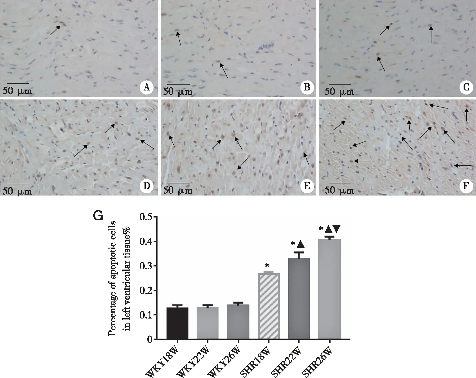
3.3 Apoptosis of left ventricular cardiomyocyte in SHRs TUNEL method was applied to evaluate the apoptosis of left ventricular cardiomyocyte.In WKY groups,the cardiomyocytes apoptosis were not evidently observed,even with the increase of the week-age.Compared with the age-matched WKY group ,the apoptosis of cardiomyocytes in the SHR group was significantly aggravated and the pathologic changes were age-related (P<0.05).

A: WKY18W group;B: WKY22W group; C: WKY26W group;D: SHR18W group; E: SHR22W group;F:SHR26W group.*:P<0.05 versus the matched-age WKY group;▲:P<0.05 versus SHR18W group;▼:P<0.05 versus SHR22W group (mean±S.D,n=4),(Magnification 40×).Fig 3 Change of left ventricular cardiomyocyte apoptosis in SHR
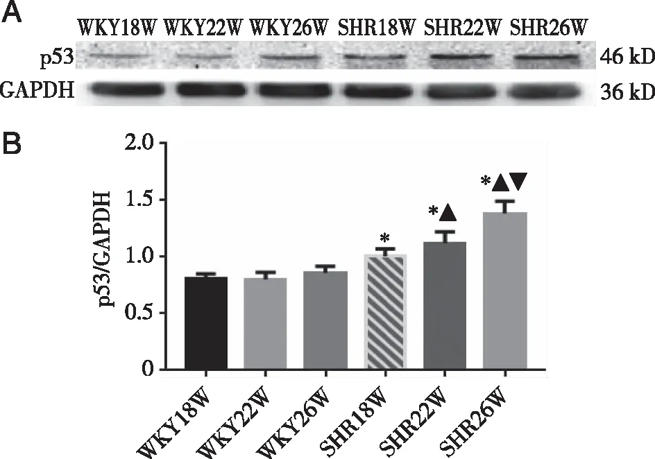
3.4 Change of p53 protein in left ventricular As shown in Figure 4,the expression level of p53 in WKY group did not change with the increase of age.Compared with the age-matched WKY group ,the expression of p53 protein in SHR was significantly increased and the expression level was further verified as age-dependent.Besides,compared with the SHR18W group,p53 protein expression level in the SHR22W group was increased significantly(P<0.05),the expression level of SHR26W group was highest among SHR groups(P<0.05).
3.5 Changes on levels of Bax ,Bcl-2 protein and the ratio value of Bcl-2/Bax The expressions of Bcl-2(Figure 5) in WKY group had no significant changes with the increase of age.Compared with the age matched WKY rats ,the expressions of Bcl-2 protein in SHR were decreased significantly,which was age-related (P<0.05).
As shown in Figure 5,no significant changes of the expression level of Bax in WKY group with the increase of age.Compared with the age-matched rat in WKY group,the expressions of the Bax protein in SHR group were significantly increased.Compared with the SHR18W group,the expression of Bax protein in SHR22W group was higher (P<0.05),and the Bax protein expression of SHR26W group was much higher than that of SHR22W group (P<0.05).
the ratio of Bcl-2/Bax(Figure 5) in WKY group did not change with the increase of age.While the ratio of Bcl-2/Bax in SHR group was significantly reduced than that of WKY group.The ratio of Bcl-2/Bax of SHR was closely related with age (P<0.05).

*:P<0.05 versus the matched-age WKY group;▲:P<0.05 versus SHR18W group;▼:P<0.05 versus SHR22W group (mean ±S.D.,n=4).Fig 4 Change on levels of p53 protein in SHRs

*:P<0.05 versus the matched-age WKY group;▲:P<0.05 versus SHR18W group;▼:P<0.05 versus SHR22W group (mean±S.D.,n=4).Fig 5 Changes on levels of Bax ,Bcl-2 protein and the ratio value of Bcl-2/Bax
4 Discussion
Heart ventricular remodeling refers that the cardiomyocytes,extracellular matrix and collagen fibres are corresponding changed,making the ventricular cavity size,shape and thickness transformed.These negative changes always are called ventricular remodeling,the basic pathological mechanism of the development of heart failure,eventually leading to irreversible end stage[1,10].Early detection and inhibition of ventricular remodeling can reduce the incidence and delay the progression of heart failure,reduce the death rate and improve the quality of life and prognosis of patients as well[11].Through measurement of the blood pressure and observing of the left ventricular pathological morphology changes and myocardial appoptosis in rats.We found that the above indexes in the SHR group were significantly aggravated compared with WKY group at the same age.In addition,compared all above indexes of SHR rats in different week-age,we found that the blood pressure of rats in SHR group gradually increased with the increase of age.Cardiomyocyte hypertrophy,disturbance,myofilament rupture and inflammatory of cell infiltration were obviously observed.We also found that the collagen was deposition and cells apoptosis was gradually aggravated.These parameters were significantly worse in the SHR26W group compared with SHR18W and SHR22W groups.it was found that the initial heart ventricular remodeling was emerged at the age of 18 weeks and gradually aggravated with the age in SHRs.
Apoptosis is the most well-known form of programmed cell death,and the mitochondrial pathway is one of the three major apoptotic pathways[4].After myocardial cells are stimulated by DNA-damaged signal,p53 could down-regulate the expression of Bcl-2 and up-regulate Bcl-2-related X protein (Bax) to decrease the ratio of Bcl-2/Bax and mitochondrial transmembrane potential.Bcl-2 and Bax are 45% homologues[12-13].Bcl-2 is an anti-apoptotic protein containing multiple BH domains,which can regulate the homeostasis of Ca2+,and the permeability of mitochondria is changed,then pores on the outer membrane of mitochondria will appear,which can make pro-apoptotic substance such as cytochrome C flow out of mitochondria to initiate apoptosis[14].Bax is one of the pro-apoptotic proteins in the Bcl-2 family,which can inhibit the activity of Bcl-2 or act on the permeability transition pore of mitochondrial membrane.Permeability transition pore of mitochondrial membrane is a polyprotein complex located at the junction of mitochondrial inner and outer membranes mediates the transport of substances[15].The abnormal opening of pores play a critical role during the induction of apoptosis.Bcl-2 is an anti-apoptotic protein and Bax is a pro-apoptotic protein,which can promote the survival of cells when they form homologous or heterodimers.Therefore,the regulation of the formation of Bcl-2/Bax dimer is a very important step in regulating apoptosis[16-17].Up-regulating the ratio of Bcl-2/Bax can effectively reduce the apoptosis of cardiomyocytes[18-20].Western blot analysis showed that,the levels of p53 and Bax protein were increased significantly,while the levels of Bcl-2 protein and the ratio of Bcl-2/Bax were decreased significantly in SHR group compared with age-matched of WKY group.Compared with that of the SHR18W group,the level of Bcl-2 and Bcl-2/Bax ratio were evidently reduced,protein expression of p53 and Bax and other index in the SHR22W and SHR26W group were significantly increased.Compared with the SHR22W group,the above index of SHR26W were worst.Aforementioned results indicated that the abnormal expression of p53 leads the unbalance of Bcl-2 and Bax,which further activated the mitochondrial apoptosis in SHR rat.It was further confirmed the occurrence and development of cardiomyocyte apoptosis.
Based on the above experimental results ,it was found that with the week-age increasing,the left ventricular remodeling and apoptosis of cadiomyocytes gradually were aggravated in SHRs,and the changes of expression levels of p53,Bcl-2 and Bax were age-related.In summary,the mechanism of left ventricular remodeling may be related to p53-Bcl-2/Bax pathway mediated mitochondrial apoptosis in SHRs.

