心周脂肪体积的影响因素及与冠心病的相关性
李佳凝 孔维敏 冯纪涛 袁晓丹 杨丽红 楼青青


【摘要】目的 采用64排CT測量中国大陆人群心周脂肪(pericardial adipose tissue, PAT)体积,分析PAT体积相关危险因素以及评估PAT与冠心病(通过冠状动脉造影)之间的关系。方法 选择从2014年09月~2015年12月在南京中医药大学附属中西医结合医院心血管内科行冠状动脉CT检查及冠状动脉造影的住院患者158例进行分析。进行临床数据、生化指标、PAT体积的测定和采集,行统计学分析。结果 本研究共纳入158名患者,男性49.37%,平均年龄为(60.66±9.45)岁。多元线性回归分析显示,年龄,男性,BMI是PAT体积的独立危险因素(R2=0.463,P<0.05)。在多因素Logistic回归模型中,校正年龄、性别后,冠心病患者的PAT体积为未患冠心病人群的1.005倍(95%CI1.001-1.009);校正年龄、性别、BMI后,冠心病患者的PAT体积为未患冠心病人群的1.006倍 (95%CI1.001-1.011);校正年龄、性别、BMI、高血压治疗、高血脂治疗、糖尿病和吸烟后,冠心病患者的PAT体积为未患冠心病人群的1.006倍(95%1.001-1.011),且P<0.05,说明PAT体积是冠心病非常重要的独立相关危险因素。结论 64排双源CT可对PAT体积进行准确定量,是一个新的预测冠心病发生的危险因素,对冠心病危险监测具有重要意义。年龄、男性、BMI是PAT体积的独立相关危险因素。因此,高龄、超重或肥胖、男性人群需要更重视PAT的测定,为进一步研究冠心病、心血管综合征及其他代谢性疾病提供重要的临床参考意义。
【关键词】心周脂肪;冠心病;CT检查;冠状动脉造影
【中图分类号】R541.4 【文献标识码】A 【文章编号】ISSN.2095.6681.2019.31..04
【Abstract】Objective To investigate the risk factors of pericardial adipose tissue (PAT) volume and assess its relationship with coronary heart disease (through the examination of coronary arteriography) using multidetector computed tomography (MDCT) scanning inmainland China.Methods MDCT and the examination of coronaryarteriography were performed on 158 hospitalized patients at department of cardiology,affiliated hospital of integrated traditional Chinese and Western Medicine,Nanjing University of Chinese Medicine, from September 2014 to December 2015.The clinical data, biochemical indexes, PAT volume determination were analyzed retrospectively.Results A total of 158 patients were enrolled in this study,participants were 49.37%male and had a mean age of 60.66 (SD 9.45)years. Multiple regression revealed that age, male gender,BMI were significant predictors of PAT volume (R2=0.463,P<0.05).The relationship between PAT volume and prevalence of CAD was found to be significant in an age- and gender-adjusted model (OR=1.005 [95%CI 1.001-1.009]); P=0.018);with the additional adjustment forage, gender and BMI(OR=1.006 [95%CI1.001–1.011];P=0.012); and with further adjustment for age, gender,BMI,hypertension treatment,lipid treatment, diabetes and smoking(OR=1.006[95%CI1.001-1.011];P=0.011),indicating that PAT volume was a very important independent risk factor for coronary heart disease.Conclusion MDCT can accurately measure the volume of PAT, which was a new risk factor for predicting the occurrence of coronary heart disease and of great significance for monitoring the risk of coronary heart disease.Age, men, and BMI were independent risk factors for PAT volume. Therefore,the elderly, overweight or obesity,male population need to pay more attention to the measurement of PAT in order to provide an important reference for clinical further study of coronary heart disease, cardiovascular syndrome and other metabolic diseases.
【Key words】Pericardial adipose tissue;Coronary heart disease;CT examination; Coronary arteriography
心周脂肪组织(Pericardial adipose tissue,PAT)是包绕于心脏周围的腹腔内脏脂肪,为心包膜内及心包膜外脂肪含量的总和[1]可以分泌多种生物活性蛋白,包括脂联素、抵抗素、各种炎症因子和游离脂肪酸[2],被当作一个新的心血管代谢指标[3]。由于其特有的解剖优势和代谢活性,日益受到人们的关注[4-5]。众所周知,心血管疾病的发生不只是单独的心血管危险因素促成的,往往有多种心血管危险因素的并存,如高。血压、吸烟、血脂异常、糖尿病、超重或肥胖、体力活动不足、不合理膳食等[6]。PAT与心血管危险因素相关,这些多个危险因素产生的累积效应,将使心血管病的危险成倍增加[7]。最近的研究[8-11]表明PAT体积可能会促使冠状动脉斑块,颈动脉硬化,心房颤动以及冠状动脉钙化的产生。因此,本研究中通过64排螺旋CT测量PAT体积,探讨影响PAT的危险因素以及PAT与冠心病的相关性,对冠心病的预防和治疗提供科学依据。
1 资料与方法
1.1 硏究对象
选取从2014年09月~2015年12月在南京中医药大学附属中西医结合医院心血管内科行冠状动脉CT检查及冠状动脉造影的住院患者158例进行分析。纳入标准:①年龄≧18周岁;②有胸闷、胸痛疑似冠心病的患者或已有冠心病需行冠状动脉CT检查的患者;③知情同意并愿意参加本项研究。排除标准:①妊娠或哺乳期妇女;②有严重肝肾功能损害;③有严重的心瓣膜病、心肌病和心包积液患者;④严重心律失常;⑤主动脉夹层、主动脉瘤;⑥有严重的呼吸系统疾病如:哮喘。研究对象均采用Judkins法行冠脉造影术,并经两位及以上心血管介入专业医师结合冠心病诊断标准(即至少有1支主要冠状动脉管径狭窄≥50%)共同诊断。糖尿病诊断标准符合2013年中国2型糖尿病防治指南诊断标准[12]。吸烟[13]指每天至少吸1支,并且连续吸烟>1年,现在仍在吸烟或者纳入本研究时戒烟时间<6个月。
1.2 研究方法
1.2.1 PAT体积的测量方法
使用64排螺旋CT(multidetector computed tomography,MDCT)掃描,从轴位测量心周脂肪,上至肺动脉分叉处,下至膈肌,前至胸壁,后至食管和降主动脉。扫描时,嘱患者屏气至扫描结束,保证尽可能小的呼吸运动伪影。具体方法:由2名有经验的医生在不知晓患者冠状动脉粥样硬化表现的情况下,逐层手动跟踪心包,提取心脏,描画扫描范围内PAT轮廓,脂肪阈值范围设定为-250~-30Hu,然后通过Volume软件自动计算出PAT体积。所有数据由2名医师独立测量,意见不同时经商议得出一致结论。
1.2.2 测量指标
登记入组患者的年龄及性别,于次日在空腹、排便、排尿的状态下,脱鞋、摘帽测量身高、体重及腰腹围。腰围在髂嵴顶部的水平面上进行[14]。测量时患者保持直立,腹部放松,双臂自然下垂,双脚合并,腰围测量时平缓呼吸,不要收腹或屏气;臀围的测量方法是经臀部最隆起部位测得身体水平周径。测量仪器采用国家技术监督部认可并经计量检验合格的身高计、体重秤和软皮尺测量,每次测量的人员固定,连续测量3次数据,取平均值为测量值。
1.2.3 相关公式
BMI=体重/身高 2(kg/m2)
1.2.4 生化指标
所有入组患者需禁饮禁食10小时,次日早晨空腹抽取静脉血。采血前一天晚上宜清淡饮食,避免摄入大量高热量、高脂食物,禁酒。测定空腹血糖、空腹血清胰岛素、总胆固醇(total cholesterol,TC)、高密度脂蛋白(high density lipoprotein,HDL)、低密度脂蛋白(low density lipoprotein,LDL)和甘油三酯(triglyceride,TG)。葡萄糖耐量试验,患者在清晨抽取静脉血后,3-5分钟喝完75g葡萄糖溶液,2h后再次抽血,测定餐后2h血糖。
1.3 统计学方法
采用 SPSS 22.0统计软件进行统计学分析。计量资料以“x±s”或中位数(四分位间距)[Median(Q)]表示。计数资料以例数(n),百分数(%)表示。正态分布的组间计量资料比较采用t检验,非正态分布的采用秩和检验;计数资料采用x2检验。影响PAT的危险因素采用多元线性回归分析,PAT体积对冠心病发生的影响采用Logistic多元回归分析。P<0.05,有统计学意义。
2 结果
2.1 一般资料
本研究共纳入158例患者,其中男性78例,女性80例。男性与女性的年龄、收缩压、BMI、腰臀比、空腹胰岛素、TC、LDL、TG组间比较差异无统计学意义(P>0.05),具有可比性。男性患者PAT体积、舒张压、腰围、臀围、空腹血糖水平,吸烟、高血压、糖尿病和冠心病比例均高于女性患者, HDL水平低于女性患者,且差异有统计学意义(P<0.05)。见表1
2.2 PAT体积危险因素多元线性回归分析结果
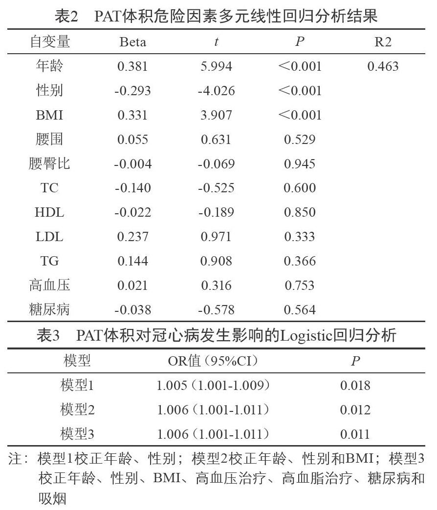
以年龄、性别(男1、女2)、BMI、腰围、腰臀比、TC、HDL、LDL、TG、高血压、糖尿病为自变量,以PAT体积为因变量,进行多元线性回归分析。结果显示年龄,男性,BMI是PAT体积的独立危险因素(P<0.05,见表2),该回归模型的R2为0.463。
2.3 PAT体积对冠心病发生影响的Logistic回归分析
在多因素Logistic回归模型中,以有无冠心病作为二分类因变量,PAT作为自变量带入回归模型。校正年龄、性别后,冠心病患者的PAT体积为未患冠心病人群的1.005倍(95%CI1.001-1.009);校正年龄、性别、BMI后,冠心病患者的PAT体积为未患冠心病人群的1.006倍 (95%CI1.001-1.011);校正年龄、性别、BMI、高血压治疗、高血脂治疗、糖尿病和吸烟等潜在混杂因素后,冠心病患者的PAT体积为未患冠心病人群的1.006倍(95%1.001-1.011),且P<0.05,说明PAT体积是冠心病非常重要的独立相关危险因素。
3 讨 论
本研究发现,年龄、男性、BMI是PAT体积的独立相关危险因素。随着年龄增长,PAT体积呈上升趋势。研究报道显示[15],>70岁的患者与<40岁的患者相比,PAT体积明显增加,分别为252±118 cm2和148±77 cm2。国内王璟[16]等研究结果与此一致。本研究中,男性PAT体积大于女性,这与Framingham心脏研究[17]和Miao[18]等研究结果一致,可能与男性的身高、体重、心脏体积及体表面积较女性大,以及男女脂肪分布的差异性有关[19]。本研究显示PAT体积与BMI呈明显正相关,与Framingham心脏研究[17]基本一致,但Hafakhi[20]等未证实BMI是PAT体积的独立相关危险因素,两个研究结果不一致,可能是Hafakhi等研究样本量不足,共74名受试者,其中男性占68%,平均(54.0±8.0)岁,均为伊拉克人;而本研究选择158名受试者,男性占49.37%,平均(60.66±9.45)岁,与伊拉克研究相比,本研究样本量和受试者年龄大。在相关干预性研究中,有证据表明降低患者的BMI可能有利于减少PAT体积。最近一项澳大利亚为期一年的干预研究[21]发现69名超重或肥胖的心脏病患者在体重减轻后,其PAT体积显著减少。Vasques[22]等报道了68名巴西人在完成减肥手术后1年,伴随着体重减轻,BMI和PAT体积明显下降。因此,高龄、超重或肥胖、男性人群需要更重视PAT的测定,这为进一步研究冠心病、心血管综合征以及其他代谢性疾病提供重要的临床参考价值。
本研究显示,平均PAT体积为(232.23±97.35)cm3。王璟[16]等对310例临床疑有冠心病患者进行64排双源CT测量PAT体积,脂肪组织CT值定为-250~-30Hu,结果显示平均PAT体积为(233.95± 75.34)cm3,这与本研究结果基本一致。相反,一个大群体,多种族研究(MESA)[23]报道华裔美国人最大PAT体积平均值约70 cm3,远远低于本研究结果,可能包括以下两个原因:首先,MESA研究纳入的对象为华裔美国人,而不是生活在中国环境下的中国人;其次,CT扫描时脂肪阈值范围设定不一致。
本研究中另一個重要结果发现PAT体积是冠心病非常重要的独立相关危险因素。PAT为心包外脂肪和心外膜脂肪之和[3],胚胎学上心包脂肪与腹腔内脏脂肪同源,这种PAT和邻近心肌组织的密切解剖学关系,通过由外向内的信号途径,使得它能通过局部旁分泌作用于心肌,提示PAT能够局部调节心脏的功能和形态、改变冠状动脉的平衡[4-5]。此外,PAT还是一个沉积在心脏周围的局部脂肪库,是一个代谢旺盛的器官,可以分泌多种生物活性蛋白,包括脂联素、抵抗素、各种炎症因子和游离脂肪酸,这些细胞因子与粥样硬化和血栓形成有关,从而促使冠状动脉粥样硬化的发生和发展[24-26]。近来国内外多项研究证明PAT体积与冠心病发病相关。2007-2009年国外的Jackson Heart Study(JHS)[27]研究调查了1414名非洲裔美国人,通过多探测器计算机断层扫描测量他们的腹部内脏脂肪组织(VAT)和PAT体积,回归分析结果显示PAT体积是冠状动脉钙化的独立危险因素。一项多民族动脉粥样硬化(MESA)[28]研究发现PAT与冠状动脉钙化的存在和严重程度相关联,且不因种族/民族而异。Nafakhi[20]和Okura[29]等研究结果与此一致,故PAT的测量对临床研究心血管疾病具有重要价值。
由上述可知,年龄、男性、BMI是PAT体积的独立相关危险因素。64排双源CT可对PAT体积进行准确定量,是一个新的预测冠心病发生的危险因素,对冠心病危险监测具有重要意义。但本研究是一个横断面的调查,抽样人群的数量有限,仅来自一家医院。因此,有必要进行更大规模的多中心临床研究,更深入地探讨PAT体积对冠心病事件的影响。
参考文献
[1] 刘笑非.赵莹佳.殷小平.心周脂肪组织与冠心病相关性研究进展[J].医学研究与教育,2017,34(6):53-59.
[2] 孔维敏,杨圣楠,陈玉凤,等.心周脂肪与心血管危险因素相关性研究进展[J].中国热带医学,2016,16(2):190-194.
[3] Ding J,Hsu FC,Harris TB,et al.The association of pericardial fat with incident coronary heart disease: the Multi-Ethnic Study of Atherosclerosis (MESA) [J].American Journal of Clinical Nutrition,2009,90(3):499-504.
[4] Mazurek T,Zhang L,Zalewski A,et al.Human epicardial adipose tissue is a source of inflammatory mediators [J].Circulation,2003,108(20):2460-2466.
[5] Lim S,Meigs JB.Ectopic fat and cardiometabolic and vascular risk [J].Int J Cardiol, 2013, 169(3):166-176.
[6] 陈伟伟,高润霖,刘力生,等.《中国心血管病报告2015》概要[J].中国循环杂志,2016,31(06):521-528.
[7] Greenland P,Knoll MD,Stamler J,et al.Major risk factors as antecedents of fatal and nonfatal coronary heart disease events[J].JAMA,2003,290(7):891-897.
[8] Konishi M,Sugiyama S,Sugamura K,et al.Association of pericardial fat accumulation rather than abdominal obesity with coronary atherosclerotic plaque formation in patients with suspected coronary artery disease [J].Atherosclerosis,2010,209(2):573-578.
[9] Brinkley TE,Hsu FC,Carr JJ,et al.Pericardial fat is associated with carotid stiffness in the Multi-Ethnic Study of Atherosclerosis [J].Nutr Metab Cardiovasc Dis,2011, 21(5):332-338.
[10] AI Chekakie MO,Welles CC,Metoyer R,et al. Pericardial fat is independently associated with human atrial fibrillation [J].J Am Coll Cardiol,2010,56(10):784-788.
[11] Yun CH,Lin TY,Wu YJ,et al.Pericardial and thoracic peri-aortic adipose tissues contribute to systemic inflammation and calcified coronary atherosclerosis independent of body fat composition,anthropometric measures and traditional cardiovascular risks [J].Eur J Radiol, 2012, 81(4):749-756.
[12] 中華医学会糖尿病学分会.中国2型糖尿病防治指南(2013年版)[J].中国糖尿病杂志, 2014,8:2-42.
[13] 国家“十五”攻关“冠心病、脑卒中综合危险度评估及干预方案的研究”课题组.国人缺血性心血管病发病危险的评估方法及简易评估工具的开发研究[J].中华心血管病杂志,2003,12:16-24.
[14] Grundy SM.Metabolic Syndrome Scientific Statement by the American Heart Association and the National Heart,Lung,and Blood Institute [J].Arteriosclerosis Thrombosis and Vascular Biology,2005,25(11):2243.
[15] Greif M,Becker A,von Ziegler F,et al.Pericardial adipose tissue determined by dual source CT is a risk factor for coronary atherosclerosis[J].Arterioscler Thromb Vasc Biol,2009,29(5):781-786.
[16] 王 璟,宫剑滨,张龙江,等.心周脂肪组织体积与传统心血管疾病危险因素相关性研究[J].中国实用内科杂志,2011,31(4):294–297.
[17] Rosito GA,Massaro JM, Hoffmann U,et al.Pericardial fat, visceral abdominal fat, cardiovascular disease risk factors,and vascular calcification in a community-based sample: the Framingham Heart Study[J].Circulation,2008,117(5):605-613.
[18] Miao C,Chen S,Ding J,et al.The association of pericardial fat with coronary artery plaque index at MR imaging:the MultiEthnic Study of Atherosclerosis (MESA) [J].Radiology,2011,261(1):109-115.
[19] Kotani K,Tokunaga K,Fujioka S,et al.Sexual dimorphism of age-related changes in whole-body fat distribution in the obese[J].Int J Obes Relat Metab Disord,1994,18(4):202-207.
[20] Nafakhi H,Al-Mosawi A,Al-Nafakh H,et al.Association of pericardial fat volume with coronary atherosclerotic disease assessed by CT angiography[J].Br J Radiol,2014,87(1038):20130713.
[21] Abed HS,Nelson AJ,Richardson JD,et al.Impact of weight reduction on pericardial adipose tissue and cardiac structure in patients with atrial fibrillation [J].Am Heart J,2015,169(5):655-662.e2.
[22] Vasques AC, Pareja JC, Souza JR,et al.Epicardial and pericardial fat in type 2 diabetics: favorable effects of biliopancreatic diversion [J].Obes.Surg,2015,25(3):477–485.
[23] McClain J, Hsu F, Brown E, et al. Pericardial adipose tissue and coronary artery calcification in the Multi-Ethnic Study of Atherosclerosis (MESA)[J].Obesity,2013,21(5):1056-1063.
[24] Iacobellis G,Pistilli D,Gucciardo M, et al. Adiponectin expression in human epicardial adipose tissue in vivo is lower in patients with coronary artery disease [J].Cytokine,2005,29(6):251-255.
[25] Baker AR,Silva NF, Quinn DW,et al. Human epicardial adipose tissue expresses a pathogenic profile of adipocytokines in patients with cardiovascular disease[J].Cardiovasc Diabetol,2006,5:1.
[26] Marchington JM,Pond CM.Site-specific properties of pericardial and epicardial adipose tissue:the effects of insulin and high-fat feeding on lipogenesis and the incorporation of fatty acids in vitro [J].Int J Obes,1990,14(12):1013-1022.
[27] Liu J,Fox CS,Hickson D,et al.Pericardial Adipose Tissue,Atherosclerosis,and Cardiovascular Disease Risk Factors [J].Diabetes Care,2010,33(7):1635-1639.
[28] McClain J,Hsu F,Brown E.Pericardial Adipose Tissue and Coronary Artery Calcification in the Multi-Ethnic Study of Atherosclerosis (MESA) [J].Obesity,2013,21(5):1056-1063.
[29] Okura K,Maeno K,Okura S.Pericardial fat volume is an independent risk factor for the severity of coronary artery disease in patients with preserved ejection fraction [J].J Cardiol,2015,65(1):37-41.
本文編辑:李 豆

