Clinicopathological characterization of long bone non-union:a prospective cross-sectional study
Avelino Colín-Vázquez,Luis Dario Bernal-Fortich,Joel Galindo-Avalos ,Juan López-Valencia,Rafael Grajales-Ruiz,Adrián Miguel-Pérez,Jorge Quiroz-Williams,Elizabeth Pérez-Hernández
1 Department of Orthopedics,UMAE “Dr.Victorio de la Fuente Narváez” IMSS-UNAM,Ciudad de México,México
2 Department of Orthopedics,Arthroscopic Surgery,Centro de Ortopedia y Medicina del Deporte,Centro Médico Puerta de Hierro,Zapopan,Jalisco,México
3 Osteoarticular Rescue Department,UMAE “Dr.Victorio de la Fuente Narváez” IMSS,México
4 Division Head of the Health Research Division,UMAE “Dr.Victorio de la Fuente Narváez” IMSS,México
5 Division Head of the Health Education and Research Division,UMAE “Dr.Victorio de la Fuente Narváez” IMSS,México
Abstract
Key words:bone non-union; pathohistology; atrophy; hypertrophy; long bone; cross-sectional study
INTRODUCTION
Epidemiology
Non-union(pseudarthrosis)is defined as the formation of a false joint where afibrocartilaginous cavity is lined with synovium producing synovial fluid.Non-union of the fracture is defined as the cessation of all reparative processes of healing without bone union.Unless there is bone loss,a nonunion is usually declared between 6 and 8 months following the fracture.1
The incidence of non-union ranges from 5-10% of all fractures.2Approximately 53% of non-union occurs in the lower limbs,the tibia being the most affected bone,followed by the femur,3,4then the humerus,the bones of the forearm and the clavicle.5Risk factors of non-union include high energy trauma,open fractures,multifragmentation,bone loss,postreduction instability,diabetes,obesity,alcoholism,peripheral vascular disease and scleroderma.6,7
Revision surgery with bone grafts,usually from the iliac crest,8is currently the best way to stimulate bone regeneration.However,several approaches have been described to promote and improve bone tissue regeneration,such as extracorporeal shockwave therapy(ESWT),ultrasound,bone morphogenic proteins(BMPs)and platelet-rich plasma(PRP).9
Cellular repair process in the bone tissue
Whenever there is a fracture,direct bone healing occurs only with absolute stability and is a biological process of osteonal bone remodeling.10,11Indirect bone healing depends on the formation offibrocartilaginous calluses and occurs in most of the cases.10This type of bone healing follows a specific biological pathway.It involves an acute in flammatory response,the recruitment of mesenchymal stem cells to generate a primary cartilaginous callus,which later undergoes revascularization and calcification,and isfinally remodeled to fully restore a normal bone structure.10-12
Macroscopic and microscopic structure
In a histological study on non-union tissue,it was found that tissue vessels often appear occluded by thrombotic material,concluding that the cells that populate non-union tissue can induce mineralization of the cell matrix,but have an insufficient blood supply to provide them with a normal amount of calcium,which is the real cause for non-union development.13Fibrocartilaginous tissue that contains occasional bony islands has been found in atrophic non-union; in hypertrophic nonunion,areas of new bone formation by both endochondral and intramembranous ossification have been observed.13,14The exact biological process that leads to a non-union remains obscure and it is well accepted that any intervention to reverse this process must be timely and well directed to re-establish both biological and mechanical deficiencies.
Objective
The purpose of the present work was to identify the possible morphological patterns of bone tissue in a state of non-union and in the presence or absence of infection.
SUBJECTS AND METHODS
Study design
This prospective,observational,cross-sectional study was conducted at Hospital de Ortopedia UMAE “Dr.Victorio de la Fuente Narváez” from May 2017 to February 2018.All procedures involving human participants were in accordance with the ethical standards of Comité Local de Investigación y Ética en Investigación en Salud at UMAE “Dr.Victorio de la Fuente Narváez”(R-2017-3401-8)on July 19,2017(Additionalfile 1)and with the 1964Helsinki Declarationand its later amendments or comparable ethical standards.This study followed the Strengthening the Reporting of Observational Studies in Epidemiology(STROBE)statement(Additionalfile 2).
Bone tissue sample harvesting
Pathological bone tissue(in a state of non-union diagnosed radiographically)was obtained from patients who underwent revision surgery in our hospital after they had provided written informed consent(Additionalfile 3).The conditions of aseptic and septic non-union were determined by clinical and laboratory means(biopsy culture).A total of 34 samples of bone tissue were collected under such conditions.To avoid any disruption in tissue morphology,the samples were collected using an osteotome instead of a bone saw.
Fixation methods
The samples werefixed in 10% neutral buffered formalin,then decalcified with 7% nitric acid and neutralized.Afterwards,the excess chemical was washed off,and samples were dehydrated by immersing tissue in a series of ethanol solutions of increasing concentrations until 100% water-free alcohol was reached.Then ethanol was replaced with xylol and the samples were immersed in liquid paraffin to form blocks.The blocks were then cut into 3 μm-thick sections.The tissue sections were then mounted on a glass microscope slide and treated with a conventional hematoxylin-eosin stain.Subsequently,they were covered with epoxy resin for visualization using conventional optical microscopy(Nikon Instruements Inc.,Melville,NY,USA).
Histopathological observation and semiquantitative evaluation
Histopathological changes of bone tissue sample were observed and then descriptive and semiquantitative evaluation was performed.
RESULTS
Baseline data
Thirty-four bone tissue samples were obtained from 34 patients aged 40(range 23-57 years)years.The male to female ratio was 2.4:1.The mean time from clinical diagnosis to surgery was 17(range 9-36 months)months.Among these patients,26 were classified as aseptic(76.5%)and 8 as septic(23.5%).In the septic non-union,men were more affected than women with a 7:1 ratio.
Bone segments
The most affected bone segments were the tibia and femur(32.3%),followed by the humerus(20.5%),the radius and the ulna(11.7%),andfinally the clavicle(3.2%).The femur was the most commonly affected in aseptic non-union and the tibia in septic non-union.
Bacteria
The most common infectious agents associated with septic non-union wereStaphylococcus aureusin 75% of the cases,andEscherichia coliin 25% of the cases.
Clinicopathological classification
Regarding the clinicopathological evolution,we found 24 patients with oligotrophic non-union(70.6%),6 patients atrophic non-union(17.6%),and 4 patients with hypertrophic non-union(11.8%).A higher incidence of infection was found in oligotrophic non-union(6 patients),followed by atrophic non-union(2 patients).Hypertrophic non-union was not found in any patient.
Disease severity
Regarding the severity of the disease,11 patients were classified as Paley A(32.4%),that is,with a bone defect of less than 1 cm; and 23 patients as Paley B(67.6%),that is,with a bone defect bigger than 1 cm.From the cases of septic non-union,7 of them were classified as Paley B(87.5%),and only 1 as Paley A(12.5%).
Microscopic description
The histological sections evaluated showed heterogeneity of the morphological patterns,with variability in the proportion,however,no specific statistical significance was observed for the types of non-union.These characteristics are summarized in Table 1.
Cell population was characterized byfibroblasts of normal morphology,arranged in short beams,with the formation of nuclear palisades and with variable proportions of extracellular matrix(collagen),including densely collagenized,hyalinized areas(Figure 1A).
Two types of cartilage,reparativefibrocartilage,and hyaline cartilage were identified(Figure 1B-E).Thefibrous cartilage was mostly associated with the hypertrophic variety of non-union,as a part of the exuberant bone callus.Likewise,cystic degenerative changes were identified,especially in the oligotrophic type.The presence of hyaline cartilage arranged in islands or lobes between thefibrous tissues was observed in the atrophic as well as in the hypertrophic forms,however,in the latter,the arrangement of the chondrocytes and their morphological characteristics simulate the hypertrophic cartilage of the growth plate.
New bone formation was a common feature in all types of non-union,either from endochondral ossification(predominantly in the atrophic and oligotrophic varieties)and/or from areas of intramembranous ossification(more frequent in the hypertrophic form)(Figure 1F-G).
Areas of bone necrosis were more prevalent in the atrophic and oligotrophic forms.These changes were also observed in addition to fragmentation of bone spicules and identification of osteosclerosis lines(Figure 1H and I).

Table 1:Histomorphological comparison of the types of non-union

Figure 1:General histological characteristics(hematoxylin-eosin staining).
Osteoblastic activity was observed in some samples in both the oligotrophic and hypertrophic forms,whereas the osteoclastic activity was predominantly identified in the atrophic form(Figure 2A and B).
Vascularization was observed in different types of nonunion,with minimal variations in the vascular proportion(Figure 2C and D),and in some samples,it was associated with lymphoplasmacytic in flammatory cell aggregates,with diffuse and perivascular disposition,as well as with areas of edema and proliferation of connective tissuefibers(Figure 2E-G).
Another change associated with the hypertrophic form was synovial non-union characterized by vascularizedfibrotic tissue with cell proliferation similar to synoviocytes and pseudomembrane formation(Figure 2H and I).
The septic and aseptic forms of non-union also showed morphological heterogeneity and variable proportions of morphological patterns.These characteristics are summarized in Table 2 and Figure 3A-F.

Figure 2:Histological characteristics of hypertrophic and oligotrophic non-union(hematoxylin-eosin staining).

Figure 3:Histological characteristics of septic and aseptic non-union(hematoxylin-eosin staining).

Table 2:Morphological comparison of aseptic and septic non-union
DISCUSSION
Non-union or pseudoarthrosis represents a public health problem with adverse consequences in a patient's quality of life.The mechanisms that lead to non-union are multifactorial and therefore the treatment has evolved since the prolonged immobilizations in the 1950's.15-20
This study has some limitations,the most important of them being that,because of the small sample size,a histopathological classification could not be reached,which is the primary objective of this study.
Few articles in the scientific literature examine the global characterization of non-union that includes radiographic,clinical and histological aspects,likewise,the current classification systems do not include histopathologicalfindings,which could be determinant for treatment of the disease,together with the biomolecular aspects.
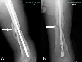
As described in the literature,the Weber-Cech classification includes both radiographic observations andfixation stability.Based on this characterization,different types of non-union are recognized:hypertrophic(Figure 4),where there is adequate curative potential due to abundant callus formation and it is associated with hypervascularity; oligotrophic(Figure 5),which is vascularized but with minimal callus formation;and atrophic(Figure 6),in which there is absence of callus formation and no bone vascularity.1

Figure 4:X-ray lateral(A)and anteroposterior(B)views of the left femur.

Figure 5:X-ray anteroposterior(A)and lateral(B)views of the right humerus.

Figure 6:X-ray lateral(A)and anteroposterior(B)views of the left knee.
In this study,the histopathological characteristics of different forms of non-union were described,from the atrophic,oligotrophic to hypertrophic variants.As observed,there is a broad,heterogeneous morphological spectrum,which correlated proportionally with the formation of scarce to exuberant bone callus,however,histological patterns characteristic of a specific subtype were not identified,which leads to difficulty in the integration of scales or associated morphological patterns.
However,variable proportions of cellularity,as well as the components of the extracellular matrix,participate in the phenomenon of non-union.In this regard,as mentioned in some publications,the elements of the connective tissue can be targeted by growth factors to stimulate bone consolidation effectively and in a controlled manner.The use ofin vitrobiological models,such as cultures and cell cocultures,construction of monolayers,use of stimulating and inhibiting factors during the healing process,etc.,are still in research.
Likewise,the in fluence of concomitant factors and demographic variables,such as comorbidities,especially in populations like ours,are also the research areas of this study.However,the association with infectious processes,the type of etiological agents and their mechanisms of antimicrobial resistance,etc.,are also important aspects to be considered in bone consolidation.They are emphasized in the education of the personnel that intervenes in the healthcare process and in the education to the community to prevent associated complications.
To conclude,long bone non-union is currently one of the major orthopedic challenges.This is due not only to the complexity of the disease but also to its devastating effects when it is not treated effectively and timely.
There is heterogeneity in the histopathological characterization of non-union,which correlates proportionally with the formation of bone callus.However,it is not possible to integrate specific morphological patterns for classification purposes.
The association of non-union with an infectious process(septic non-union)showed a higher degree of tissue lysis,which clinically delays and interferes with the process of bone consolidation.
Additionalfiles
Additionalfile 1:Ethics committee approval.
Additionalfile 2:STROBE checklist.
Additionalfile 3:Model consent form.
Author contributions
Study design,manuscript drafting and English translation:ACV,LDBF,JGA,and JLV; sample collection:AMP and RGR; sample processing and biostatistics review:JQW and EPH.All authors approved thefinal version of this manuscript.
Conflicts of interest
None declared.
Financial support
None.
Institutional review board statement
All procedures performed in studies involving human participants were in accordance with the ethical standards of Comité Local de Investigación y Ética en Investigación en Salud at UMAE “Dr.Victorio de la Fuente Narváez”(R-2017-3401-8)on July 19,2017 and with the 1964Helsinki Declarationand its later amendments or comparable ethical standards.
Declaration of patient consent
The authors certify that they have obtained all appropriate patient consent forms.In the form the patients have given their consent for their images and other clinical information to be reported in the journal.The patients understand that their names and initials will not be published and due efforts would be made to conceal their identity.
Reporting statement
This study followed the STrengthening the Reporting of OBservational studies in Epidemiology(STROBE)statement.
Biostatistics statement
The statistical methods of this study were reviewed by Dr.Pérez-Hernández and Dr.Quiroz-Williams at UMAE “Dr.Victorio de la Fuente Narváez” IMSS,México.
Copyright license agreement
The Copyright License Agreement has been signed by all authors before publication.
Data sharing statement
For data sharing,individual participant data will not be available.
Plagiarism check
Checked twice by iThenticate.
Peer review
Externally peer reviewed.
Open access statement
This is an open access journal,and articles are distributed under the terms of the Creative Commons Attribution-NonCommercial-ShareAlike 4.0 License,which allows others to remix,tweak,and build upon the work non-commercially,as long as appropriate credit is given and the new creations are licensed under the identical terms.
 Clinical Trials in Orthopedic Disorder2018年4期
- Clinical Trials in Orthopedic Disorder的其它文章
- Ultrasound-guided supine lumbar plexus block versus iliac fascia block for analgesia in older adult patients undergoing hip replacement:a randomized controlled trial
- Efficacy of arthroscopic surgery for discoid lateral meniscus injury in knee joint:a self-control study
- Local anesthetic infiltration before open reduction and internalfixation for ankle fracture:a single-blind randomized controlled study
- Efficiency of tight monitoring by nurse practitioners in rheumatoid arthritis patients in remission after treatment with rituximab:study protocol for a randomized,open-label,controlled trial
- Hemostasis following local versus intravenous tranexamic acid in patients undergoing posterior open reduction and internalfixation of thoracolumbar fractures:study protocol for a parallel-group,randomized controlled trial
- Efficacy of silver needle acupuncture combined with muscle relaxation in the treatment of capulohumeral periarthritis:study protocol for a prospective,single-center,randomized,parallel-controlled trial
