
Comparative foliar anatomy of three Khaya species(Meliaceae) used in Nigeria as antisickling agent
2018-09-07 06:46:26OloladeOyedapoJosephAgbedahunsiIllohAkinwumiAkinloye
Ololade A. Oyedapo , Joseph M. Agbedahunsi , H. C Illoh , Akinwumi J. Akinloye
1. Drug Research and Production Unit, Faculty of Pharmacy, Obafemi Awolowo University, Ile Ife, Nigeria
2. Department of Botany, Obafemi Awolowo University, Ile Ife, Nigeria
ABSTRACT Khaya belongs to the family Meliaceae. In Nigeria the genus is represented by three species viz; K. senegalensis A. Juss.,K. grandifoliola C. DC. and K. ivorensis A. Chev. Comparative foliar anatomy of the three Khaya species was carried out to identify and describe distinctive anatomical characters that could possibly be used to delimit the three taxa. Transverse section, epidermal peels and cleared leaves of these three species were made. Characteristic similarity and disparity in the tissues arrangement as well as cell inclusions were noted for description and delimitation. The three Khaya species studied had essentially the same anatomical features, e.g., venation pattern having open polygonal areoles and the veins terminals biforkated. However, there were characters that seem to be species specific, e.g., vien termination number and areole width. The leaf epidermal studies of the three species revealed similarities in stomatal type which are generally staurocytic,epidermal cells and undulating anticlinal cell walls but stomata density varied. Hexacytic stomata is only observed in the abaxial surface of K. grandifoliola which distinguished this species from the others. The leaf petiole shape of the three species are round and difficult to distinguish into adaxial and abaxial surfaces. The cuticle is striated, vascular bundles are heart shape, conjoint, concentric and amphivasal, but are different in epidermal and collenchyma cell layer numbers. The leaf transverse sections of the three Khaya species studied have conjoint, concentric and amphicribral bundles while the leaf cuticle of K. senegalensis and K. grandifoliola are striated but that of Khaya ivorensis is non-striated.
Keywords: Khaya; foliar; anatomy; epidermal; stomata; vascular
1 Introduction
Khaya is a genus of six species of trees in the family Meliaceae (common name mahogany), native to tropical Africa and Madagascar. It is also found along the Western Africa sub-region. It is found in Nigeria,Angola, Cameroon, Cote d, ivoire, Gabon, Ghana and Liberia where it grows primarily in lowland tropical rainforest. The three species found in Nigeria are Khaya senegalensis A. Juss., Khaya grandifoliola C.DC. and Khaya ivorensis A. Chev. It is called Oganwo by the Yoruba speaking people of southwestern Nigeria (Arbonier, 2004). In spite of the plants being morphologically different they are all commonly called African mahogany. They are used to treat different types of diseases. Earlier research by Awe et al.(1991) ranked the anti-plasmodial activities of the three species as K. ivorensis>K. grandifoliola>K. senegalensis. Fall et al. (1999) reported the antisickling activity of K. senegalensis.
Odugbemi et al. (2007) and Olowokudejo et al.(2008) presented a series of ethnomedicinal uses of Khaya species. Khaya seeds, leaves and bark when made into a concoction are used for the treatment of headache and convulsion. The bitter bark decoction is used for cough and whooping cough, stomach ache,fever, threatened abortion, as a lotion for rheumatism,dermatomycosis and malaria fever in Nigeria. When mixed with black pepper corn it is used to treat diarrhea and dysentery. Bark decoction is used as a drink or bath for back pains (Odugbemi et al., 2007;Olowokudejo et al., 2008).
WAC (2004) also presented a series of ethnomedicinal uses of Khaya species; the very bitter bark has a considerable reputation in its natural range as a fever remedy. The bark is also used as a vermifuge, taenicide, depurative and for treating syphilis. Bark extract is used for treating jaundice, dermatoses, scorpion bite, allergies, infection of the gums, hookworm,bleeding wounds (disinfectant), and as a laxative.Seeds and leaves are used for treating fever, headache; roots against sterility, for the treatment of mental illness, against syphilis, leprosy and as an aphrodisiac. Crushed stem bark and seeds are regarded as emmenagogue. Bark can also be used in traditional veterinary practice, e.g., for cattle suffering from liver fluke, for ulcers in camels, donkeys and horses, and in horses for internal ailments associated with mucous diarrhea (WAC, 2004).
The lower strata of the population living in developing countries rely heavily on traditional medicine due to their cultural alignment as well as their inability to afford the cost of treatment offered by orthodox medical practitioners. Considering the fact that medicinal plants have been a source of succor in the management of many diseases in developing countries,Khaya species is no exception especially in the management of malaria and sickle cell diseases. Traditional medical practitioners found it difficult to identify these three species from one another because phenotypically they look alike, they have the same common name (Mahogany) and the Yoruba speaking population in Nigeria call all three species Oganwo. It is therefore expedient to determine the micro morphological similarities and differences among the three species for ease of identification by traditional medical practitioners using the leaves of K. senegalensis,K. grandifoliola and K. ivorensis.
2 Materials and methods
2.1 Collection and preparation of plant samples
Fresh leaf samples of the three Khaya species were collected; K. senegalensis from the Botanical Garden (7°31'20.42''N, 4°31'20.93"E) of Obafemi Awolowo University, Ile-Ife, Osun State, K. grandifoliola from Abata Egba village (7°19'11.18''N,4°36'04.89"E), Osun State and K. ivorensis from Oke Ado (Okomu Forest Reserves) (6°20'00''N,5°16'00"E), in Ovia Local Government, Okada, Edo State. The samples were identified and authenticated by the curator at the IFE herbarium located in the Department of Botany, Obafemi Awolowo University,Ile-Ife where voucher specimens were deposited of K.senegalensis (IFH No. 16289), K. grandifoliola (IFH No. 16289) and K. ivorensis (IFH No. 16343). The collected samples were preserved in 50% ethanol(Akinloye et al., 2015).
2.2 Sectioning
Sections were acquired from mature leaves of K.senegalensis, K. grandifoliola and K. ivorensis.Transverse section (TS) of the leaf and petiole of each plant were cut at 10 micron thickness using a sledge microtome (Reichert, Austria) (Akinloye et al., 2015).
2.3 Leaf peeling
Mature leaves were cut into sizeable portions and kept in concentrated nitric acid until the surfaces become swollen under sunlight. Pieces of leaves with swollen surfaces were transferred into water in a glass petri dish. The swollen surfaces were peeled off using fine tip forceps. The peels were preserved in 50%ethanol (Akinloye et al., 2015).
2.4 Leaf clearing
Matured leaves were cut into sizeable portions and boiled in absolute ethanol for forty minutes, rinsed in water twice and soaked in 5% sodium hydroxide for 16 hours. After rinsed in water twice, they were transferred into 5% domestic bleach (JIK). They remained in the bleach until the whole leaves became completely white. They were then rinsed in water thrice and preserved in 50% ethanol (Akinloye et al., 2015).
2.5 Staining and microscopic observation of sections, peels and clear leaves
Leaf and petiole sections were stained for five minutes in Safranin O, rinsed in water twice and counter stained for five minutes in Alcian blue and again rinsed in water twice. The sections were treated thereafter in a series of graded ethanol solution (50%,70%, 80%, 90%, and 100%) to remove water and excess stain. The sections were transferred into absolute xylene in two series to remove the last trace of water,to clear the sections (making it more transparent) and to remove the last trace of ethanol. Each section was then mounted on a glass slide in DPX(R) Mountant(Akinloye et al., 2015).
Epidermal peels and cleared leaves of the Khaya species were stained for five minutes in Safranin O.They were rinsed thrice in water and mounted in 25%glycerol on a glass slide and covered with a glass cover slip, whose edges were sealed with nail polish. Microscopic observation of each slide was made and recorded. Photo micrographs of the slides were made using an Accu-scope trinocular microscope(ACCU-scope 3001 LED Trinocular microscope with 3.2 MP CMOS digital camera). Tissue and cell identification and description were performed according to Esau (1977), Bilgramiks et al. (1982), and Metcalfe and Chalk (1989).
3 Results
3.1 Leaf venation
All three species have multiple open areoles that are polygonal, with bi-forked terminal veins (Table 1,Figures 1a-1c). Vein islets are lacking. The vein terminals of K. senegalensi, K. grandifoliola and K.ivorensis are 2-50 (plate 15a-15c). Areole length in the three species varied between 0.011-0.165 µm,while average length varied between 0.058-0.078 µm.Areole width varied between 0.007-0.091 µm, while average width varied between 0.035-0.045 µm (Table 1).

Figure 1 Venation patterns of Khaya species showing multiple areoles and areole shape. (a) K. senegalensis, (b) K. grandifoliola,(c) K. ivorensis (VT: veinlet termination, BFT: bi-forked terminals, ARL: areoles, PGS: polygonal shape, MOA: multiple areoles)
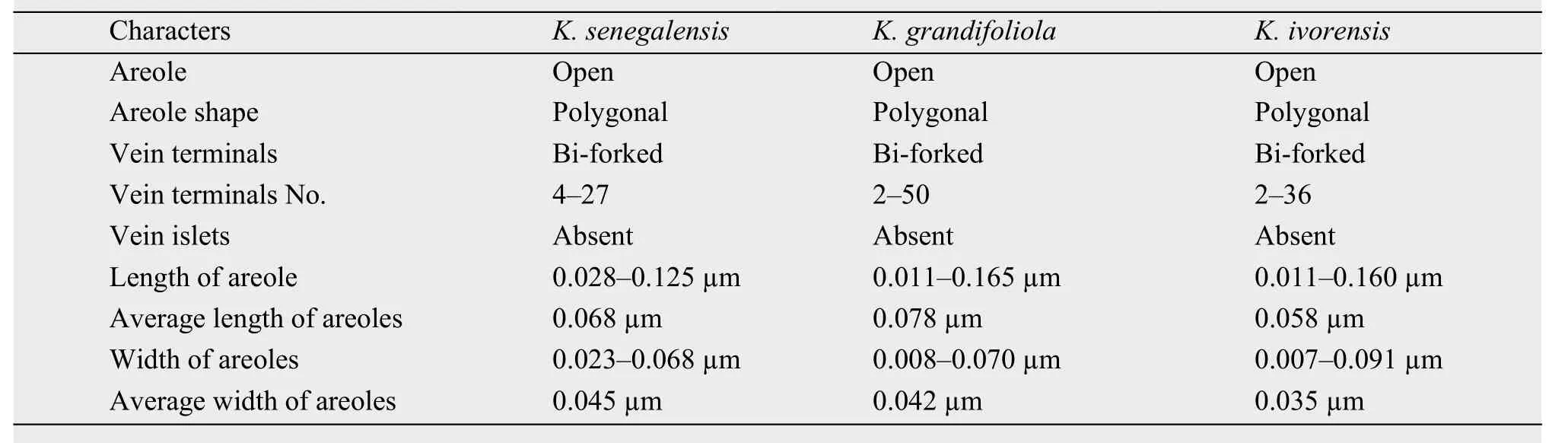
Table 1 Comparative architectural features of leaves from three Khaya species
3.2 Leaf epidermal studies
The epidermal cell shape of Khaya species generally varies from polygonal, rectangular to triangular for adaxial and abaxial surfaces. The anticlinal walls of the three species are straight and undulating on both surfaces (Figures 2 and 3). Stomata are present in the three species on both the adaxial and abaxial surfaces (amphistomatic). Stomata present on the adaxial surfaces of K. senegalensis and K. ivorensis(hypoamphistomatic) are scanty when compared with that of K. grandifoliola (epiamphistomatic). Stomata type is generally staurocytic on both surfaces but on the abaxial surface of K. grandifoliola are hexacytic(Table 3). Stomata density on the adaxial surface of K. senegalensis is 0.3 stomata/mm2, K. grandifoliola 3 stomata/mm2and K. ivorensis 0.2 stomata/mm2. On the abaxial surfaces of the three species, stomata density is as follows: K. senegalensis 2 stomata/mm2,K. grandifoliola 3 stomata/mm2and K. ivorensis 4 stomata/mm2. K. ivorensis has the highest stomata density on the abaxial surface while K. senegalensis has the least. Crystal druses are present within the epidermal cells on both surfaces (Tables 2 and 3).
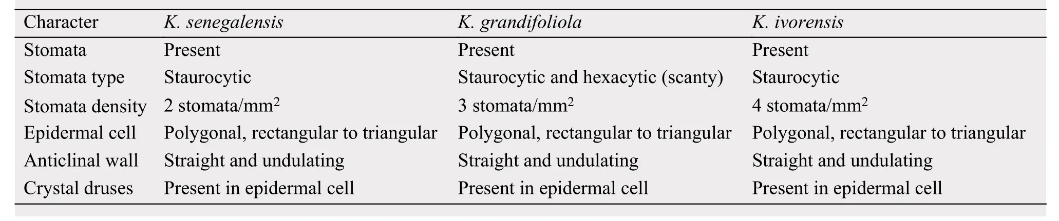
Table 3 Leaf abaxial epidermal and epidermal appendage features of three Khaya species
3.3 Leaf transverse section
The cuticle of K. senegalensis and K. grandifoliola are striated while that of K. ivorensis is non-striated. Adaxial epidermis of the three species is a single layer, and epidermal cell shape varies from circular,rectangular, to oval. The adaxial epidermis is thicker than the abaxial epidermal layer. The mesophyll is distinctive, divided into palisade and spongy layers.The palisade layer is made up of tightly packed elongated, cylindrical shape parenchyma cells which vary from 1-2 layers. The spongy mesophyll varies in shape from short cylindrical, circular, oval, to polygonal, with loosely packed parenchyma cells. Crystal druses are present in the mesophyll within the parenchyma cells. Vascular bundle is conjoint, concentric and amphicribral.
4 Discussion
Foliar venation has been used by Levin (1986) to provide insight into relationships within the subfamily Phyllathoideae, and also in the tribe Euphobiceae by Seghal and Paliwal (1974). Venation pattern is useful in delimiting the species of Khaya considering the variations in the number of vein terminals, average length and areole width.
Vein terminals of the three species varies, K. senegalensis is 4-27, K. grandifoliola 2-50, and that of K.ivorensis 2-36. Areole length varies, K. senegalensis is between ±0.028 and ±0.125 µm, K. grandifoliola±0.011 to ±0.165 µm, and K. ivorensis ±0.011 to±0.160 µm. Average areole length for K. senegalensis is ±0.068 µm, K. grandifoliola ±0.078 µm, and K.ivorensis ± 0.058 µm. Areole width varies, K. senegalensis is between ±0.023 and ±0.068 µm, K. grandifoliola ±0.008 to ±0.070 µm, and K. ivorensis ±0.007 to±0.091 µm. Average areole width for K. senegalensis is ±0.045 µm, K. grandifoliola ±0.042 µm, and K.ivorensis ±0.035 µm (Table 1). Therefore, the number of vein terminals, length and width of areoles can be used as a diagnostic character to differentiate the three Khaya species studied.
The epidermal cell shape of Khaya species generally varies from polygonal, rectangular to triangular on adaxial and abaxial surfaces. Stomata present on the adaxial surfaces of K. senegalensis and K. ivorensis are scanty when compared with that of K. grandifoliola. Stomata type is generally staurocytic at both surfaces but at the abaxial surface of K. grandifoliola hexacytic stoma is present which serve as a diagnostic feature that differentiate it from the other two species viz K. senegalensis and K. ivorensis. Stomata density on the adaxial surface of K. senegalensis is 0.3 stomata/mm2, K. grandifoliola 3 stomata/mm2,and K. ivorensis 0.2 stomata/mm2. On the abaxial surfaces of the three species, stomata density is as follows: K. senegalensis 2 stomata/mm2, K. grandifoliola 3 stomata/mm2, and K. ivorensis 4 stomata/mm2.K. ivorensis has the highest stomata density on the abaxial surface while K. senegalensis has the least.Isawumi (1986) suggested in his research on epidermal studies in the specie of Jatropha L. (Euphorbi-aceae) has also established the use of stomata frequency and density in delineating taxa. Ergastic substances are present on both surfaces; crystal druses are present within the cells on both surfaces (Tables 2 and 3). Therefore, the presence of hexacytic stoma on the abaxial surface of K. grandifoliola and the stomata density of the three Khaya species study can be used in their identification.
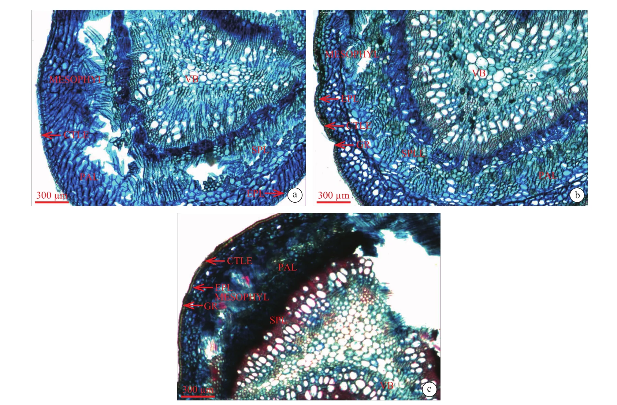
Figure 4 Leaf anatomy in mid-transverse section of Khaya species. (a) K. senegalensis,(b) K. grandifoliola, (c) K. ivorensis (EPL: epidermal layer, MEL: mesophyll layer,VB: vascular bundle, PAL: palisade layer, SPL: spongyl layer)
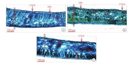
Figure 5 Transverse section showing leaf lamina. (a) K. senegalensis, (b) K. grandifoliola, (c) K. ivorensis (EPL: epidermal layer,MEL: mesophyll layer, VB: vascular bundle, PAL: palisade layer, SPL: spongyl layer, CTLE: cuticle, GR: grove)
Leaf anatomy according to Carliquist (1961)provides a variety of features that could be used for taxonomic purposes. Many researchers have utilized leaf anatomy for solving taxonomic problems in many species of plants, for example Illoh (1995) on genus Celosia, Adedeji (2004) on Emilia as well as Adedeji and Illoh (2004) on Hibiscus. Though there is uniformity in the leaf anatomical characters like palisade shape and spongy mesophyll cells and types of vascular bundles which possibly point to the generic traits in these species, however, variations in cuticular striation could be utilized in delimiting the members of this genus as observed in this study. This study revealed that the cuticle of two of the species, K. senegalensis and K. grandifoliola are striated while that of K. ivorensis is non-striated. The adaxial epidermis is a single layer in the three species having various cell shapes, e.g., circular, rectangular, oval, and are thicker than the abaxial epidermal layer.
5 Conclusion
From the study of the leaf anatomy of K. senegalensis, K. grandifoliola and K. ivorensis, it can be concluded that the three Khaya species present some similar micro morphology, however there are some differences in the features presented. The number of vein terminals, average length and width of areoles varies in the three species studied and are of useful taxonomic tools for their delimitation.
The stomata frequency and density on the adaxial and abaxial surfaces also varies in the three species and can be used in delineating the species. Khaya grandifoliola presented some features that distinguished it from K. senegalensis and K. ivorensis in the presence of hexacytic stomata in its leaf which is not present in the other two species.
There is also variation in the cuticular striations of the three species especially for K. ivorensis whose cuticle is non-striated while the cuticle of K. senegalensis and K. grandifoliola are striated.
 Sciences in Cold and Arid Regions2018年4期
- Sciences in Cold and Arid Regions的其它文章
- International Workshop on Cryospheric Changes and Their Regional& Global Impacts Successfully Held in Gansu Province
- Cultivated-land change in Mu Us Sandy Land of China before and after the first-stage grain-for-green policy
- Comparisons of plant calcium fraction between two different vegetation zones in semi-arid region
- Effects of N:P ratio of Artemisia ordosica on growth influenced by soil calcium carbonate
- Seasonal dynamics of N:P ratio stoichiometry and Ca fraction for four dominant plants in the Alxa Desert
- Change in summer daily precipitation and its relation with air temperature in Northwest China during 1957-2016