Kaempferol attenuates cognitive deficit via regulating oxidative stress and neuroinflammation in an ovariectomized rat model of sporadic dementia
Somayeh Kouhestani , Adele Jafari, Parvin Babaei ,
1 Cellular and Molecular Research Center, Faculty of Medicine, Guilan University of Medical Sciences, Rasht, Iran
2 Department of Physiology, Faculty of Medicine, Guilan University of Medical Sciences, Rasht, Iran
3 Neuroscience Research Center, Guilan University of Medical Sciences, Rasht, Iran
Funding: This study was supported by a grant from Research and Technology Chancellor of Guilan University of Medical Sciences, Iran (No.IR.GUMS. REC. 1936.51).
Abstract Alzheimer’s disease (AD) is associated with oxidative stress, and ultimately results in cognitive deficit. Despite existing literature on the pathophysiology of AD, there is currently no cure for AD. The present study investigated the effects of kaempferol (Kmp) isolated from the extract of Mespilus germanica L. (medlar)leaves on cognitive impairment, hippocampal antioxidants, apoptosis, lipid peroxidation and neuro-inflammation markers in ovariectomized (OVX) rat models of sporadic AD. Kaempferol, as the main flavonoid of medlar extract has been previously known for anti-oxidative, anti-inflammatory and anti-neurotoxic effects.Thirty-two female Wistar rats were ovariectomized, and randomly divided into four groups: sham, OVX +saline, OVX + streptozotocin (STZ) + saline, OVX + STZ + Kmp. Animals received intracerebroventricular injection of STZ (3 mg/kg, twice with one day interval) to establish models of sporadic AD. Intraperitoneal injection of Kmp (10 mg/kg) for 21 days was performed in the OVX + STZ + Kmp group. Spatial learning and memory of rats were evaluated using a Morris water maze. Finally, brain homogenates were used for biochemical analysis by enzyme-linked immunosorbent assay. The results showed a significant improvement in spatial learning and memory as evidenced by shortened escape latency and searching distance in Morris water maze in the OVX + STZ + Kmp group compared with the OVX + STZ group. Kmp also exhibited significant elevations in brain levels of antioxidant enzymes of superoxide dismutase and glutathione, while reduction in tumor necrosis factor-α and malondialdehyde. Our results demonstrate that Kmp is capable of alleviating STZ-induced memory impairment in OVX rats, probably by elevating endogenous hippocampal antioxidants of superoxide dismutase and glutathione, and reducing neuroinflammation. This study suggests that Kmp may be a potential neuroprotective agent against cognitive deficit in AD.
Key Words: Alzheimer’s disease; oxidative stress; neuroinflammation; Mespilus germanica L.; Kaempferol;neural regeneration
Introduction
Alzheimer’s disease (AD) is a complex neurodegenerative disorder with cognitive and behavioral disturbances that remains the most protracted disease. In 2015, 46.8 million people worldwide lived with AD, and this population will reach 131.8 million by the end of 2050 (Aliev et al., 2016). AD is characterized by the accumulation of senile plaques and neurofibrillary tangles in the brain (Kamat et al., 2014). Senile plaques consist of protein amyloid beta (Aβ), microglia and pro-inflammatory cytokines,particularly tumor necrosis factor-α (TNF-α) (Kar et al., 2004;Batarseh et al., 2016). The most recent studies confirmed the influence of reactive oxygen species (ROS) on the development of different disorders, including AD. Oxidative stress leads to reduction in glutathione (GSH) content, which is one of the important endogenous antioxidants capable to scavenge reactive oxygen species (Wojtunik-Kulesza et al., 2016). Therefore,oxidative stress leads to lipid peroxidation and formation of malondialdehyde (MDA) (Reyazuddin et al., 2016) and TNF-α(Kumar et al., 2017). Finally, production of ROS triggers the release of cytochrome c from mitochondria, and the activation of caspase-3 and apoptosis (Redza-Dutordoira and Averill-Batesa,2016).
There is currently no definite treatment for AD, because the signaling pathways of AD are complex and definite initial causative factors is unknown. The existing therapies improve some behavioral symptoms, but hardly mitigate cognitive deficit. Moreover, at least half of AD patients do not respond to current medications (Farlow et al., 2008). Therefore, it is of great importance to find reagents capable of combating cognitive deficit in AD. Targeting oxidative stress and increasing antioxidant defenses of the brain would be an important strategy for maintaining survival of brain structure.
Recently, there has been an upsurge of interest in the therapeutic potential of flavonoids, a group of natural compounds in plants (Kumar and Pandey, 2013). Flavonoids as free radical scavengers and inhibitors of lipid peroxidation, are capable of passing the blood-brain barrier (Youdim et al., 2004; Banjarnahor and Artanti, 2014), and are involved in synaptic plasticity(Spencer, 2010). Considering the fact that the brain is rich in peroxidizable fatty acids, elevation in brain free radicals nega-tively influences physiological functions. Consequently, flavonoids are important in the prevention of neuronal damages by balancing oxidative and anti-oxidative status.
Vegetables and fruits are two important flavonoids which are rich in diet sources. For example, medlar (Mespilus germanica L.) is a small tree from the Rosaceae family which has been cultivated for many years in Europe, Turkey, and Iran;and it consists of bioactive antioxidant compounds of flavonoids (Ercisli et al., 2012 ). Our recent study has demonstrated that alcoholic medlar leaves extract consisted of Kaempferol(Kmp), chrysin, luteolin, myricetin, naringenin, quercetin and rutin, and administration of extract increased passive avoidance learning in rat models of metabolic syndrome (Kouhestani et al., 2017).
Kmp is an anti-inflammatory compound which has the potent iron-chelating capability and ROS scavenging property(Kim et al., 2012). In vitro assays showed protective effects of Kmp on oxidative stress-induced cytotoxicity in PC12 cells(Kim et al., 2010). In addition, Kmp administration in parkinsonian mouse models improved motor coordination, raised striatal dopamine level, and increased superoxide dismutase(SOD) activity (Li and Pu, 2011).
Considering above-mentioned results from the previous studies, we assumed that Kmp might have the capability to alleviate cognitive deficit in the animal models of sporadic AD. Therefore, this study was designed to evaluate the effect of Kmp on spatial learning and memory, endogenous antioxidants SOD and glutathione (GSH), inflammatory marker TNF-α, lipid peroxidation marker MDA and apoptosis marker cytochrome c. To approach this, we performed intracerebroventricular (ICV) injection of streptozotocin (STZ) in female ovariectomized (OVX) rats to establish rat models of sporadic AD.
Materials and Methods
Extraction and isolation of Kmp
Mespilus germanica L. (medlar) leaves were collected from Guilan province of Iran in spring and were confirmed (Herbarium code of 6157) by a specialist from the herbarium center of the Guilan University. Extraction and purification of medlar flavonoids were carried out according to our previous work (Kouhestani et al., 2017). Briefly, 5 g of the dried leaves was dissolved in 104 mL of 70% ethanol and kept on a 40°C heater at a speed of 40 m/s. Then 2 M of hydrochloric acid and ethyl acetate were mixed and transferred to a rotary evaporator to achieve pure flavonoids. Two-dimensional paper chromatography was used for detecting all components of extract (at 366 and 254 nm). Then Kmp was isolated by thin layer chromatography and high-performance thin layer chromatography.
Ethics statement
The study protocol was approved by the National Institutes of Health guide for the care and use of Laboratory Animals (NIH Publications No. 8023, revised 1978) modified by the Ethics Committee of Guilan University of Medical Sciences, Rasht,Iran (IR.GUMS.REC.1396.51).
Animals
Thirty-two adult female Wistar rats, aged 3 months, weighing 200 to 250 g, were provided by Guilan University of Medical Sciences, Rasht, Iran. Animals were anesthetized with a mixture of 75 mg/kg ketamine (Rotexmedica GmbH, Trittau,Germany) and 5 mg/kg of xylazine (SciENcelab, Houston,Texas, USA), and ovaries were removed to cease estrus cycle(Babaei et al., 2017). All animals were housed four per cage at standard conditions (22 ± 2° C, 12 hour light/dark cycle,light on 7:00 a.m.) and fed standard-pellet rat chow and tap water ad libitum.
Rats were randomly divided into four groups (n = 8 per group): sham, OVX + saline, OVX + STZ + saline, OVX +STZ + Kmp. After 3 weeks, animals were bilaterally cannulated under anesthesia, using stereotaxic apparatus (Stoelting,Chicago, IL, USA) according to ventricular coordinates: anterior-posterior = –0.8; medio-lateral = ± 1.5; and dorso-ventral = –3.4 (Paxinos and Watson) (Pourmir et al., 2016;Agrawal et al., 2011).
Drug administration
Animals received ICV injection of STZ (3 mg/kg, 10 μL on each side) (Sigma-Aldrich, St. Louis, MO, USA) on days 1 and 3 after surgery. The OVX + STZ + Kmp group received a daily injection of Kmp (10 mg/kg, intraperitoneal), and the control group received 10 μL of saline (0.9%) for 21 days (Ramezani et al., 2016).
Learning and memory test
Spatial learning and memory of animals were assessed using Morris water maze (MWM) test. The protocol used for MWM included five blocks (four blocks of working memory and one reference memory). Each block consisted of four trials and each trial lasted 90 seconds with an interval of 20 minutes.The apparatus consisted of a circular water tank (148 cm diameter and 60 cm high) with a rectangular platform (10 cm) at a fixed position in the target quadrant, 1.5 cm below the water level. The water temperature was maintained at 26°C. Also,visible test was carried out after the probe test to examine the animal’s vision. Total time spent in the target quadrant (TTS),escape latency time to reach the platform and swimming speed were recorded using camera and “Ethovision 11 Noldus”tracking system (Netherland) (Babaei et al., 2017).
Tissue preparation
Following the behavioral test, animals were decapitated under ether anesthesia and their brains were quickly removed,cleaned with ice-cold saline and stored at –80° C. To prepare the homogenized tissue of the brain, lysis buffer (containing Tris-HCl, pH 8.0, NaCl, sodium deoxycholate, SDS,EDTA, Triton X-100, 1 mL diluted protease inhibitor) was used, and supernatants collected after 10-minute centrifugation process (at 4000 r/min) were kept for biochemical analysis (Asadi et al., 2015).Biochemical analysis
The hippocampal SOD activity was analyzed using Super Oxide Dismutase Assay Kit (Zellbio GmbH, Ulm, Germany).

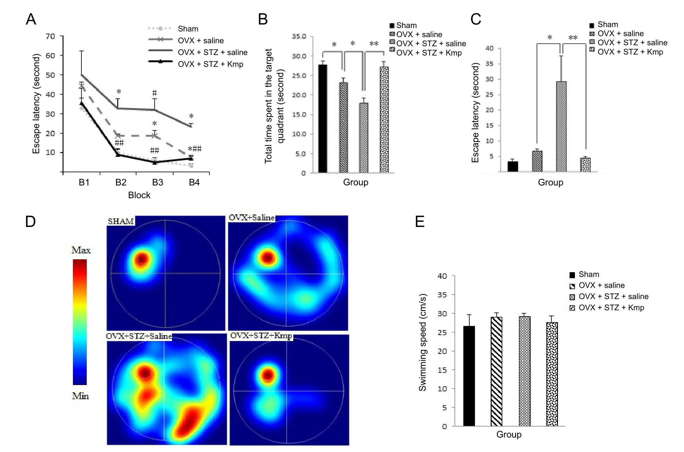
Figure 1 Eあects of kaempferol (Kmp) on cognitive function of an ovariectomized (OVX) rat model of sporadic dementia.
After adding reagents, samples, and standards into the wells,absorbance was measured at 0 and 2 minutes with a microplate reader (Awareness Technology Inc, Palm city, FL, USA)at 532 nm. The concentration of SOD was expressed as U/mg protein. Then SOD activity was calculated based on the below formula:

As an antioxidant enzyme, GSH was determined using Glutathione Assay Kit (Zellbio GmbH, Ulm, Germany), a microplate reader at 412 nm.
TNF-α, an inflammation marker, was assayed using a rat TNF-α ELISA kit (Diaclone SAS, Besancon, France) with a microplate reader at 540 nm.
The MDA as a marker of lipid peroxidation was determined using an MDA assay kit (Zellbio GmbH, Ulm, Germany), and absorbance at 532 nm was measured using a spectrophotometer (UNICCO Inc., Houston, TX, USA).
The level of cytochrome c was analyzed using cytochrome c ELISA Kit (Abcam, Cambridge, UK) with a microplate reader at 450 nm.
Statistical analysis
Normality of variables was estimated by Kolmogorov-Smirnov and Shapiro-Wilk test, and then behavioral data related to working memory were analyzed using repeated measure analysis of variance (ANOVA) followed by Tukey’s posthoc test. Molecular variables and reference memory data were analyzed using one-way ANOVA with the Tukey’s post-hoc test. Results are expressed as the mean ± standard error (SE),and values of P < 0.05 were considered statistically significant.SPSS software was used for statistical analysis (version 22,IBM Cooperation, Chicago, IL, USA)
Results

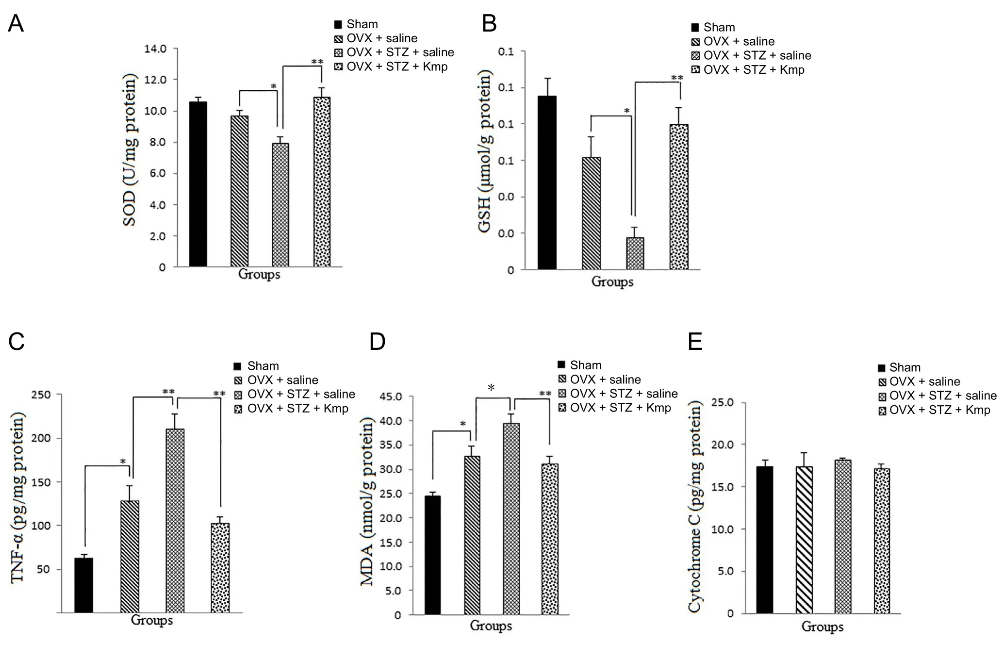
Figure 2 Eあect of kaempferol (Kmp) on hippocampal SOD, GSH, TNF-α, MDA, cytochrome c levels in an ovariectomized rat model of sporadic dementia.
Effects of Kmp on learning and memory function of AD rats Reduction in escape latency during acquisition trials indicates that all animals successfully learned to find the hidden platform in MWM. Results showed a significant difference between groups in escape latency in acquisition phase in Block 1 (B1): [F (3,12) =1.06, P = 0.042]; B2: [F(3,12) =13.31, P =0.001]; B3: [F (3,12) =16.31, P = 0.001] and B4: [F(3,12) =48.22, P = 0.001]. Escape latency was significantly increased in B2, B3 and B4 in the OVX + STZ+ saline group compared with the OVX + saline group (P = 0.029, 0.050, 0.001), while the OVX + STZ + Kmp group demonstrated significant reduction in B2, B3 and B4 compared with the OVX + STZ +saline group (P = 0.001) (Figure 1A).
Moreover, there were significant differences in TTS [F(3,28)= 15.111, P = 0.001] and escape latency [F(3,28) = 9.11, P =0.001]) in probe test between groups. As shown in Figure 1B,D, TTS was significantly increased in the OVX + STZ + Kmp group compared with the OVX + STZ + saline group (P =0.001). In addition, escape latency was significantly increased in the OVX + STZ + saline group compared with the OVX +saline group (P = 0.003), while that was significantly decreased in the Kmp receiving group compared with the OVX + STZ+ saline group (P = 0.001) (Figure 1C). Results did not show significant difference in swimming speed between experimental groups [F(3,28) = 0.462, P = 0.711] (Figure 1E).
Effects of Kmp on oxidative stress in the brain of AD rats
Significant differences were found in the brain levels of SOD[F(3,26) = 9.61, P = 0.001] and GSH [F(3,26) = 11.62, P =0.001] between groups. SOD and GSH levels were significantly increased in the OVX + STZ + Kmp group compared with the OVX + STZ + saline group (P = 0.001; Figure 2A, B).
There was significant change in the brain level of TNF-α[F(3,26)=21.48, P = 0.001] between groups. Brain level of TNF-α was significantly increased in the OVX + STZ + saline group compared with OVX + saline group (P = 0.001), while it was significantly decreased in the OVX + STZ + Kmp group (P = 0.001) compared with the OVX + STZ + saline group (Figure 2C).
A significant difference was found in the brain level of MDA [F(3,26) = 11.32, P = 0.001] between groups. Brain level of MDA level was significantly increased in the OVX +STZ + saline group compared with the OVX + saline group(P = 0.049), while it was significantly decreased in the OVX +STZ + Kmp group compared with the OVX + STZ + saline group (P = 0.009) (Figure 2D).
Cytochrome c level did not change significantly in either of the groups [F(3,19) = 1.501, P = 0.264]; however, it was decreased slightly, but insignificantly, in the OVX + STZ + Kmp compared with the OVX + STZ + saline group (Figure 2E).
Discussion
In the present study, we investigated the effect of Kmp on STZ-induced memory impairment and hippocampal endogenous antioxidants SOD and GSH. In addition, TNF-α, MDA,and cytochrome c levels were measured because of their roles in the inflammation, lipid peroxidation, and apoptosis respectively.
The obtained data clearly demonstrated that: (1) OVX and ICV injection of STZ produced learning and memory deficits,decreased SOD and GSH levels in the hippocampus, while elevated MDA, TNF-α, and cytochrome c levels. (2) Kmp(10 mg/kg per day) reversed the STZ-induced cognitive dysfunction significantly and enhanced hippocampal SOD and GSH levels. (3) Kmp also reduced the levels of inflammatory markers MDA and TNF-α, but not the apoptotic factor of cytochrome c.
Consistent with previous studies (Su et al., 2012; Parker et al., 2014; Solmaz et al., 2015), results of the current study showed that OVX increases the risk of cognitive decline, parallel with enhancement in lipid peroxidation and neuroinflammation. Moreover, ICV injection of STZ doubled memory impairment, induced oxidative stress, lipid peroxidation, and neuroinflammation. STZ has been known to induce neuronal damage by producing free radicals, Aβ deposits (Veerendra Kumar and Gupta, 2003; Huang et al., 2016), and lipid peroxidation (Rai et al., 2014). Santos et al. (2012) previously reported disruption of working memory 3 hours after STZ injections, which was followed by degenerative processes in the hippocampus (Santos et al., 2012). Elevation in MDA(a marker of lipid peroxidation), and TNF-α in the present study reflects oxidative stress and inflammatory status (Gustaw-Rothenberg et al., 2010). TNF-α, as an inflammatory marker, plays an essential role in Aβ plaque accumulation, cell death, and cognitive deficits (Chang et al., 2017).
On the other hand, treatment with Kmp for 21 days significantly reversed STZ-induced cognitive dysfunction, increased hippocampal SOD and GSH levels, and reduced MDA and TNF-α levels. Many studies including ours, suggest that flavonoids have the potential to enhance memory and learning function (Veerendra Kumar and Gupta, 2003; Kouhestani et al., 2017). These components have been known to activate intracellular signaling pathways of memory storage such as mitogen-activated protein kinases, phosphoinositide 3-kinase,protein kinase B (Krishnaveni, 2012), and cAMP response element-binding protein (CREB) (Spencer, 2010).
Elevation in SOD and GSH levels in the present study reflects improvement in brain endogenous antioxidants. SOD and GSH are the most important first-line antioxidant defense systems against toxicity of ROS (Wojtunik-Kulesza et al.,2016). GSH works via scavenging ROS and removing hydrogen and lipid peroxidase (Halliwell and Gutteridge, 1989), and improves neurological functions.
Kmp, the essential flavonoid of medlar leaves extract, has been shown to augment cellular antioxidant defense capacity(Kampkotter et al., 2007; Hong et al., 2009). Kmp also scavenges hydroxyl radical and peroxynitrite increases the activity of antioxidant enzymes and prevents lipid peroxidation(Ozgova et al., 2003; Kampkotter et al., 2007).
Furthermore, the present study showed that Kmp caused 21% reduction in hippocampal MDA level, 52% reduction in hippocampal TNF-α level, and only 6% reduction in hippocampal cytochrome c level. These findings reflect the ability of Kmp to alleviate neurotoxicity induced by STZ.
In line with our findings, Sharoar et al. (2012) and Li and Pu(2011) reported that chronic administration of Kmp leads to attenuating neurotoxicity induced by Aβ and 1-methyl-4-phenyl-1,2,3,6-tetrahydropyridine respectively. Also Kmp exhibited anti-parkinsonian activity which was related to increases in SOD and glutathione peroxidase activities and reduction in MDA level. In addition, Chen et al. (2012) reported that Kmp attenuated lung injury in mice. Considering no significant change in cytochrome c in our study explains that Kmp might not be effective on the mitochondrial intrinsic pathway of apoptosis. However, the effect of Kmp on other pro-apoptotic factors remains to be elucidated in future studies.
Taken together, findings from this study suggest potential important clinical relevance of considering anti-inflammatory and antioxidant properties of Kmp, as it was reported in metabolic syndrome (Hoang et al., 2015), Parkinson (Li and Pu,2011) and cardiovascular diseases (Perez-Vizcaino and Duarte,2010).
In conclusion, this study demonstrated for the first time that Kmp ameliorates STZ-induced cognitive dysfunction possibly through regulating antioxidants and neuroinflammation in ovariectomized rat hippocampus, however, the underlying mechanism remains unclear.
Author contributions: PB designed this study, performed experiments,wrote and revised the paper for important intellectual content. SK performed experiments and statistical analysis and participated in paper writing. AJ performed ELISA assay and revised the manuscript for important intellectual content. All authors approved the final version of the paper.Conflicts of interest: The authors declare that they have no conflicts of interest.
Financial support: This study was supported by a grant from Research and Technology Chancellor of Guilan University of Medical Sciences,Iran (No. IR.GUMS. REC. 1936.51). The funder did not participate in data collection and analysis, paper writing or submission.
Institutional review board statement: The study protocol was approved by the National Institutes of Health guide for the care and use of Laboratory Animals (NIH Publications No. 8023, revised 1978) modified by the Ethics Committee of Guilan University of Medical Sciences, Rasht, Iran.IR.GUMS.REC.1396.51.
Copyright license agreement: The Copyright License Agreement has been signed by all authors before publication.
Data sharing statement: Datasets analyzed during the current study are available from the corresponding author on reasonable request.
Plagiarism check: Checked twice by iThenticate.
Peer review: Externally peer reviewed.
Open access statement: This is an open access journal, and articles are distributed under the terms of the Creative Commons Attribution-Non-Commercial-ShareAlike 4.0 License, which allows others to remix, tweak,and build upon the work non-commercially, as long as appropriate credit is given and the new creations are licensed under the identical terms.
Open peer reviewer: Rodolfo Pinto-Almazán, Hospital Regional de Alta Especialidad de Ixtapaluca, Mexico.
Additional file: Open peer review report 1.
- 中国神经再生研究(英文版)的其它文章
- Validity and reliability of the Ocular Motor Nerve Palsy Scale
- The combination of induced pluripotent stem cells and bioscaffolds holds promise for spinal cord regeneration
- Melatonin for the treatment of spinal cord injury
- Depression following a traumatic brain injury: uncovering cytokine dysregulation as a pathogenic mechanism
- Loss of canonical Wnt signaling is involved in the pathogenesis of Alzheimer’s disease
- The balance between efficient anti-inflammatory treatment and neuronal regeneration in the olfactory epithelium

