
Traumatic optic neuropathy secondary to acupuncture treatment for glaucoma: A case report
2018-07-26 09:59:50WenYeeLeeWeeMinTehNorlinaMohdRamliAhmadMtSaad
Journal of Acute Disease 2018年3期
Wen Yee Lee, Wee Min Teh, Norlina Mohd Ramli, Ahmad Mt Saad
1 Department of Ophthalmology, Hospital Sultanah Bahiyah, Alor Setar, Kedah, Malaysia
2 Department of Ophthalmology, Universiti of Malaya, Petaling Jaya, Selangor, Malaysia
Keywords:Acupuncture Traumatic optic neuropathy Glaucoma Vitreous hemorrhage
ABSTRACT Internal organ injuries have been recognized as a major complication of acupuncture. Reported ocular adverse events include traumatic cataract, oculomotor nerve injury, endophthalmitis and retinal puncture. We report a case of traumatic optic neuropathy and self-sealed globe perforation following acupuncture. A Chinese gentleman with primary open angle glaucoma presented with sudden loss of vision in the right eye after acupuncture therapy. The vision dropped to 2/60 from 6/6 premorbid. Relative afferent pupillary defect was present. Fundus examination showed hemorrhage from the optic disc into the vitreous. It is likely that the optic nerve injury occurred when the acupuncture needle was advanced deep into locations near the optic nerve. Main acupoints used in acupuncture treatment of glaucoma include Tongziliao GB-1, Jingming BL-1 and Chengqi ST-1. It is crucial to have a good understanding of ocular anatomy to avoid potentially blinding complications.
1. Introduction
Acupuncture is a system of complementary medicine in which fine needles are inserted in the skin at specific points to treat various conditions. It is a key component of traditional Chinese medicine,but it is now also gaining popularity and acceptance in Western countries. Glaucoma is a chronic, progressive optic neuropathy with characteristic patterns of optic nerve damage and visual field loss.Although research and inroads have been made in the treatment of glaucoma, lowering of intraocular pressure remains the mainstay of treatment currently. Acupuncture has also been used in the treatment of glaucoma. However, it is also known to cause various ocular complications. We would like to report a case of traumatic optic neuropathy and self-sealed globe perforation following acupuncture.
2. Case report
A 63-year-old Chinese gentleman with bilateral advanced primary open angle glaucoma was on long-term follow-up since 2012, and had left eye trabeculectomy done in August 2016. He presented with sudden loss of vision in the right eye after three sessions of acupuncture therapy for consecutive days. During each session, he claimed that the needles were inserted though the lateral canthal region of both eyes. The needles were advanced until he felt a pricking sensation. On the second day of therapy, he developed acute blurred vision in his right eye. However, he was told that this commonly occurred during acupuncture, and was reassured to continue the session. He visited the hospital three days after the third session of acupuncture as his symptoms remained.
On examination, the right eye vision dropped from 6/6 to 2/60.Clinically, there was possible penetration of the globe as suggested by presence of localized subconjunctival hemorrhage over the superotemporal part of the right eye. The cornea was clear and the anterior chamber was deep. There was no hyphema in the anterior chamber and intraocular pressure was 16 mmHg. Traumatic optic neuropathy was suspected based on the findings of marked relative afferent pupillary defect and localized hemorrhage over the right optic disc (Figure 1). There were no signs of infection noted. There were no retinal breaks seen during fundus examination.

Figure 1. Fundus photo of right eye after acupuncture treatment for glaucoma shows localized hemorrhage over the optic disc.
As the eyeball integrity appeared intact, no surgical intervention was planned. He was also not given any systemic steroids for the possible traumatic optic neuropathy due to the late presentation. He was followed up weekly for the initial two weeks, and there was resolution of the hemorrhage at the optic disc and vitreous at the end of week 2 (Figure 2). Unfortunately, his right eye vision did not improve despite resolution of the hemorrhage.

Figure 2. Fundus photo of right eye after vitreous hemorrhage resolved demonstrating fibrovascular membrane on the optic disc.
For the treatment of glaucoma, several acupoints have been identified. Among them are (Figure 3): Acupoint Tongziliao GB-1-lateral to the lateral canthus; Acupoint Jingming BL-1-above the medial canthus; Acupoint Chengqi ST-1-inferior border of the orbit.
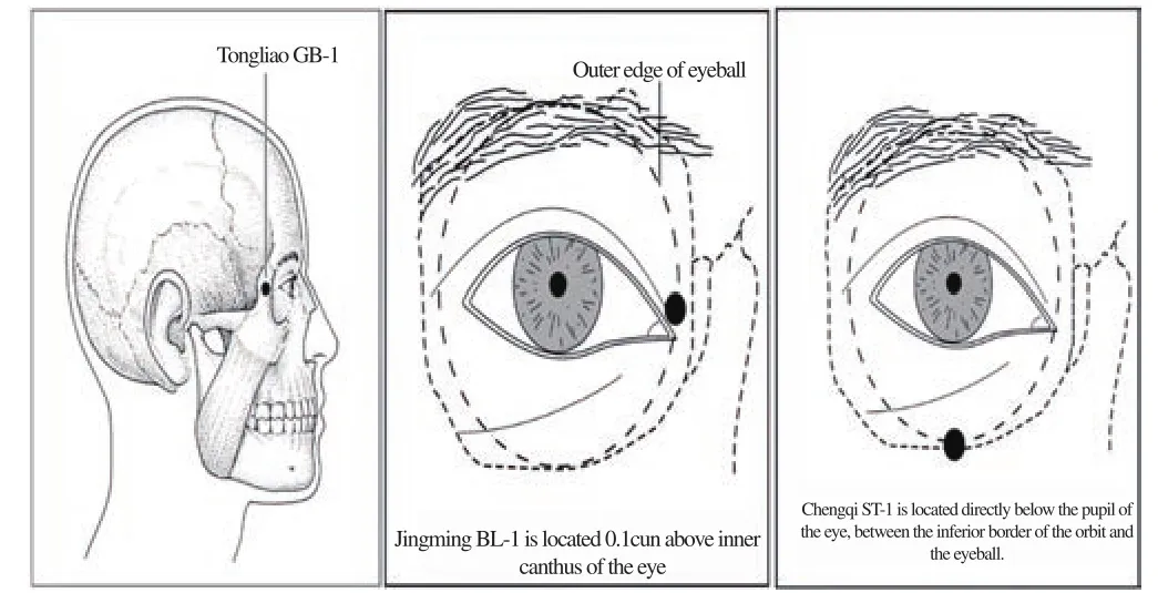
Figure 3. The common acupoints used in glaucoma.
3. Discussion
Acupuncture features prominently in traditional Chinese medicine.Its application dates as far back as the 6thcentury BC, when the medical profession in China at that time had already included the practice of acupuncture[1]. Acupuncture is now gaining popularity and also acceptance in many countries, including the West. Because of its widespread use, it is important to establish the safety and efficacy of the treatment.
There have been various types of acupuncture-related adverse events identified in the literature. Serious systemic adverse events such as pneumothorax, cardiac tamponade, spinal cord injury and viral hepatitis have been reported[2-5].
Application of acupuncture in eye conditions is also wellestablished. Some of the symptoms and conditions treated using acupuncture include squint, cataract, optic neuritis, optic atrophy,myopia, glaucoma, redness of the eyes, watering of the eyes, itchy eyes, swollen and painful eyes, and eyelid spasm[6].
During the procedure, acupuncture needles are advanced deep into the acupoint to achieve an ache radiating to behind of the eye as describe in the Chinese literature[6]. All these points are anatomically predisposed to optic nerve injury and globe perforation due to the proximity of the structures. Other reported adverse events include traumatic cataract, oculomotor nerve injury, endophthalmitis and retinal puncture[7-8].
Our patient sought treatment for his glaucoma at an acupuncture center. From his description, the likely acupoint used in his treatment was acupoint Tongziliao GB-1. The needle was advanced from the area of lateral canthus of the eye and directed medially. The direction of this needle entry poses a very high risk of optic nerve injury. The pain felt by this patient during the acupuncture treatment likely arose from the penetration into the sclera, while the acute loss of vision was due to vitreous hemorrhage and traumatic optic nerve injury.
Fortunately for the patient, he did not develop any signs of infection as the globe perforation appeared to be self-sealing. The vitreous hemorrhage resolved spontaneously, revealing an area of retinal scar just adjacent to optic nerve.
In one of the cases reported in the literature, patient had to undergo vitrectomy surgery with endolaser and intravitreal antibiotics for similar presentation as in our patient[8]. However, comparing the visual outcome of our patient, his vision remained poor because of the optic nerve injury.
To be concluded that this case report wishes to highlight the dangers of acupuncture treatment for the eye. It is important for the acupuncturist to recognize and differentiate between the pain of imminent globe puncture and/or nerve injury from desired therapeutic effect of the procedure. However, this is a very subjective assessment tool as pain threshold varies amongst patients.
Conflict of interest statement
The authors report no conflict of interest.
 Journal of Acute Disease2018年3期
- Journal of Acute Disease的其它文章
- Cost analysis and characteristics of the patients admitted to emergency service with poisoning
- A comparison of culture and PCR methods for identification of Aggregatibacter actinomycetemcomitans isolated from acute necrotizing ulcerative gingivitis
- Scoring systems in prediciting mortality rate of patients applying emergency department
- Earthquake planning and crisis management with an emphasis on the facilities, utilities, and services of the health care centers of Tiran and Karvan County, Isfahan Province, Iran: A case study
- A survey on the epidemiology of trauma and China trauma care training in subtropical regions of Hainan Province
- Traditional Chinese Medicine and its protective function over braininjured patients