
Reorganization of injured anterior cingulums in a hemorrhagic stroke patient
2018-07-23 02:59:02SungHoJang,ChulHoonChang,HanDoLee
中国神经再生研究(英文版) 2018年8期
In this study, we reported on a patient who showed a new neural tract between the injured anterior cingu‐lums and the basal forebrain, as shown by diffusion tensor tractography (DTT).
A 62‐year‐old female underwent coiling for a rup‐tured aneurysm in the left middle cerebral artery (M1)and decompressive craniectomy for intracerebral hem‐orrhage in the left fronto‐temporal lobe, intraventricu‐lar hemorrhage, and subarachnoid hemorrhage (Figure 1A, B). At three weeks after a hemorrhagic stroke,she showed bilateral motor weakness (right side: 2‐/5 and left side: 2/5) and severe cognitive impairment(decreased alertness, Mini‐Mental State Exam score:uncheckable). She began to recover gradually after two months of rehabilitation.
Diffusion tensor imaging was performed at three weeks after onset using a 6‐channel head coil on a 1.5T Philips Gyroscan Intera (Hoffman‐LaRoche Ltd,Best, The Netherlands) with single‐shot echo‐planar imaging and navigator echo. Sixty contiguous slices(acquisition matrix = 96 × 96; reconstruction matrix= 192 × 192; field of view = 240 × 240 mm2; repetition time = 10,726 ms; echo time = 76 ms,b= 1000 s/mm2,number of excitations = 1, slice gap = 0 mm and thick‐ness = 2.5 mm) were acquired for each of the 32 non‐collinear diffusion‐sensitizing gradients. Fiber tracking was performed using the fiber assignment continuous tracking (FACT) algorithm implemented within the diffusion tensor imaging task card software (Philips Extended MR Work Space 2.6.3). For reconstruction of the cingulum, the seed region of interest (ROI) was placed in the middle portion of the cingulum and the target ROI was placed in the posterior portion of the cingulum on the colored coronal images. Termination criteria were fractional anisotropy (FA) < 0.15 and an angle change > 27° (Yoo et al., 2014).
On 3‐week DTT images, both cingulums showed discontinuations between the anterior cingulum and the basal forebrain. However, the discontinued right anterior cingulum was connected to the left basal fore‐brainviathe genu of the corpus callosum. In addition,the discontinued left anterior cingulum was connect‐ed to an unusual neural tract from the right anterior cingulum connected to the left basal forebrain (Figure 1C).
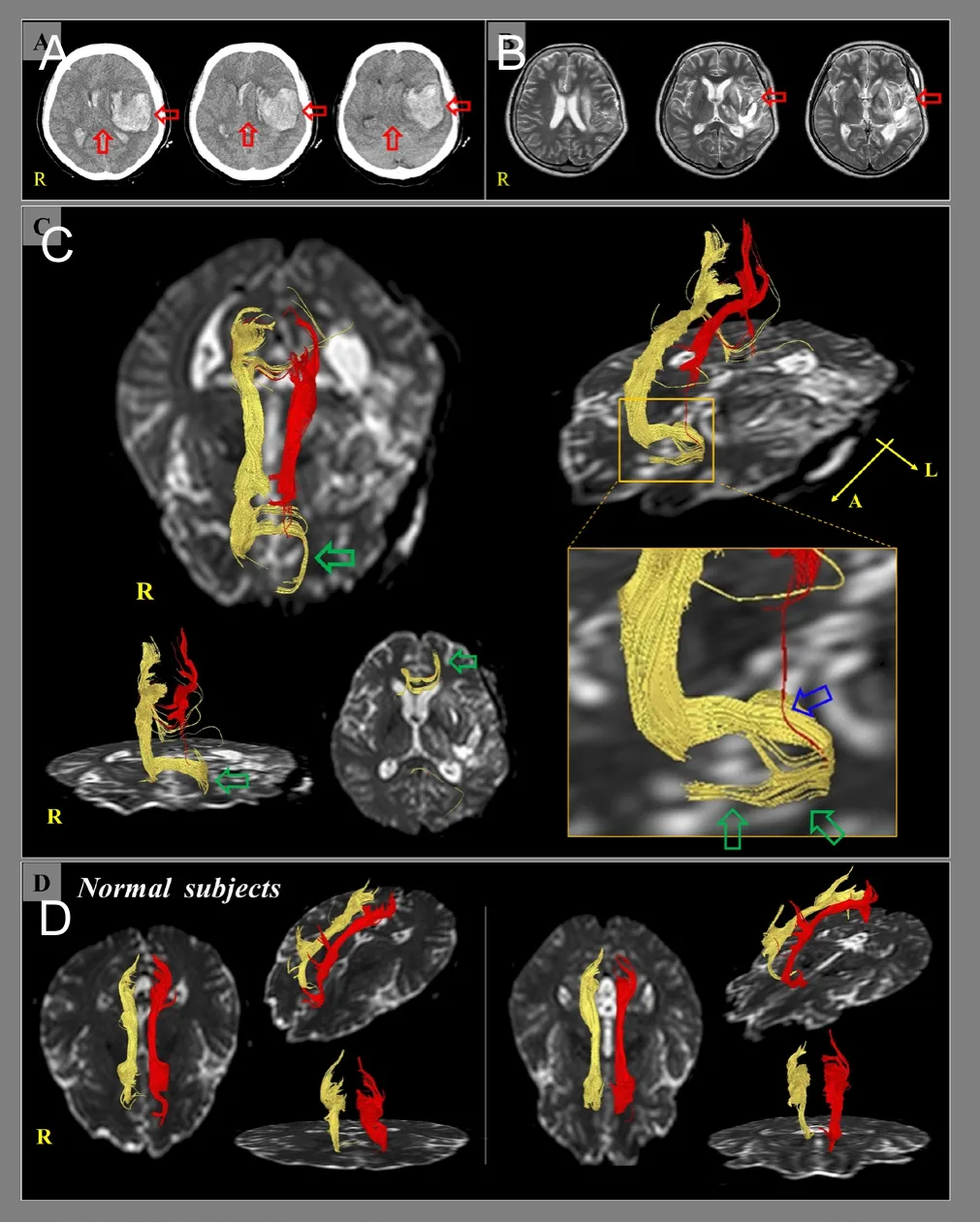
Figure 1 Brain computed tomography (CT), magnetic resonance(MR) imaging, and diffusion tensor tractography (DTT) of a 62-year-old female patient with a hemorrhagic stroke who showed a new neural tract between the injured anterior cingulums and the basal forebrain.
In this patient, discontinuations of both anterior cingulums appeared to be ascribed to the left intracere‐bral hemorrhage and subsequent subfalcine herniation(Figure 1A, B). We observed an unusual neural tract between the discontinued right anterior cingulum and the left basal forebrainviathe genu of the corpus callo‐sum, and the discontinued left anterior cingulum was connected to this unusual neural tract.
The cingulum, which connects the orbitofrontal cor‐tex and the medial temporal lobe, plays an important role in cognition. In particular, it is associated with memory function because the cingulum is the passage of cholinergic innervation from the cholinergic nuclei(the medial septal nucleus [Ch1], the vertical nucleus of the diagonal band [Ch2], and the nucleus basalis of Meynert [Ch4]) in the basal forebrain to the cerebral cortex (Woolf and Butcher, 1986; Selden et al., 1998;Nieuwenhuys et al., 2008; Naidich and Duvernoy,2009). Therefore, the unusual neural tract between the left basal forebrain and the injured anterior cingulums likely contributes to a compensatory phenomenon to obtain cholinergic innervations from cholinergic nuclei in the left basal forebrain after interruption of cholinergic innervations by complete injury of both anterior cingulums (Woolf and Butcher, 1986; Selden et al., 1998; Nieuwenhuys et al., 2008; Naidich and Du‐vernoy, 2009).
Discontinuations of both anterior cingulums indi‐cate blockage of cholinergic innervation from the basal forebrain to the cerebral cortex. Therefore, we believe that the development of this unusual neural tract be‐tween the basal forebrain and injured cingulums after interruption of cholinergic innervation from the basal forebrain by complete injury of the anterior cingulum might have resulted in the reorganization of choliner‐gic innervations after stroke (Yeo et al., 2012; Seo and Jang, 2013, 2014; Yoo et al., 2014; Jang et al., 2015).
In conclusion, we reported a stroke patient who showed a new neural tract between the injured ante‐rior cingulums and the basal forebrain. This finding appears to suggest the reorganization of cholinergic innervations after stroke.
Sung Ho Jang, Chul Hoon Chang, Han Do Lee*Department of Physical Medicine and Rehabilitation,College of Medicine, Yeungnam University, Namku, Daegu,Republic of Korea (Jang SH, Lee HD)Department of Neurosurgery, College of Medicine,Yeungnam University, Namku, Daegu, Republic of Korea(Chang CH)
*Correspondence to:Han Do Lee, M.S.,lhd890221@hanmail.net.
orcid:0000-0002-1668-2187 (Han Do Lee)
Accepted:2018-01-03
doi:10.4103/1673-5374.235308
Author contributions:Study conception and design: SHJ, CHC,and HDL; data acquisition: CHC, HDL; data analysis: HDL; paperpreparation and writing: SHJ; paper authorization: HDL. All authors approved the final version of this paper for publication.
Conflicts of interest:None declared.
Financial support:This work was supported by the National Research Foundation (NRF) of Korea Grant funded by the Korean Government(MSIP) (2015R1A2A2A01004073). The funding body played no role in the study conception design, in the collection, analysis and interpretation of data, in the preparation and writing of the report, and in the decision to submit the article for publication.
Copyright license agreement:The Copyright License Agreement has been signed by all authors before publication.
Institutional review board statement:This study was approved by the Institutional Review Board of Yeungnam University Hospital, Republic of Korea (approval No. YUMC-2017-06-020).
Data sharing statement:Datasets analyzed during the current study are available from the corresponding author on reasonable request.
Plagiarism check:Checked twice by iThenticate.
Peer review:Externally peer reviewed.
Open access statement:This is an open access journal, and articles are distributed under the terms of the Creative Commons Attribution-Non-Commercial-ShareAlike 4.0 License, which allows others to remix, tweak,and build upon the work non-commercially, as long as appropriate credit is given and the new creations are licensed under the identical terms.
- 中国神经再生研究(英文版)的其它文章
- In Memoriam: Ray Grill (1966–2018)
- A novel chronic nerve compression model in the rat
- Analgesic effect of AG490, a Janus kinase inhibitor, on oxaliplatin-induced acute neuropathic pain
- Three-dimensional visualization of the functional fascicular groups of a long-segment peripheral nerve
- Novel conductive polypyrrole/silk fibroin scaffold for neural tissue repair
- Local inhibition of matrix metalloproteinases reduced M2 macrophage activity and impeded recovery in spinal cord transected rats after treatment with fibroblast growth factor-1 and nerve grafts