血清胱抑素C水平与血管性认知功能障碍的相关性研究
刘海涛 张淑玲△ 白宏英
1)郑州人民医院神经内科一病区,河南 郑州 450000 2)郑州大学第二附属医院,河南 郑州 450014
血管认知功能障碍是由血管疾病引起的异常认知状态,好发于老年人群,患者认知缺陷严重不足,但未达到痴呆标准,基本功能不会丧失。早期识别和治疗血管性认知障碍尤为重要[1-3]。研究显示,寻找准确的生物标志物以及危险因素,用于早期诊断以及预防血管性认知障碍,对临床探索血管性认知障碍发病机制、疾病特点以及临床治疗具有重要意义[4-5]。血清胱抑素C(Cys-C)是一种由Cys C(CST3)基因编码,由人体组织分泌的半胱氨酸蛋白酶抑制剂,近年来,其在血管认知功能障碍以及阿尔茨海默病中的研究越来越多[6-8]。作为肾功能的生物标志物,其也是血管认知功能障碍的有效预测因子[9-10]。多种证据表明,Cys C具有抗炎功能,并对年龄相关疾病发挥保护作用[11-13]。目前,临床对于血管性认知功能障碍的影响因素结论并不统一。本研究探讨血清Cys-C水平与血管性认知功能障碍的相关性,并分析其在发生血管性认知功能障碍中的意义。
1 资料与方法
1.1研究对象选择2014-02-30—2017-07-30于郑州人民医院神经内科住院的110例血管性认知功能障碍患者(认知障碍组),选取同期90例健康体检者为对照组。男127例,女73例,年龄35~77(53.8±6.4)岁。记录2组基础资料(年龄、吸烟史、饮酒史、性别、高血压史等)。本研究经过我院伦理委员会审核,患者或其家属对研究内容知情且签订书面同意书。
1.2纳入排除标准纳入标准:(1)认知障碍组患者均接受蒙特利尔认知评估量表(MoCA,共7个项目,满分30分,≥26分为正常)评估认知功能[14],且MoCA评分<26分;(2)认知障碍组患者头颅MRI或CT提示存在脑缺血病灶;(3)对照组MoCA评分≥26分。排除标准:(1)先天性疾病或精神病引起的身体残疾;(2)合并阿尔茨海默病;(3)疼痛综合征、晚期糖尿病、恶性肿瘤、肾衰竭、肝功能衰竭、严重贫血者;(4)甲状腺功能异常者;(5)正在接受认知障碍治疗者;(6)严重交流障碍者。
1.3检测方法入院后清晨抽取空腹5 mL静脉血,样品收集1 h内以3 000 r/min离心10 min,分离血浆。实验室检测前,将分离的血浆储存在-30 ℃冰箱内。使用Behring BN ProSpec分析仪(Dade Behring)检测Cys C(免疫比浊法)、血糖、甘油三酯(TC)、血肌酐、低密度脂蛋白(LDL-C)、高密度脂蛋白(HDL-C)等水平。所有检测结果重复3次。
1.4观察指标比较2组一般资料以及Cys C水平,分析血管性认知功能障碍的独立危险因素。

2 结果
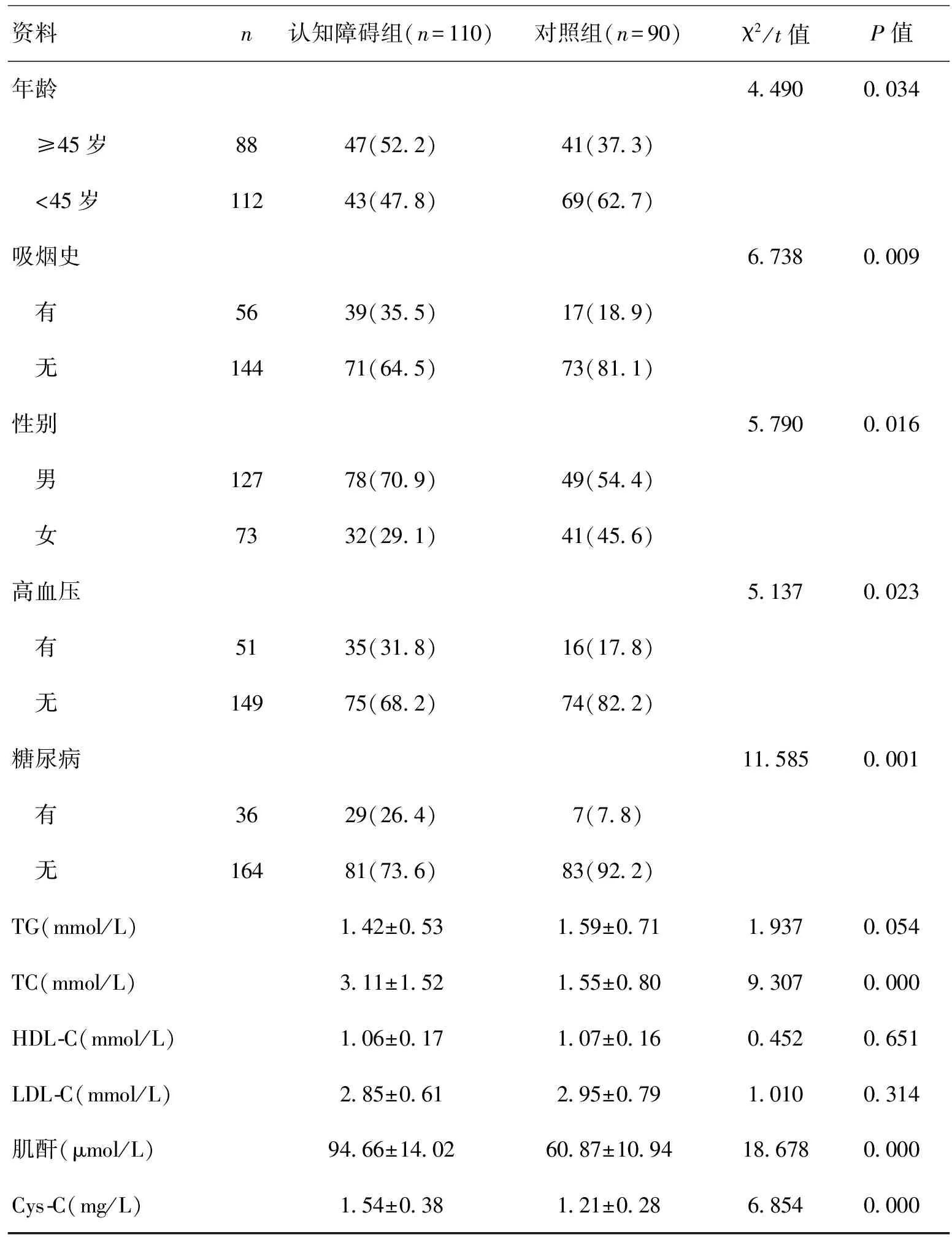
2.1 2组临床资料比较2组年龄、性别、吸烟史、高血压史、糖尿病史、TC、血肌酐、血糖、Cys-C水平差异有统计学意义(P<0.05)。血管性认知功能障碍患者年龄、TC、血肌酐、血糖、Cys-C水平,以及男性比例、吸烟史、高血压史、糖尿病史比例高于对照组。见表1。
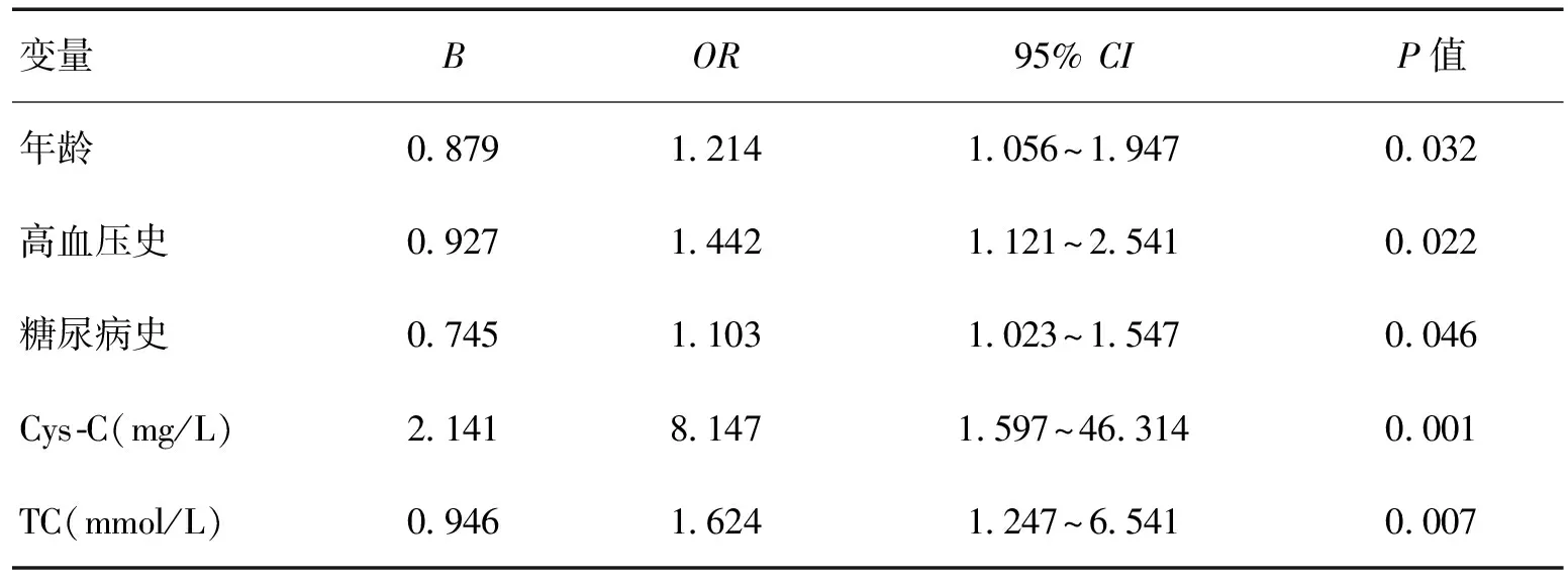
2.2多因素分析Logistic回归分析示,Cys-C、TC、高血压史、糖尿病史、年龄是血管性认知功能障碍的独立影响因素(P<0.05)。高水平Cys-C、TC、高龄以及合并高血压史、糖尿病史是血管性认知功能障碍的独立危险因素。见表2。
3 讨论
认知功能障碍是老年群体高发疾病。近年来,随着人口老龄化进程加快,发病率呈现上升趋势,是引起老年痴呆的第2大疾病,严重影响其健康和生活质量[15-16]。大多数认知功能障碍患者存在多发性脑梗死或脑白质缺血,如未得到及时有效治疗,多数患者最终发展为痴呆[17-23]。血管性认知功能障碍包括各种脑血管疾病引起的认知障碍,如脑出血、脑栓塞、脑血流灌流异常等。Cys-C是由Cys C(CST3)基因编码的Cys超家族成员,属于内源性组织蛋白酶抑制物,可抑制蛋白酶B,具有平衡血管壁内组织蛋白酶、抗蛋白酶活性的作用[18]。早期研究重点为Cys-C作为肾功能的生物标志物,在肾脏疾病中起作用。近年来,其在血管性认知功能障碍以及阿尔茨海默病中的研究越来越多[19]。已有研究证实,Cys-C与脑血管病变具有显著相关性,Cys C不仅可调节痴呆,还与血管功能相关,并介导血管舒张、炎症和氧化应激等反应[24-30]。

表1 2组临床资料比较

表2 血管性认知功能障碍的Logistic回归分析结果
ZHANG等[31]分析了阿尔茨海默病脑组织中淀粉样沉积物成分时发现Cys-C,提示Cys-C可能参与了神经组织损伤。已有研究证实,Cys-C基因多态性与阿尔茨海默病、认知功能障碍具有相关性[5,20]。本研究显示,血管性认知功能障碍患者Cys-C水平高于正常体检者;此外,2组年龄、性别、吸烟史、高血压史、糖尿病史、TC、血肌酐、血糖差异有统计学意义(P<0.05)。血管性认知功能障碍患者年龄、TC、血肌酐、血糖、Cys-C水平,以及男性比例、吸烟史、高血压史、糖尿病史比例高于正常体检者。Logistic回归分析示,Cys-C、TC、高血压史、糖尿病史、年龄是血管性认知功能障碍的独立影响因素(P<0.05)。高水平Cys-C、TC、高龄以及合并高血压史、糖尿病史是血管性认知功能障碍的独立危险因素。ZUO等[32-35]研究不同类型痴呆症(血管性认知功能障碍和阿尔茨海默病)患者的Cys C和HDL水平,调查Cys C和HDL是否与不同类型痴呆的严重程度和患病率相关,结果显示,与健康对照组相比,血管性认知功能障碍患者血浆Cys C水平较高,HDL水平较低,血浆Cys C/HDL对痴呆具有诊断价值。本研究中血管性认知功能障碍Cys C水平较健康体检者高,与相关研究[36-38]一致。HDL是存在于全身循环和脑中脂蛋白颗粒的异质组的一部分,主要分别促进脂质和脂质相关分子从体内和整个身体的清除和递送。有证据表明,血浆HDL及其主要蛋白质组分ApoA-Ⅰ也具有有效的血管保护特性,如促进血管功能改善,抑制炎症,抑制内皮修复,防止脂质氧化和刺激内皮修复[10,39]。本研究中,2组HDL差异无统计学意义,可能与纳入患者种族差异、检测方法以及纳入标准差异有关。研究认为,Cys C可能在痴呆发病机制中起重要作用,血浆Cys C水平可能是区分血管性认知功能障碍与健康受试者的有效筛选工具[17]。
在排除混杂影响因素后,本研究显示,高水平Cys-C、TC、高龄以及合并高血压史、糖尿病史是血管性认知功能障碍的独立危险因素。刘利红等[24]分析了411例血管性认知功能障碍的危险因素,结果显示,Cys-C是血管性认知功能障碍的独立危险因素,与本研究一致。此外,RAFAILIDIS等[40]研究认为,血管危险因素,如年龄、TC、高血压史、糖尿病史等增加血管性认知功能障碍的发病率,并促进其向痴呆发展。血清Cys-C水平异常可促进动脉粥样硬化发生,动脉硬化狭窄影响脑组织血供;此外,动脉损伤促进炎性细胞因子释放,造成脑组织损伤;Cys-C可通过血脑屏障参与脑部炎症反应,促进认知功能障碍发展[41-43],本研究进一步证实了Cys-C在血管性认知功能障碍中作用。
[1] FRANCES A,SANDRA O,LUCY U.Vascular cognitive impairment,a cardiovascular complication[J].World J Psychiatry,2016,6(2):199-207.
[2] HORSBURGH K,WARDLAW J M,VAN AGTMAEL T,et al.Small vessels,dementia and chronic diseases-molecular mechanisms and pathophysiology[J].Clin Sci (Lond),2018,132(8):851-868.
[3] ANDRIUTA D,ROUSSEL M,BARBAY M,et al.Differentiating between Alzheimer's Disease and Vascular Cognitive Impairment:Is the "Memory Versus Executive Function" Contrast Still Relevant?[J].J Alzheimers Dis,2018,63(2):625-633.
[4] LEE M J,SEO S W,NA D L,et al.Synergistic effects of ischemia and beta-amyloid burden on cognitive decline in patients with subcortical vascular mild cognitive impairment[J].JAMA Psychiatry,2014,71(4):412-422.
[5] BARBAY M,TAILLIA H,NEDELEC-CICERI C,et al.Prevalence of Poststroke Neurocognitive Disorders Using National Institute of Neurological Disorders and Stroke-Canadian Stroke Network,VASCOG Criteria (Vascular Behavioral and Cognitive Disorders),and Optimized Criteria of Cognitive Deficit[J].Stroke,2018,49(5):1 141-1 147.
[6] BREINING A,SILVESTRE J S,DIEUDONNE B,et al.Biomarkers of vascular dysfunction and cognitive decline in patients with Alzheimer's disease:no evidence for association in elderly subjects[J].Aging Clin Exp Res,2016,28(6):1 133-1 141.
[7] MATSUOKA D,WATANABE H,SHIMIZU Y,et al.Synthesis and evaluation of a novel near-infrared fluorescent probe based on succinimidyl-Cys-C(O)-Glu that targets prostate-specific membrane antigen for optical imaging[J].Bioorg Med Chem Lett,2017,27(21):4 876-4 880.
[8] BENLI E,AYYILDIZ S N,CIRRIK S,et al.Early term effect of ureterorenoscopy (URS) on the Kidney:research measuring NGAL,KIM-1,FABP and CYS C levels in urine[J].Int Braz J Urol,2017,43(5):887-895.
[9] HEYWOOD W E,GALIMBERTI D,BLISS E,et al.Identification of novel CSF biomarkers for neurodegeneration and their validation by a high-throughput multiplexed targeted proteomic assay[J].Mol Neurodegener,2015,10:64.
[10] ZHONG X M,HOU L,LUO X N,et al.Alterations of CSF cystatin C levels and their correlations with CSF Alphabeta40 and Alphabeta42 levels in patients with Alzheimer's disease,dementia with lewy bodies and the atrophic form of generalparesis[J].PLoS One,2013,8(1):e55328.
[11] LIU Y,LI J,WANG Z,et al.Attenuation of early brain injury and learning deficits following experimental subarachnoid hemorrhage secondary to Cystatin C:possible involvement of the autophagy pathway[J].Mol Neurobiol,2014,49(2):1 043-1 054.
[12] WAHEED S,MATSUSHITA K,ASTOR B C,et al.Combined association of creatinine,albuminuria,and cystatin C with all-cause mortality and cardiovascular and kidney outcomes[J].Clin J Am Soc Nephrol,2015,8(3):434-442.
[13] YANG F,LI D,DI Y,et al.Pretreatment Serum Cysta-tin C Levels Predict Renal Function,but Not Tumor Characteristics,in Patients with Prostate Neoplasia[J].Biomed Res Int,2017,2017:7 450 459.
[14] 阿拉腾巴根,刘相辰,赵丽珍.MoCA量表的临床应用研究进展[J].内蒙古医学杂志,2014,46(4):429-432.
[15] WALLIN A,NORDLUND A,JONSSON M,et al.Alzhei-mer's disease-subcortical vascular disease spectrum in a hospital-based setting:Overview of results from the Gothenburg MCI and dementia studies[J].J Cereb Blood Flow Metab,2016,36(1):95-113.
[16] SHANG J,YAMASHITA T,FUKUI Y,et al.Different Associations of Plasma Biomarkers in Alzheimer's Disease,Mild Cognitive Impairment,Vascular Dementia,and Ischemic Stroke[J].J Clin Neurol,2018,14(1):29-34.
[17] CHU C S,TSENG P T,STUBBS B,et al.Use of statins and the risk of dementia and mild cognitive impairment:A systematic review and meta-analysis[J].Sci Rep,2018,8(1):5 804.
[18] CAI J J,ZHOU J,HAN T,et al.Diagnostic value of serum cystatin C for acute kidney injury in patients with liver cirrhosis[J].Zhonghua Gan Zang Bing Za Zhi,2017,25(5):360-364.
[19] GOMPOU A,PERREA D,KARATZAS T,et al.Relationship of Changes in Cystatin-C With Serum Creatinine and Estimated Glomerular Filtration Rate in Kidney Transplantation[J].Transplant Proc,2015,47(6):1 662-1 674.
[20] STUKAS S,ROBERT J,WELLINGTON C L.High-density lipoproteins and cerebrovascular integrity in Alzheimer's disease[J].Cell Metab,2014,19(4):574-591.
[21] ZOU J,CHEN Z,WEI X,et al.Cystatin C as a potential therapeutic mediator against Parkinson's disease via VEGF-induced angiogenesis and enhanced neuronal autophagy in neurovascular units[J].Cell Death Dis,2017,8(6):e2854.
[22] KAUR G,LEVY E.Cystatin C in Alzheimer's disease[J].Front Mol Neurosci,2014,5:79.
[23] WANG R,CHEN Z,FU Y,et al.Plasma Cystatin C and High-Density Lipoprotein Are Important Biomar-kers of Alzheimer's Disease and Vascular Dementia:A Cross-Sectional Study[J].Front Aging Neurosci,2017,9:26.
[24] 刘利红,张正伟.血清胱抑素C水平与血管性认知功能障碍的相关性[J].临床医学,2017,37(10):14-16.
[25] SAITO S,YAMAMOTO Y,IHARA M.Mild Cognitive Impairment:At the Crossroad of Neurodegeneration and Vascular Dysfunction[J].Curr Alzheimer Res,2015,12(6):507-512.
[26] SVEINSSON O A,KJARTANSSON O,VALDIMARSSON E M.Cerebral ischemia/infarction - epidemiology,causes and symptoms[J].Laeknabladid,2014,100(5):271-279.
[27] JABBARLI R,REINHARD M,NIESEN W D,et al.Predictors and impact of early cerebral infarction after aneurysmal subarachnoid hemorrhage[J].Eur J Neurol,2015,22(6):941-947.
[28] ZHANG Y B,SU Y Y,HE Y B,et al.Early Neurological Deterioration after Recanalization Treatment in Patients with Acute Ischemic Stroke:A Retrospective Study[J].Chin Med J (Engl),2018,131(2):137-143.
[29] NISHIKAWA H,SUZUKI H.Possible Role of Inflammation and Galectin-3 in Brain Injury after Subara-chnoid Hemorrhage[J].Brain Sci,2018,8(2):E30.
[30] EBENDAL T.Function and evolution in the NGF family and its receptors [J].Neurosci Res,1992,32(4):461-470.
[31] ZHANG W,HUANG Y,YING L,et al.Efficacy and Safety of Vinpocetine as Part of Treatment for Acute Cerebral Infarction:A Randomized,Open-Label,Controlled,Multicenter CAVIN (Chinese Assessment for Vinpocetine in Neurology) Trial[J].Clinical Drug Investigation,2016,36(9):1-8.
[32] ZUO F T,LIU H,WU H J,et al.The effectiveness and safety of dual antiplatelet therapy in ischemic cerebrovascular disease with intracranial and extracranial arteriostenosis in Chinese patients[J].Medicine,2017,96(1):e5497.
[33] OH P C,AHN T,KIM D W,et al.Comparative effect on platelet function of a fixed-dose aspirin and clopidogrel combination versus separate formulations in patients with coronary artery disease:A phase IV,multicenter,prospective,4-week non-inferiority trial[J].Int J Cardiol,2016,202(7):331-335.
[34] HU W,TONG J,KUANG X,et al.Influence of proton pump inhibitors on clinical outcomes in coronary heart disease patients receiving aspirin and clopidogrel:A meta-analysis[J].Medicine,2018,97(3):e9638.
[35] HOSHINO T,SISSANI L,LABREUCHE J,et al.Prevalence of Systemic Atherosclerosis Burdens and Overlapping Stroke Etiologies and Their Associations With Long-term Vascular Prognosis in Stroke With Intracranial Atherosclerotic Disease[J].JAMA Neurol,2018,75(2):203-211.
[36] WU L,WANG A,WANG X,et al.Factors for short-term outcomes in patients with a minor stroke:results from China National Stroke Registry[J].BMC Neurol,2015,15:253.
[37] KERNAN W N,OVBIAGELE B,BLACK H R,et al.Guidelines for the prevention of stroke in patients with stroke and transient ischemic attack:a guideline for healthcare professionals from the American Heart Association/American Stroke Association[J].Stroke,2014,45(7):2 160-2 236.
[38] RANGEL-CASTILLA L,RAJAH G B,SHAKIR H J,et al.Management of acute ischemic stroke due to tandem occlusion:should endovascular recanalization of the extracranial or intracranial occlusive lesion be done first?[J].Neurosurg Focus,2017,42(4):E16.
[39] YEO L L,ANDERSSON T,YEE K W,et al.Synchronous cardiocerebral infarction in the era of endovascular therapy:which to treat first?[J].J Thromb Thrombolysis,2017,44(1):104-111.
[40] RAFAILIDIS V,CHRYSSOGONIDIS I,TEGOS T,et al.Imaging of the ulcerated carotid atherosclerotic plaque:a review of the literature[J].Insights Imaging,2017,8(2):213-225.
[41] 闫纪琳.脑梗死后血管性认知障碍与血清胱抑素C水平的相关性分析[J].中西医结合心脑血管病杂志,2016,14(14):1 669-1 670.
[42] 张筱英,刘萍,罗本燕.急性腔隙性脑梗死患者血清胱抑素C与认知功能的相关性研究[J].中国神经精神疾病杂志,2017,43(1):8-12.
[43] 刘健伟,王雁,隋爱华.梗死后血管性认知障碍与血清胱抑素C及APOE基因多态性的关系[J].中风与神经疾病杂志,2015,32(6):487-489.

