下颌前牙与牙槽骨的位置关系对种植治疗设计的影响
于甜甜 蒲必双 刘金 夏玉兰 王罡 兰晶
1.山东大学口腔医院种植科,山东省口腔组织再生重点实验室;2.修复科,济南 250012;3.合肥市口腔医院种植科,合肥 230000;4.山东省立医院口腔颌面外科,济南 250012
下颌前牙与牙槽骨的位置关系对种植治疗设计的影响
于甜甜1蒲必双1刘金2夏玉兰3王罡4兰晶1
1.山东大学口腔医院种植科,山东省口腔组织再生重点实验室;2.修复科,济南 250012;3.合肥市口腔医院种植科,合肥 230000;4.山东省立医院口腔颌面外科,济南 250012
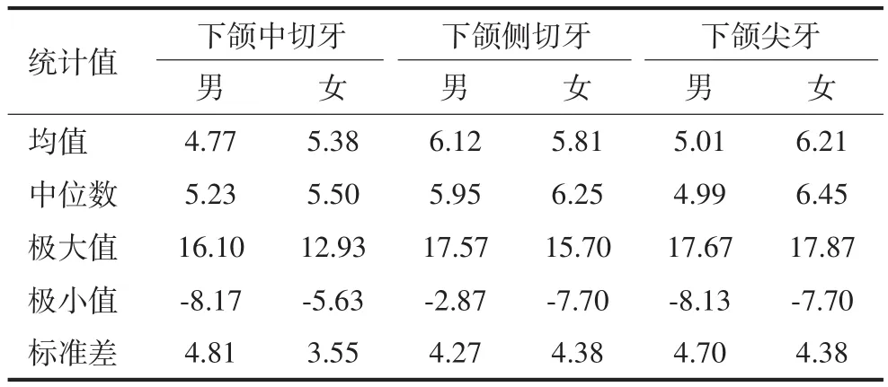
目的 利用锥形束CT(CBCT)研究正中矢状面下颌前牙与牙槽骨的相对位置关系,为下颌前牙区的种植修复设计提供参考。方法 从CBCT影像库中选取下颌恒牙列完整的影像学资料150例,按照性别、牙位分为6组,以受测牙正中矢状面的牙槽骨长轴为基准,测量下颌前牙牙体长轴相对于牙槽骨长轴的倾斜角度(偏舌侧为正,偏唇侧为负)。使用SPSS 19.0软件对数据进行统计学分析。结果 男性下颌中切牙、下颌侧切牙、下颌尖牙牙体长轴相对于牙槽骨长轴倾斜角度的平均值分别为4.77°(-8.17°~16.10°)、6.12°(-2.87°~17.57°)、5.01°(-8.13°~17.67°),女性倾斜角度的平均值依次为5.38°(-5.63°~12.93°)、5.81°(-7.70°~15.70°)、6.21°(-7.70°~17.87°)。其中,男性下颌中切牙、下颌侧切牙、下颌尖牙倾斜角度在-10°~10°区间的百分比依次为87.34%、80.67%、88.00%,女性依次为90.67%、82.66%、82.66%。无论男性还是女性,下颌前牙牙体长轴相对于牙槽骨长轴倾斜角度在(-10°~10°)的百分比均超过80%,且牙体长轴偏向唇侧均不超过10°,偏向舌侧均不超过20°。结论 绝大多数下颌前牙牙体长轴与牙槽骨长轴的方向基本一致,临床上可以参考二者的位置关系制定合理的种植治疗方案。
下颌前牙; 牙体长轴; 牙槽骨长轴; 正中矢状面
牙齿按一定的规律生长在牙槽窝内,形成弧形排列的上、下牙弓,进而行使功能。对前牙区牙齿的唇舌向倾斜角度来说,上下颌切牙均向唇侧倾斜,上下颌尖牙位置则相对较正;但牙体长轴与牙槽骨长轴方向是否具有一致性,目前的观点尚未达成一致。有学者[1]认为,上下颌前牙与颌骨前端牙槽突的倾斜方向一致;还有学者[2-5]通过对上颌前牙锥形束CT(cone beam computed tomography,CBCT)影像进行测量分析发现,上颌前牙与牙槽骨存在一定的倾斜角度。目前对上颌前牙与牙槽骨位置关系的文献报道[2-5]较多,但对下颌前牙区的研究较少。本研究利用CBCT对下颌前牙与牙槽骨的位置关系进行研究,以便为下颌前牙区的种植修复设计提供数据参考。
1 材料和方法
1.1 研究对象的选择
从山东大学口腔医院CBCT影像库中,筛选出150例符合标准的影像学资料,男女各75例,年龄19~48岁,其中19~28岁、29~38岁、39~48岁男女各25例。所选择的CBCT影像均应符合以下标准:1)牙列完整的下颌恒牙列;2)牙齿无扭转、错位,无明显牙列拥挤或牙间隙;3)牙齿萌出正常且牙根发育无异常;4)无龋坏或非龋性牙体缺损,无充填体及修复体;5)无牙周炎、根尖周炎、牙根内外吸收等病理性改变;6)无外伤史或正畸、正颌治疗史;7)CBCT影像清晰,无伪影。
1.2 测量方法
使用Galileos自带的SIDEXIS XG2.53软件进行图像的重建与处理。选取受测牙水平横断面,将横断面调整至受测牙牙颈部(图1A),选取平分受测牙近远中的平面作为矢状向平面,并调整角度使其通过牙体长轴(图1B)。该矢状向平面为受测牙的测量平面,即受测牙的正中矢状面[5](图1C)。
在受测牙的正中矢状面上定点[5],如图2所示。A点:下颌前牙切缘中点;B点:下颌前牙根尖点;C点:下颌前牙唇侧牙槽嵴顶点;D点:下颌前牙舌侧牙槽嵴顶点;E点:位于下颌唇侧骨壁上,与下颌前牙根尖点的连线为最短距离的点;F点:位于下颌舌侧骨壁上,与下颌前牙根尖点的连线为最短距离的点;G点:C、D连线中点;H点:E、F连线中点。β角:AB连线(牙体长轴)与GH连线(牙槽骨长轴)的夹角;相对于GH连线,AB连线偏舌侧时β角设为正值,偏唇侧时β角设为负值。所有数据均由同一位测量者进行测量,每颗牙齿测量3次,取平均值作为最终测量值。

图1 下颌前牙测量平面的定位Fig1 Orientating the measurement plane of the mandibular anterior teeth
1.3 统计学分析
使用SPSS 19.0统计软件包进行统计分析。对测量所得数据按不同性别、不同牙位(12个组,每组75颗牙齿)进行正态性检验,均符合正态性分布;再对相同性别同名牙位进行t检验,结果无统计学差异,因此将相同性别同名牙位的数据合并,最终有6个组,每组150颗牙齿,对所得数据进行统计学描述。

图2 定点及β角的测量Fig 2 Localization of landmark points and the measurement of the angle β
2 结果
以受测牙正中矢状面牙槽骨长轴为基准,下颌前牙牙体长轴倾斜角度β的测量结果见表1。下颌前牙区牙体长轴舌向倾斜的角度均不超过20°,唇向倾斜角度均不超过10°。

表 1 下颌前牙牙体长轴相对于牙槽骨长轴唇舌向倾斜角度β的测量值Tab1 The internal angle β formed by the long axis of the mandibular anterior teeth and the alveolar bone °, n=150
根据表1结果,将倾斜角度β划分为-10°≤β<0°、0°≤β≤10°、10°<β≤20° 3大类,分别记为第1类、第2类、第3类。下颌前牙区牙体长轴倾斜角度β在各区间分布百分比见表2。

表 2 下颌前牙牙体长轴相对于牙槽骨长轴倾斜角度β分布百分比Tab 2 Frequency distribution of angle β formed by the long axis of the mandibular anterior teeth and the alveolar bone %
由表2可见,下颌前牙区牙齿倾斜角度较小,在10°以内者占80%以上。
3 讨论
下颌前牙区易受外伤、牙周病等影响,是种植修复的常见区域。下颌前牙区颌骨骨质致密,具有双层皮质骨,种植体易获得理想的初期稳定性;但是下颌骨的血供相对较差,不利于种植体的骨结合;同时,下颌骨正中的营养孔处有舌下动脉与颏下动脉吻合,如果术中受到损伤,可能引发口底出血,处理不及时,可导致窒息。
下颌前牙区单颗牙齿缺失,尤其是单颗下颌中切牙缺失后,往往近远中向骨宽度不足;此外,下颌骨唇侧骨板均由牙槽嵴顶向颏隆凸呈凹形弯曲,舌侧骨板形态多样,可呈凸型、平坦型及凹形。下颌骨的这种解剖结构决定了种植体植入位置的微小偏差,即有可能导致出现邻牙损伤,唇舌侧骨板穿通等不利后果,影响种植成功率。同时,下颌前牙亦属于美学区,种植时应综合考虑软硬组织的状况,从而保证长期的美学效果。由此可见,下颌前牙种植并不属于种植的简易区,术前需利用CBCT对种植术区的条件进行充分评估。
下颌前牙拔除后长时间的缺牙状态和软硬组织的变化会带给患者美观、发音和社交上的困扰。同时,前牙唇侧骨板多为束状骨,动物实验[6]显示,天然牙拔除后,牙周膜纤维被切断,阻断了束状骨的血供,同时固有牙槽骨失去了功能性刺激,导致骨板吸收,牙槽骨唇舌向骨厚度降低,增加了种植的难度。即刻种植可以明显缩短治疗疗程,减少手术次数,从而减少患者的痛苦程度,具有无可取代的优势。即刻种植虽然不能阻止种植体唇侧骨板的吸收,但可以有效减少骨吸收量[7],同时在严格把握适应证的前提下,即刻种植的成功率几乎可以与延期种植相媲美[8]。对于前牙区,即刻种植一般应选择厚牙龈生物型且唇侧骨壁完整、骨壁厚度超过1 mm的病例,最大程度地降低牙龈退缩的风险,减少美学并发症的发生。由于解剖生理学因素的限制,即刻种植一般不作为前牙区的常规治疗方案,而是建议行早期种植[9]。
无论即刻种植还是早期种植,种植体植入在理想的三维位置是获得良好的临床修复效果的必备因素之一。本研究的目的即是探讨下颌前牙牙体长轴与牙槽骨长轴的位置关系,指导种植体植入理想的三维位置,从而为下前牙即刻或早期种植获得最优的临床效果提供数据参考。本研究发现,绝大多数下前牙牙体长轴与牙槽骨长轴方向基本一致,有将近20%的下颌前牙与牙槽骨方向存在一定的倾斜角度。笔者依据倾斜方向以及倾斜角度β将其分为3类。
相对于牙槽骨长轴来说,第1、2类人群的牙体长轴唇舌侧倾斜角度在10°以内(80%以上的下颌前牙),倾斜角度相对较小,可视为牙齿与牙槽骨倾斜方向基本一致。微创拔除患牙后,当唇侧骨板保存完整且厚度大于1 mm时,可采用小翻瓣或无翻瓣技术,将种植体沿牙体长轴方向略偏舌侧植入,既可以获得最大的骨接触面积,获得良好的初期稳定性,又可以避免种植体对唇侧骨板的压力,防止产生微裂纹而导致骨质丧失[10];同时于种植体与唇侧骨板间植入骨粉,进行骨增量,维持唇侧骨板的高度[9],降低牙龈退缩的风险。
第3类人群牙体长轴与牙槽骨长轴的倾斜角度较大,唇侧根方骨板菲薄,即刻种植时,顺着牙体长轴方向植入,存在唇侧骨板穿通的风险。为了规避对唇侧骨板的损伤,同时获得良好的初期稳定性,通常需要行大翻瓣手术,暴露术区唇侧骨板后,种植体与牙体长轴成一定角度植入,后期采用角度基台调整方向,但角度不应超过20°,否则将出现修复困难[11];同时于唇侧骨板外进行骨增量,代偿皮质骨的吸收,维持唇侧丰满度,保证软硬组织长期稳定性。当唇侧骨板缺损,初期稳定性差时,建议对拔牙窝进行位点保留结合延期种植,临床修复效果更为可靠[12]。
对于下颌前牙区进行延期种植的患者,应通过CBCT对缺牙区牙槽骨状况及对侧同名牙牙槽骨与牙体长轴位置关系进行综合评估,设计种植体型号、植入位置及方向角度,依据下前牙唇舌向骨厚度选择小直径种植体[13]、引导骨再生或块状骨移植等骨增量技术,以获得良好的临床治疗效果。
[1] 王美青. 口腔解剖生理学[M]. 7版. 北京: 人民卫生出版社, 2012:75-76.Wang MQ. Oral anatomy and physiology[M]. 7th ed. Beijing: People’s Medical Publishing House, 2012:75-76.
[2] Lau SL, Chow J, Li W, et al. Classification of maxillary central incisors-implications for immediate implant in the esthetic zone[J]. J Oral Maxillofac Surg, 2011, 69(1):142-153.
[3] Kan JY, Roe P, Rungcharassaeng K, et al. Classification of sagittal root position in relation to the anterior maxillary osseous housing for immediate implant placement: a cone beam computed tomography study[J]. Int J Oral Maxillofac Implants, 2011, 26(4):873-876.
[4] 朱一博, 邱立新. 上颌切牙解剖分型与种植方案的选择[J]. 中华口腔医学杂志, 2013, 48(4):223-225.Zhu YB, Qiu LX. Anatomical classification of maxillary incisorsand options of implant strategy[J]. Chin J Stomatol, 2013, 48(4):223-225.
[5] 刘金, 蒲必双, 夏玉兰, 等. 上颌前牙倾斜角度对种植治疗设计的影响[J]. 华西口腔医学杂志, 2016, 34(6):611-616.Liu J, Pu BS, Xia YL, et al. Evaluation of maxillary anterior teeth by cone-beam computed tomography[J]. West Chin J Stomatol, 2016, 34(6):611-616.
[6] Araújo MG, Lindhe J. Dimensional ridge alterations following tooth extraction. An experimental study in the dog[J]. J Clin Periodontol, 2005, 32(2):212-218.
[7] Araújo MG, Wennström JL, Lindhe J. Modeling of the buccal and lingual bonewalls of fresh extraction sites following implant installation[J]. Clin Oral Implants Res, 2006, 17(6):606-614.
[8] Esposito M, Grusovin MG, Polyzos IP, et al. Timing of implant placement after tooth extraction: immediate, immediatedelayed or delayed implants? A Cochrane systematic review[J]. Eur J Oral Implantol, 2010, 3(3):189-205.
[9] 李德华. 前牙区种植选择即刻种植还是早期种植[J]. 中华口腔医学杂志, 2013, 48(4):200-202.Li DH. Immediate or early implant placement in the anterior region[J]. Chin J Stomatol, 2013, 48(4):200-202.
[10] Kohal RJ, Hürzeler MB, Mota LF, et al. Custom-made root analogue titanium implants placed into extraction sockets. An experimental study in monkeys[J]. Clin Oral Implants Res, 1997, 8(5):386-392.
[11] Almog DM, Torrado E, Meitner SW. Fabrication of imaging and surgical guidesfor dental implants[J]. J Prosthet Dent, 2001, 85(5):504-508.
[12] Hämmerle CH, Chen ST, Wilson TG Jr. Consensus statements and recommendedclinical procedures regarding the placement of implants in extraction sockets[J]. Int J Oral Maxillofac Implants, 2004, 19(Supp l):26-28.
[13] 高岩, 徐淑兰, 周磊, 等. Osstem MS一段式种植体用于下前牙小间隙种植修复的临床回顾研究[J]. 实用口腔医学杂志, 2015, 31(5):639-643. Gao Y, Xu SL, Zhou L, et al. A clinical retrospective study on Osstem MS one-stage implant restoration of small edentulous space in the mandibular anterior region[J]. J Pract Stomatol, 2015, 31(5):639-643.
Influence of positional relationship between the long axis of the mandibular anterior teeth and the alveolar bone on the treatment design of dental implants
Yu Tiantian1, Pu Bishuang1, Liu Jin2, Xia Yulan3, Wang Gang4, Lan Jing1.
(1. Dept.of Implantology, School of Stomatology, Shandong University, Shandong Provincial Key Laboratory of Oral Tissue Regeneration, Jinan 250012, China; 2. Dept. of Prosthodontics, School of Stomatology, Shandong University, Shandong Provincial Key Laboratory of Oral Tissue Regeneration, Jinan 250012, China; 3. Dept. of Implantology, Hefei Stomatology Hospital,Hefei 230000, China; 4. Dept. of Oral and Maxillofacial Surgery, Shandong Provincial Hospital, Jinan 250012, China)
Correspondence: Lan Jing, E-mail: kqlj@sdu.edu.cn.
Objective This study aimed at investigating and measuring the positional relationship between the long axis of the mandibular anterior teeth and the alveolar bone using cone beam computed tomography (CBCT) to provide reference data for implant treatment. Methods From the CBCT image database, 150 cases of radiographic data were selected according to the inclusion criteria and then were divided into six groups: males’ mandibular central incisors, males’ mandibular lateral incisors, males’ mandibular canines, females’ mandibular central incisors, females’ mandibular lateral incisors, and females’ mandibular canines. The angle (β) formed by the long axis of the mandibular anterior teeth and the corresponding alveolar bone was measured and recorded. Based on the long axis of alveolar bone, if the teeth incline to the lingual side, the value of the angle (β) was positive; otherwise, the value was negative. The resultant data were analyzed by SPSS 19.0. Results The β of the mandibular central incisors presented a mean value of 4.77° (range: -8.17°-16.10°) for male subjects and 5.38° (range: -5.63°-12.93°) for female subjects. The β of the mandibular lateral incisors exhibited a mean value of 6.12° (range: -2.87°-17.57°) for male subjects and 5.81° (range: -7.70°-15.70°) for female subjects. Finally, the β of the mandibular canines presented a mean value of 5.01° (range: -8.13°-17.67°) for male subjects and 6.21° (range: -7.70°-17.87°) for female subjects. The percentages of the β between -10° and 10° of males’ mandibular incisors, mandibular lateral incisors, and mandibular canines were 87.34%, 80.67%, and 88.00%, respectively and those of females were 90.67%, 82.66%, and 82.66%, respectively. Whether male or female, the percentages of the β between-10° and 10° of the mandibular anterior teeth were more than 80%. The β that inclined to the lingual was not more than 20° and to the labial did not exceed 10°. Conclusion The long axis of the mandibular anterior teeth was almost consistent with the long axis of the alveolar bone. Therefore, the positional relationship could be referred to make reasonable implants treatment plan.
the mandibular anterior; the long axis of the tooth; the long axis of the alveolar bone; the median sagittal plane
R 783
A
10.7518/hxkq.2017.06.008
2017-05-16;
2017-08-22
于甜甜,硕士,E-mail:ytttian@126.com
兰晶,主任医师,博士,E-mail:kqlj@sdu.edu.cn
(本文编辑 吴爱华)

