窄带成像技术膀胱镜与普通白光膀胱镜诊疗非肌层浸润性膀胱肿瘤价值的meta分析
王云汉 李响
1四川大学华西医院泌尿外科 610000 成都
论 著
窄带成像技术膀胱镜与普通白光膀胱镜诊疗非肌层浸润性膀胱肿瘤价值的meta分析
王云汉1李响1
1四川大学华西医院泌尿外科 610000 成都
目的:系统探索评价窄带成像技术膀胱镜(NBI)膀胱镜相对于普通白光膀胱镜(WLI)在非肌层浸润性膀胱肿瘤(NMIBC)患者的诊疗价值。方法:收集PubMed、Medline、EMBASE,CNKI(中国国家知识基础设施数据库)和万方数字化期刊群在2016年8月前国内外公开发表的有关NBI与WLI对非肌层浸润性膀胱肿瘤的诊疗疗效比较的临床对照研究文献,使用以下搜索关键词:“bladder cancer”、“White-Light Imaging”、“Narrow Band Imaging”。此外,手工检索相关的相关的参考书目检索所有发表评论和文章,通过Revman 5.3软件进行meta分析。结果:共计20篇文献纳入研究,其中随机对照研究4篇,系统回顾性研究16篇;在非肌层浸润性膀胱肿瘤患者检出率的比较中,纳入研究对象NBI组560例和与WLI组560例,RD=0.12,[95% CI:0.091,0.12],P<0.000 01,I2=0%;在原位膀胱肿瘤患者检出率的比较中,纳入研究对象NBI组360例和与WLI组334例,RD=0.17,[95% CI:0.11,0.23],P<0.000 01,I2=66%;在TURBT术后复发的比较中,NBI组与WLI组分别有754例和764例研究对象纳入,OR=0.62,[95% CI:0.45,0.86],P=0.005,I2=55%。结论:NBI膀胱镜在非肌层浸润性膀胱肿瘤患者检出率和原位膀胱肿瘤患者检出率中相对于WLI膀胱镜具有明显优势,并且能够降低TURBT术后患者肿瘤复发率。
Meta分析;非肌层浸润性膀胱肿瘤;窄带成像技术膀胱镜;普通白光膀胱镜
膀胱肿瘤是泌尿系统常见肿瘤之一,全球每年约有150 000例新诊患者。2016年NCCN指南[1]中指出,膀胱肿瘤在亚洲国家肿瘤发病率排名14位,欧美等发达国家发病率更高;在我国,随着国民健康意识和诊疗水平的不断提高,膀胱肿瘤在我国确诊率也在逐年上升,位居我国男性肿瘤发病率第4位。而对于膀胱肿瘤的诊疗,白光膀胱镜(white-light Imaging, WLI)一直是确诊膀胱肿瘤和经尿道膀胱肿瘤电切(transurethral resection of bladder cancer, TURBT)的首选方法[2],但这种传统的内镜方法在诊断早期微小肿瘤、膀胱原位癌(carcinomainsitu, CIS)等病变时灵敏度并不高,并且在WLI-TURBT中,由于肿瘤累及范围无法有效识别,易致使切除肿瘤范围不够而造成膀胱肿瘤残留和复发。然而随着内镜技术的不断发展与提高,窄带成像膀胱镜(narrow band imaging, NBI)通过窄带成像技术辅助下,在诊断膀胱肿瘤灵敏度、特异度以及降低术后肿瘤复发率等方面有着较强的优势[3],从而在泌尿外科膀胱肿瘤诊疗中应用越来越广泛。因此,为系统评价NBI与WLI在膀胱肿瘤诊治中的临床价值,我们通过收集既往文献报道研究结果行meta分析,综合分析其结果,以此为临床医生对于膀胱肿瘤的诊治方式选择提供循证医学依据。
1 资料与方法
1.1 临床资料
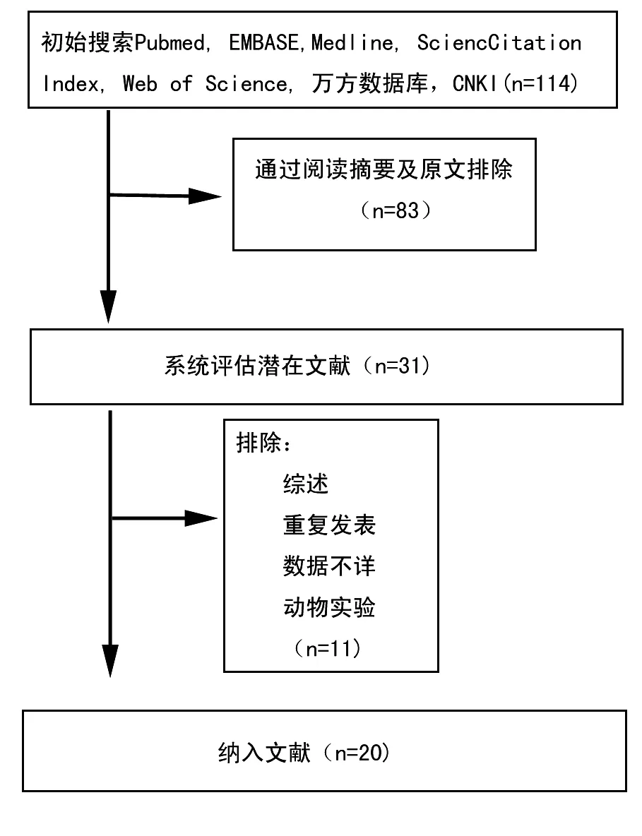
收集PubMed、Medline、EMBASE、CNKI(中国国家知识基础设施数据库)和万方数字化期刊群在2016年7月前国内外公开发表的有关NBI与WLI对非肌层浸润性膀胱肿瘤的诊疗疗效比较的临床对照研究文献。使用以下搜索关键词:“bladder cancer”、“White-Light Imaging”、“Narrow Band Imaging”。此外,手工检索相关的相关的参考书目检索所有发表评论和文章(图1)。
1.2 文献纳入与排除
纳入标准①研究类型和干预措施:比较膀胱肿瘤患者实施NBI与WLI的临床对照研究,无语种限制;②病例对象:非肌层浸润性膀胱肿瘤患者,排除肿瘤已远处和淋巴转移、既往有其他肿瘤病史患者;③终点类型:最大随访期内的肿瘤复发人数;④组织病理诊断:病理确诊为膀胱肿瘤。
剔除标准:①文摘、综述、讲座类文献;②数据不全文献;③个案报道;④动物实验;⑤只实施NBI或者WLI膀胱镜诊疗临床研究报道;⑥确诊膀胱肿瘤不是以活检或者术后病理组织学检查的研究。

图1 文献纳入与排除
1.3 统计学方法
采用Revman 5.3软件进行Meta分析,对两种内镜方式对膀胱肿瘤患者的肿瘤检测率、原位癌检测率采用Risk Difference(RD)以及TURBT术后复发率采用Odds Ratio(OR)及其95% CI为疗效分析统计量。P≤0.05为差异有统计学意义。应用 Revman 5.3对每项研究数据进行同质性检验,若研究数据能通过同质性检验,则采用固定效应模型进行分析,否则采用随机效应模型进行分析。
2 结果
2.1 纳入文献及质量评价
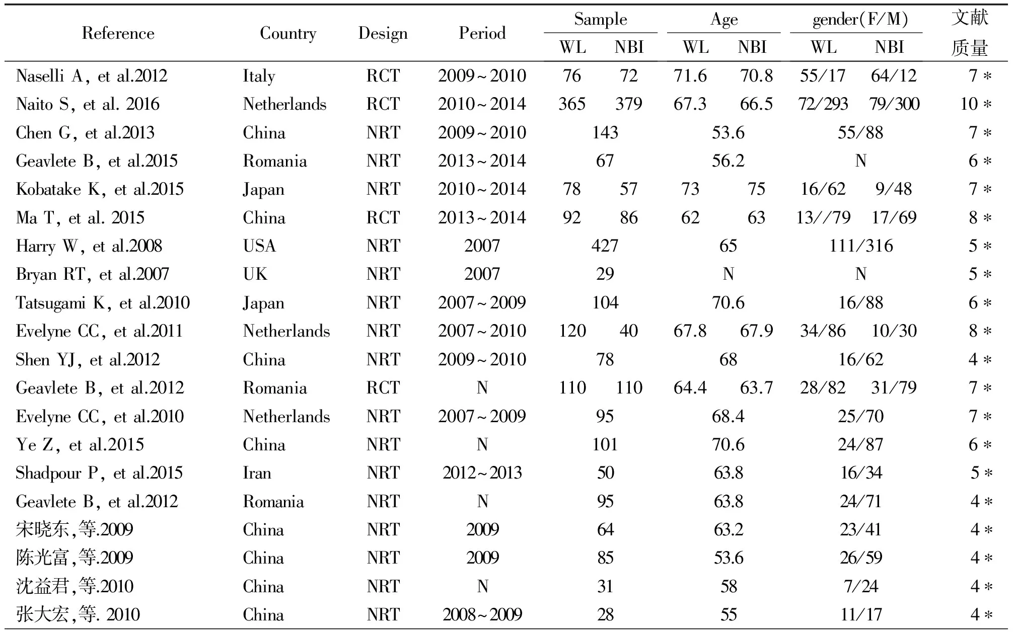
总共涉及114篇文献关于NBI与WLI诊疗非肌层浸润性膀胱肿瘤的临床文献研究,最后获得20篇[4~23]符合要求的研究,提取文献中的肿瘤患者检出率、原位癌检测率,以及TURBT术后复发人数等数据,并进行统计学处理。文献质量评价采用NOS量表[24]对所纳入的20篇文献进行质量评价,研究对象的选择有较好的代表性,具体情况见表1。
2.2 膀胱肿瘤检出率
在检出率的meta分析中,NBI组与WLI组分别有560例和520例研究对象纳入,对纳入的相关数据进行异质性检验,采用固定效应模型进行分析,RD=0.12,[95% CI:0.091,0.12],P<0.000 01,I2=0;在患者检出率中有统计学意义,NBI明显优于WLI(图2)。
2.3 原位膀胱肿瘤患者检出率
在原位膀胱肿瘤患者检出率的meta分析中,NBI组与WLI组分别有360例和334例研究对象纳入,对纳入的相关数据进行异质性检验,采用随机效应模型进行分析,RD=0.17,[95% CI:0.11,0.23],P<0.000 01,I2=66%;在原位膀胱肿瘤患者检出率中有统计学意义,NBI明显优于WLI。
2.4 TURBT术后膀胱肿瘤复发率率
在TURBT术后膀胱肿瘤复发率的meta分析中,NBI组与WLI组分别有754例和764例研究对象纳入,对纳入的相关数据进行异质性检验,采用随机效应模型进行分析,OR=0.62,[95% CI:0.45,0.86],P=0.005,I2=55%;在TURBT术后膀胱肿瘤复发率中差异有统计学意义,NBI明显优于WLI。

表1 纳入文献及质量评价

图2 膀胱肿瘤检出率(患者水平)

图3 原位膀胱肿瘤检出率(患者水平)

图4 TURBT术后膀胱肿瘤复发率率(患者水平)
2.5 偏倚
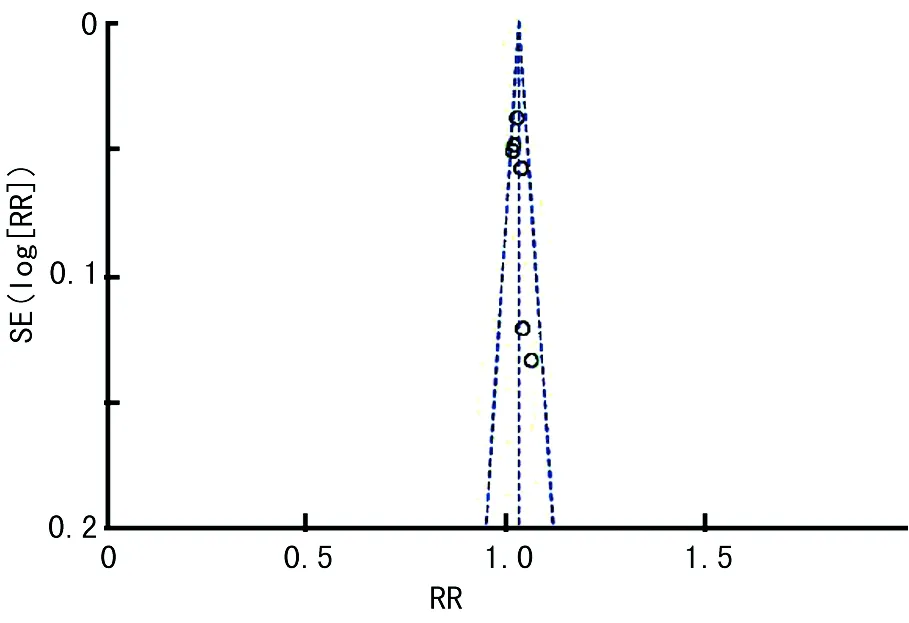
采用RevMan 5.3软件软件的漏斗图对纳入的研究分别进行测量,各指标的图形基本对称,所有点基本集中在漏斗中部,但纳入研究均为已经公开发表的文献,不能避免发表偏倚(图5)。

图5 偏倚分析
3 讨论
膀胱肿瘤是泌尿系肿瘤当中的常见肿瘤之一,其发病率在我国呈逐年上升的趋势;而长期以来,WLI膀胱镜一直作为膀胱肿瘤诊断与手术的首选诊疗方式,其在膀胱肿瘤检出率与肿瘤范围定位中,一直存在着一定的不足,泌尿外科医生一直在寻求一种更为准确的诊疗方式。而NBI膀胱镜通过将普通白光滤过分解成为540 nm的绿光光带和415 nm的蓝光光带,从而能够使得正常的膀胱黏膜与癌变组织对比度增强,从而使得黏膜形态成像更为清楚,NBI已经广泛应用于消化道疾病的内镜检查中[25],而在泌尿外科的诊疗中也逐渐受到重视。
本次研究中,NBI膀胱镜检查对膀胱肿瘤患者的检出率明显优于WLI膀胱镜[RR=1.17,(95% CI: 1.11,1.23),P<0.000 01];在原位膀胱肿瘤患者的检出率中,NBI也具有相对优势[RR=1.47,(95% CI: 1.27, 1.69),P<0.000 01]。分析其原因,我们认为在NBI膀胱镜检查中,单一的白光被分解成415~540 nm窄光带,其光波更容易被黏膜中血红蛋白吸收,膀胱肿瘤相对于正常黏膜组织有着更加丰富的血供及黏膜表面微血管改变,而在特殊光带作用下,癌变组织与正常组织对比显像更为清晰,因而肿瘤患者在NBI膀胱镜中的检出率明显优于WLI膀胱镜检查。Tatsugami等[17]在一项回顾性分析中,研究共计纳入313例病理组织活检,NBI膀胱镜在肿瘤检出中灵敏度为(98.4%vs. 85.4%),阳性率(64.5%vs. 61.2%),由此看出NBI在膀胱肿瘤诊断中明显优于WLI膀胱镜检,这与我们本次结论相符合。此外,大部分非肌层浸润性膀胱肿瘤虽然可以经TURBT治疗,但术后依然存在较高的复发率。在本次研究中,NBI膀胱镜辅助下TURBT术后患者的膀胱肿瘤复发率明显低于WLI膀胱镜[RR=0.67,(95% CI:0.50,0.89),P=0.006],究其原因,我们认为NBI膀胱镜能够更好地显示异常黏膜组织[26],有助于术者对膀胱肿瘤界限判断;而在WLI膀胱镜中,术者只能通过内镜显示将主要病变切除,肿瘤周围组织仅依据术者经验性切除,从而致使一些微小膀胱肿瘤病变残留,由此一定程度上增加的膀胱肿瘤复发概率;但是在NBI膀胱镜的辅助下,术者能够准确定位肿瘤位置以及切除可疑膀胱肿瘤癌变组织,从而降低肿瘤术后复发率。在一项多中心的随机对照研究中,Naito等[4]纳入985例患者,最长随访时间为1年,分别纳入WLI组共计481例患者和NBI组共计484例患者,其TURBT术后1年复发率为(27.3%vs. 5.6%;P=0.002),由此看出,NBI膀胱镜辅助TURBT术后肿瘤复发率明显降低,这与本次研究结果显示一致。与此同时,NBI只需要通过光学过滤即可以达到增强正常组织与肿瘤细胞的对比,但有研究指出相对于普通NBI和WLI膀胱镜检查,荧光膀胱镜或者光学动力学膀胱镜通过荧光染色如5-ALA等,更能够大幅度提高早期微小膀胱肿瘤诊断率,其检出率可达到84.4%[27],但是值得注意的是,荧光膀胱镜检查时需要膀胱灌注价格昂贵的荧光染料,由此操作繁琐、成本较高,不易推广;而NBI膀胱镜检查不仅具有较高的膀胱肿瘤检出率,还具有操作简单、学习曲线短、价格适宜的优点[28],不可否认,NBI在膀胱肿瘤诊断中更具有较强优势,值得推广。
由于meta分析属于观察性研究,其结果受到许多因素的影响,本研究仍有以下不足:①纳入的文献量和样本量不足,仍需要大样本试验研究;②文献多为回顾性研究,希望将来有更多高质量随机对照研究,尽管纳入随机对照研究4篇,但仍然相对较少;③纳入研究中样两组治疗方式患者在年龄、肿瘤大小、BMI等方面无法完全匹配,由此可能带来误差;④文献随访时间为短中期随访,期待长时间随访结果统计;以上不足都可致使结果偏倚和异质性,我们期待更多高质量、长时间随机对照试验来NBI和WLI膀胱镜对非肌层浸润性膀胱肿瘤的诊疗提供更为可靠的证据。
总之,NBI膀胱镜在非肌层浸润性膀胱肿瘤患者检出率和原位膀胱肿瘤患者检出率中相对于WLI膀胱镜具有明显优势,并且能够降低TURBT术后患者肿瘤复发率。
[1]Clark PE, Spiess PE, Agarwal N, et al. NCCN Guidelines Insights: Bladder Cancer, Version 2.2016. J Natl Compr Canc Netw, 2016,14(10):1213-1224.
[2]Hafner C, Knuechel R, Zanardo L, et al. Evidence for oligoclonality and tumor spread by intraluminal seeding in multifocal urothelial carcinomas of the upper and lower urinary tract. Oncogene, 2001,20(35):4910-4915.
[3]Gono K, Obi T, Yamaguchi M, et al. Appearance of enhanced tissue features in narrow-band endoscopic imaging. J Biomed Opt, 2004,9(3):568-577.
[4]Naito S, Algaba F, Babjuk M, et al. The Clinical Research Office of the Endourological Society (CROES) Multicentre Randomised Trial of Narrow Band Imaging-Assisted Transurethral Resection of Bladder Tumour (TURBT) Versus Conventional White Light Imaging-Assisted TURBT in Primary Non-Muscle-invasive Bladder Cancer Patients: Trial Protocol and 1-year Results. Eur Urol, 2016,70(3):506-515.
[5]Shadpour P, Emami M, Haghdani S. A Comparison of the Progression and Recurrence Risk Index in Non-Muscle-Invasive Bladder Tumors Detected by Narrow-Band Imaging Versus White Light Cystoscopy, Based on the EORTC Scoring System. Nephrourol Mon, 2016,8(1):e33240.
[6]Geavlete B, Bulai C, Ene C, et al. The Influence of NBI Vision Over the First Follow-up Cystoscopy Outcomes in Newly Diagnosed NMIBC Patients. Chirurgia (Bucur), 2015,110(2):157-160.
[7]Kobatake K, Mita K, Ohara S, et al. Advantage of transurethral resection with narrow band imaging for non-muscle invasive bladder cancer. Oncol Lett, 2015,10(2):1097-1102.
[8]Ma T, Wang W, Jiang Z, et al. [Narrow band imaging-assisted holmium laser resection reduced the recurrence rate of non-muscle invasive bladder cancer: a prospective, randomized controlled study]. Zhonghua Yi Xue Za Zhi (Chinese), 2015,95(37):3032-3035.
[9]Ye Z, Hu J, Song X, et al. A comparison of NBI and WLI cystoscopy in detecting non-muscle-invasive bladder cancer: A prospective, randomized and multi-center study. Sci Rep, 2015,5:10905.
[10]Chen G, Wang B, Li H, et al. Applying narrow-band imaging in complement with white-light imaging cystoscopy in the detection of urothelial carcinoma of the bladder. Urol Oncol, 2013,31(4):475-479.
[11]Geavlete B, Jecu M, Multescu R, et al. Narrow-band imaging cystoscopy in non-muscle-invasive bladder cancer: a prospective comparison to the standard approach. Ther Adv Urol, 2012,4(5):211-217.
[12]Geavlete B, Multescu R, Georgescu D, et al. Narrow band imaging cystoscopy and bipolar plasma vaporization for large nonmuscle-invasive bladder tumors--results of a prospective, randomized comparison to the standard approach. Urology, 2012,79(4):846-851.
[13]Naselli A, Introini C, Timossi L, et al. A randomized prospective trial to assess the impact of transurethral resection in narrow band imaging modality on non-muscle-invasive bladder cancer recurrence. Eur Urol, 2012,61(5):908-913.
[14]Shen YJ, Zhu YP, Ye DW, et al. Narrow-band imaging flexible cystoscopy in the detection of primary non-muscle invasive bladder cancer: a "second look" matters?. Int Urol Nephrol, 2012,44(2):451-457.
[15]Cauberg EC, Mamoulakis C, de la Rosette JJ, et al. Narrow band imaging-assisted transurethral resection for non-muscle invasive bladder cancer significantly reduces residual tumour rate. World J Urol, 2011,29(4):503-509.
[16]Cauberg EC, Kloen S, Visser M, et al. Narrow band imaging cystoscopy improves the detection of non-muscle-invasive bladder cancer. Urology, 2010,76(3):658-663.
[17]Tatsugami K, Kuroiwa K, Kamoto T, et al. Evaluation of narrow-band imaging as a complementary method for the detection of bladder cancer. J Endourol, 2010,24(11):1807-1811.
[18]沈益君,朱一平,叶定伟,等.窄波成像电子膀胱软镜在膀胱肿瘤诊断中的应用价值.中华泌尿外科杂志,2010,31(6):383-385.
[19]张大宏,张琦,刘峰.窄光成像膀胱镜在膀胱肿瘤诊断中的应用.中华泌尿外科杂志,2010,31(3):182-184.
[20]陈光富,王保军,马鑫,等.NBI技术结合电子软膀胱镜在膀胱肿瘤早期诊断中的应用研究.临床泌尿外科杂志,2009,24(8):603-605.
[21]宋晓东,叶章群,包世新,等.窄带光成像在膀胱癌早期诊断及复发监测中的应用研究.现代泌尿生殖肿瘤杂志,2009,1(6):325-327.
[22]Bryan RT, Billingham LJ, Wallace DM. Narrow-band imaging flexible cystoscopy in the detection of recurrent urothelial cancer of the bladder. BJU Int, 2008,101(6):702-705, 705-706.
[23]Herr HW, Donat SM. A comparison of white-light cystoscopy and narrow-band imaging cystoscopy to detect bladder tumour recurrences. BJU Int, 2008,102(9):1111-1114.
[24]Stang A. Critical evaluation of the Newcastle-Ottawa scale for the assessment of the quality of nonrandomized studies in meta-analyses. Eur J Epidemiol, 2010,25(9):603-605.
[25]Hirata I, Nakagawa Y, Ohkubo M, et al. Usefulness of magnifying narrow-band imaging endoscopy for the diagnosis of gastric and colorectal lesions. Digestion, 2012,85(2):74-79.
[26]Mariappan P, Finney SM, Head E, et al. Good quality white-light transurethral resection of bladder tumours (GQ-WLTURBT) with experienced surgeons performing complete resections and obtaining detrusor muscle reduces early recurrence in new non-muscle-invasive bladder cancer: validation across time and place and recommendation for benchmarking. BJU Int, 2012,109(11):1666-1673.
[27]Inoue K, Anai S, Fujimoto K, et al. Oral 5-aminolevulinic acid mediated photodynamic diagnosis using fluorescence cystoscopy for non-muscle-invasive bladder cancer: A randomized, double-blind, multicentre phase II/III study. Photodiagnosis Photodyn Ther, 2015,12(2):193-200.
[28]韦荣超,吴承耀,张振声,等.膀胱镜检查在膀胱癌诊断的研究进展.第二军医大学学报,2012,33(11):1257-1259.
Narrow-band imaging and white light imaging for non-muscle invasive bladder cancer: a meta-analysis
WangYunhan1LiXiang1
(1Department of Urology, West China Hospital, Sichuan Universtity, Chengdu 610000, China)
Corresponding author: Li Xiang, xiangli.87@163.com
Objective: To systematically evaluate the value of cystoscopy by narrow-band imaging (NBI) for non-muscle-invasive bladder caner. Methods: We searched through the major medical databases such as Pub Med, EMBASE, Medline, Science Citation Index, Web of Science and Chinese National Knowledge Infrastructure Database (CNKI) and Wangfang ( Database of Chinese Ministry of Science & Technology) for all published studies from 2007 until July 2016. The following search terms were used: "bladder cancer", "White-Light Imaging", "Narrow Band Imaging" . Furthermore, additional related studies were manually searched in the reference lists of all published reviews and retrieved articles. We used the Review Manager software (RevMan 5.3, Cochrane Collaboration) to carry out the meta-analysis. Results: There were 20 eligible studies included. We found the detection rate on bladder cancer patients was significantly higher in NBI group than in WLI group (RR=1.17, [95% CI: 1.11,1.23],P<0.000 01,I2=0%), and the detection rate of carcinoma in situ also was higher in NBI group than in WLI group (RR=1.47, [95% CI: 1.27,1.69],P<0.000 01,I2=66%). Meanwhile, the recurrence rate of TURBT was significantly lower in NBI group than in WLI group (RR=0.67, [95% CI: 0.50, 0.89],P=0.006,I2=52%). Conclusions: Cystoscopy by NBI was considered to detect more patients of non-muscle-invasive bladder cancer and carcinoma in situ than WLI, and it might reduce the recurrence rate after TURBT.
meta-analysis; non-muscle-invasive bladder cancer; narrow-band imaging; white light imaging.
李响,xiangli.87@163.com
2016-11-29
R737.14
A
10.19558/j.cnki.10-1020/r.2017.04.009

