三维斑点追踪成像左心室应变指标正常参考范围的Meta分析
曹 寅,黄 晶,邹燕珂,廖晴瑶,熊 波,谭 杰
(重庆医科大学附属第二医院心血管内科,重庆 400010)
三维斑点追踪成像左心室应变指标正常参考范围的Meta分析
曹 寅,黄 晶*,邹燕珂,廖晴瑶,熊 波,谭 杰
(重庆医科大学附属第二医院心血管内科,重庆 400010)
目的 采用Meta分析评估健康成人的三维斑点追踪成像(3D-STI)左心室整体纵向应变(GLS)、整体环向应变(GCS)、整体径向应变(GRS)、整体面积应变(GAS)的正常参考范围。方法 在Embase、Pubmed、Cochrane Library等英文资料库中,检索2016年8月1日前公开发表的关于3D-STI评价左心功能的病例对照研究。按照纳入和排除标准,对文献进行筛查和提取数据。以加权均数差(WMD)及95%可信区间(CI)作为合并统计量,采用的统计分析软件为STATA 12.0。结果 最终纳入27项研究,1 552名健康成年被试。GLS的WMD和95%CI为17.80、(16.27,19.33),GCS的WMD和95%CI为24.73、(22.50,26.95),GRS的WMD和95%CI为47.86、(39.52,56.19), GAS的WMD和95%CI为36.17、(34.08,38.26)。结论 通过Meta分析确定成人左心室应变指标的正常参考范围,对采用3D-STI评价疾病状态下心脏结构和功能的变化具有一定的指导意义。
三维斑点追踪成像;心室,左;应变;参考范围
近年来,心血管事件发生率显著增加。早期发现左心室结构重构和功能异常对疾病诊治和预后至关重要。通过三维斑点追踪成像(three-dimensional speckle tracking imaging, 3D-STI)技术可敏感地获得心肌应变参数,准确评估心肌功能[1],可重复性较高[2-3]。目前临床3D-STI已逐步用于左心室功能的评估[4-7]。但采用3D-STI评价成人左心室应变指标仍缺乏正常参考范围,在一定程度上限制了该技术的应用。本研究通过对健康成人3D-STI左心室应变指标进行Meta分析,获得左心室应变指标的正常参考范围,为3D-STI评估左心室功能变化提供参考依据。
1 资料与方法
1.1文献检索 检索Embase、Pubmed、Cochrane Library等数据库于2016年8月1日前公开发表的英文文献。检索词为“three-dimensional speckle tracking”、“left ventricular”;对入选文献的参考文献进行二次检索。
1.2纳入与排除标准 纳入标准:公开发表的英文文献;采用3D-STI技术评价健康个体左心室功能的观察性研究和对照组为健康人群的对照研究;包含左心室应变指标,即整体纵向应变(global longitudinal strain, GLS)、整体环向应变(global circumferential strain, GCS)、整体径向应变(global radial strain, GRS)、整体面积应变(global area strain, GAS);被试者年龄>18岁。排除标准:动物和基础实验研究;缺少左心室应变指标的研究;重复发表的文献;综述、文摘、读者来信;编辑评论、会议论文;非英文文献。
1.3文献筛选及资料提取 根据纳入和排除标准,由2名有经验的工作者对符合标准的文献进行单独筛选,并记录基本资料和左心室应变指标,判断不一致时经协商解决。提取的基本资料包括第一作者、开展研究的国家、发表年限、纳入健康人群或正常对照组样本数、健康人群或正常对照组人群的基本特征和3D-STI左心室应变指标(GLS、GRS、GCS、GAS)。
1.4统计学分析 采用STATA 12.0统计分析软件。以加权均数差(weighted mean difference, WMD)和95%可信区间(confidence interval, CI)为合并统计量。通过I2和Q检验对入选研究进行异质性检验:当I2<50%且P≥0.1为不存在异质性,采用固定效应模型;反之采用随机效应模型分析。存在异质性的研究,对可能造成异质性的因素进行亚组分析。以漏斗图和Egger检验评价发表偏倚;以排除单个研究后观察结果有无显著变化的方法判断敏感度,评价研究成果的稳定性。
2 结果
2.1文献检索及筛选结果 检索数据库初步获得619条文献,排除重复记录的文献328篇,排除综述等非试验性文献和研究对象为非成年人的文献254篇,进一步阅读剩余74篇文献,排除18篇无健康对照组的文献,以及29篇缺少3D-STI整体应变指标的文献;最终纳入27篇[8-34]。
2.2纳入文献基本特征和质量评价 入选的27篇文献中,共纳入1 552名符合条件的健康成年被试者,27篇均包含GLS数据;26篇[8-29,31-34]包含1 522名健康被试者的GCS数据;24篇[8-9,11-29,32-34]包含1 473名健康被试者的GRS数据;18篇[8-9.11-15,17-19,21-22,26-29,32-33]包含983名健康被试者的GAS数据。纳入研究的基本特征见表1。
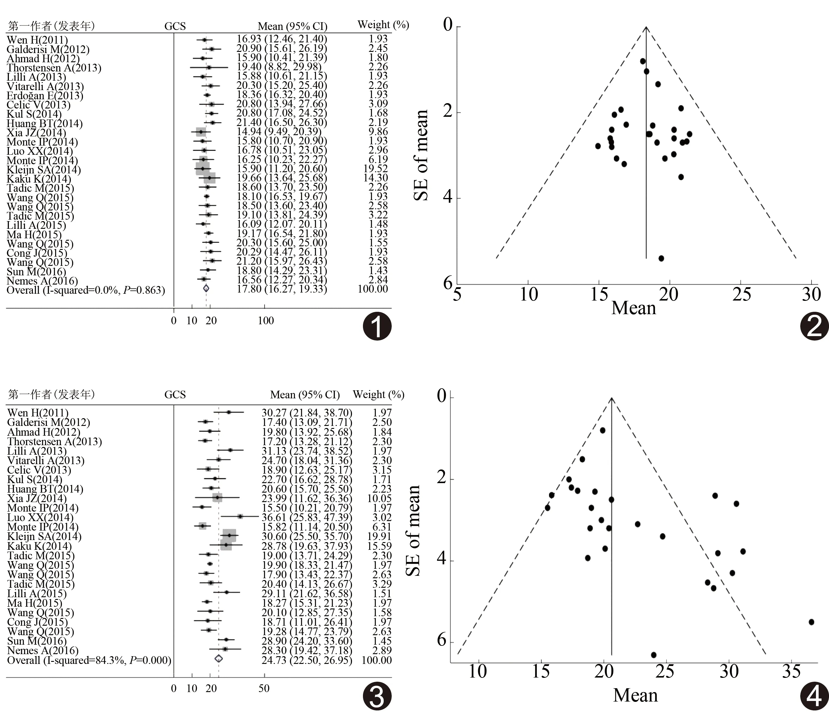
2.3 纳入文献定量数据分析 27篇文献测量GLS的异质性较小(I2=0.0%,P=0.863),选择固定效应模型分析,GLS参考范围森林图见图1[WMD=17.80,95%CI(16.27,19.33)]。根据Egger检验以及漏斗图分布(图2),认为GLS无发表偏倚(P=0.855)。
26篇文献测量的GCS存在异质性(I2=84.3%,P<0.01),选择随机效应模型分析,GCS参考范围森林图见图3[WMD=24.73,95%CI(22.50,26.95)]。根据Egger检验以及漏斗图分布(图4),认为GCS存在发表偏倚(P=0.022)。对不同设备厂家进行亚组分析,GE、Toshiba、Philips组的异质性降低(I2=58.6%、74.8%、75.0%,P=0.002、0.001、0.007),选择随机效应模型分析;3组GCS的WMD和95%CI分别为20.07和(18.98,21.16)、20.46和(18.58,22.33)、25.19和(22.21,28.17)。
24篇文献测量的GRS存在异质性(I2=45.2%,P=0.009),采用随机效应模型,GRS的WMD和95%CI为47.86和(39.52,56.19),见图5。根据Egger检验及漏斗图分布(图6),认为GRS无发表偏倚(P=0.987)。对设备生产厂家进行亚组分析,GE组(I2=38.2%,P=0.066)及Philips组(I2=78.3%,P=0.010)存在异质性,选择随机效应模型分析,Toshiba组(I2=30.8%,P=0.204)异质性减小,选择固定效应模型分析; GE组、Philips组、Toshiba组GRS的WMD和95%CI分别为46.84和(42.87,50.80)、44.73和(40.32,49.13)、42.81和(31.04,54.57)。

表1 纳入研究的基本特征
注:Y:包含;N:不包含
18篇文献测量的GAS存在异质性(I2=57.3%,P=0.001),选择随机效应模型,GAS的WMD和95%CI为36.17和(34.08,38.26),见图7。根据Egger检验结果以及漏斗图两侧分布对称(图8),认为GAS不存在发表偏倚(P=0.771)。根据设备生产厂家进行亚组分析,GE组(I2=35.4%,P=0.092)的异质性较高,选择随机效应模型分析。Toshiba组(I2=47.5%,P=0.126)的异质性较小,选择固定效应模型分析,GE组和Toshiba组GAS的WMD和95%CI分别为32.86和(30.92,34.79)、39.06和(35.59,42.52)。
2.4敏感度分析 剔除单篇纳入文献对剩余文献进行敏感度分析,发现左心室应变参数范围的合并效应指标未发生明显变化,提示入选的文献稳定性好。
3 讨论
3D-STI克服了传统2D超声技术的不足,可更准确地对心脏功能进行实时评估,提供更加准确的结果[4-5,35];相对于传统检查技术,3D-STI具有更高的效率和可重复性[6-7,35]。因其对心肌损伤早期变化和功能异常非常敏感,所以对于许多疾病初期阶段心功能异常的诊断有重要意义。通过3D-STI技术可检测到高血压早期患者左心室容量的增高[21]和肥胖人群左心室功能的早期改变[36]。此外,3D-STI对预后的评估也有重要价值[12]。目前,对心肌应变参数正常范围的研究缺乏统一标准,使该项技术的应用受到一定限制[9-10,14,20,23,37]。本研究通过对纳入的1 552名健康成年被试者的3D-STI检查结果进行分析,获得了左心室整体应变指标的正常参考范围。
本研究提示GLS相较于其他3个应变指标,具有更低的异质性。研究[21]证明,对于原发性高血压患者,GLS的降低发生在LVEF和其他指标改变前,是一个非常敏感的指标。可能由于GLS受心内膜纵向心肌状态影响,而纵向心肌功能异常多出现在病变早期。故对于心肌梗死[35]、高血压[21]、糖尿病[37-38]患者,GLS可作为一个心功能评估的敏感指标。此外,作为一个新的左心室功能评价指标,GAS结合了纵向应变与环向应变,可最大限度地降低追踪误差以及形变对左心室功能评价造成的干扰。研究[26]证实GAS与左心室射血分数有很高相关性,且相较于其他3个指标,GAS可更好地识别健康人群与早期心力衰竭患者的差异,可作为提示左心室收缩功能异常的可靠指标[21]。

图1 GLS参考范围森林图 图2 GLS发表偏倚漏斗图 图3 GCS参考范围森林图 图4 GCS发表偏倚漏斗图

图5 GRS参考范围森林图 图6 GRS发表偏倚漏斗图 图7 GAS参考范围森林图 图8 GAS发表偏倚漏斗图
GCS、GRS、GAS数据存在异质性,且提示不同设备所获得的3D-STI心肌应变参数结果可能不具有可比性[21,34],因此,本研究根据不同厂家(GE、Toshiba、Philips)检测仪器进行亚组分析,结果显示Toshiba组GRS和GAS的异质性降低,可能因单一厂家分组,降低了因仪器不同而导致的误差。GCS指标按厂家亚组分析中异质性也有降低,但降低不明显。考虑仪器来源于不同厂家对异质性有影响但可能不是最主要影响因素。
本研究的不足:仅对英文数据库进行检索,样本量过小;除GLS外,GCS、GRS、GAS等参数存在异质性;纳入的12个亚洲国家研究中11个来自中国,还需考虑人种和地域因素可能导致测量结果的差异。
总之,本研究通过Meta分析,初步确定了成人左心室应变指标的正常参考范围,对采用3D-STI评价心脏结构和功能的改变具有一定的指导意义。
[1] Pecoits-Filho R, Barberato SH. Echocardiography in chronic kidney disease:Diagnostic and prognostic implications. Nephron Clin Pract, 2010,114(4):c242-c247.
[2] Marwick TH. Measurement of strain and strain rate by echocardiography: Ready for prime time? J Am Coll Cardiol, 2006,47(7):1313-27.
[3] Ng AC, Tran DT, Newman M, et al. Comparison of myocardial tissue velocities measured by two-dimensional speckle tracking and tissue Doppler imaging. Am J Cardiol, 2008,102(6):784-789.
[4] Amundsen BH, Helle-Valle T, Edvardsen T, et al. Noninvasive myocardial strain measurement by speckle tracking echocardiography: Validation against sonomicrometry and tagged magnetic resonance imaging. J Am Coll Cardiol, 2006,47(4):789-793.
[5] Amundsen BH, Crosby J, Steen PA, et al. Regional myocardial long-axis strain and strain rate measured by different tissue Doppler and speckle tracking echocardiography methods: A comparison with tagged magnetic resonance imaging. Eur J Echocardiogr, 2009,10(2):229-237.
[6] Nesser HJ, Mor-Avi V, Gorissen W, et al. Quantification of left ventricular volumes using three-dimensional echocardiographic speckle tracking: Comparison with MRI. Eur Heart J, 2009,30(13):1565-1573.
[7] Reant P, Barbot L, Touche C, et al, Evaluation of global left ventricular systolic function using three-dimensional echocardiography speckle-tracking strain parameters. J Am Soc Echocardiogr, 2012,25(1):68-79.
[8] Monte IP, Mangiafico S, Buccheri S, et al. Early changes of left ventricular geometry and deformational analysis in obese subjects without cardiovascular risk factors: A three-dimensional and speckle tracking echocardiographic study. Int J Cardiovasc Imaging, 2014,30(6):1037-1047.
[9] Luo XX, Fang F, Lee AP, et al. What can three-dimensional speckle-tracking echocardiography contribute to evaluate global left ventricular systolic performance in patients with heart failure? Int J Cardiol, 2014,172(1):132-137.
[10] Lilli A, Tessa C, Diciotti S, et al. Simultaneous strain-volume analysis by three-dimensional echocardiography: Validation in normal subjects with tagging cardiac magnetic resonance. J Cardiovasc Med (Hagerstown), 2015,18(4):223-229.
[11] Celic V, Tadic M, Suzic-Lazic J, et al. Two- and three-dimensional speckle tracking analysis of the relation between myocardial deformation and functional capacity in patients with systemic hypertension. Am J Cardiol, 2014,113(5):832-839.
[12] Ma H, Xie RA, Gao LJ, et al. Prediction of left ventricular filling pressure by 3-dimensional speckle-tracking echocardiography in patients with coronary artery disease. J Ultrasound Med, 2015,34(10):1809-1818.
[13] Wang Q, Zhang C, Huang D, et al. Evaluation of myocardial infarction size with three-dimensional speckle tracking echocardiography: A comparison with single photon emission computed tomography. Int J Cardiovasc Imaging, 2015,31(8):1571-1581.
[14] Monte IP, Mangiafico S, Buccheri S, et al. Myocardial deformational adaptations to different forms of training: A real-time three-dimensional speckle tracking echocardiographic study. Heart Vessels, 2015,30(3):386-395.
[15] Cong J, Fan T, Yang X, et al. Structural and functional changes in maternal left ventricle during pregnancy: A three-dimensional speckle-tracking echocardiography study. Cardiovasc Ultrasound, 2015,13:6.
[16] Sun M, Kang Y, Cheng L, et al, Global longitudinal strain is an independent predictor of cardiovascular events in patients with maintenance hemodialysis: A prospective study using three-dimensional speckle tracking echocardiography. Int J Cardiovasc Imaging, 2016,32(5):757-66.
[17] Kleijn SA, Pandian NG, Thomas JD, et al. Normal reference values of left ventricular strain using three-dimensional speckle tracking echocardiography: Results from a multicentre study. Eur Heart J Cardiovasc Imaging, 2015,16(4):410-416.
[18] Wang Q, Gao Y, Tan K, et al. Subclinical impairment of left ventricular function in diabetic patients with or without obesity: A study based on three-dimensional speckle tracking echocardiography. Herz, 2015,40(Suppl 3):260-268.
[19] Nemes A, Kalapos A, Domsik P, et al. Is elite sport activity associated with specific supranormal left ventricular contractility? (Insights from the three-dimensional speckle-tracking echocardiographic MAGYAR-Sport Study). Int J Cardiol, 2016,220:77-79.
[20] Kaku K, Takeuchi M, Tsang W, et al. Age-related normal range of left ventricular strain and torsion using three-dimensional speckle-tracking echocardiography. J Am Soc Echocardiogr, 2014,27(1):55-64.
[21] Galderisi M, Esposito R, Schiano-Lomoriello V, et al. Correlates of global area strain in native hypertensive patients: A three-dimensional speckle-tracking echocardiography study. Eur Heart J Cardiovasc Imaging, 2012,13(9):730-738.
[22] Thorstensen A, Dalen H, Hala P, et al. Three-dimensional echocardiography in the evaluation of global and regional function in patients with recent myocardial infarction: A comparison with magnetic resonance imaging. Echocardiography, 2013,30(6):682-692.
[23] Ahmad H, Gayat E, Yodwut C, et al. Evaluation of myocardial deformation in patients with sickle cell disease and preserved ejection fraction using three-dimensional speckle tracking echocardiography. Echocardiography, 2012,29(8):962-969.
[24] Lilli A, Baratto MT, Del Meglio J, et al. Left ventricular rotation and twist assessed by four-dimensional speckle tracking echocardiography in healthy subjects and pathological remodeling: A single center experience. Echocardiography, 2013,30(2):171-179.
[25] Vitarelli A, Capotosto L, Placanica G, et al. Comprehensive assessment of biventricular function and aortic stiffness in athletes with different forms of training by three-dimensional echocardiography and strain imaging. Eur Heart J Cardiovasc Imaging, 2013,14(10):1010-1020.
[26] Wen H, Liang Z, Zhao Y, et al. Feasibility of detecting early left ventricular systolic dysfunction using global area strain: A novel index derived from three-dimensional speckle-tracking echocardiography. Eur J Echocardiogr, 2011,12(12):910-916.
[27] Tadic M, Ilic S, Cuspidi C, et al. Subclinical hyperthyroidism impacts left ventricular deformation: 2D and 3D echocardiographic study. Scand Cardiovasc J, 2015,49(2):74-81.
[28] Wang D, Sun JP, Lee AP, et al. Evaluation of left ventricular function by three-dimensional speckle-tracking echocardiography in patients with myocardial bridging of the left anterior descending coronary artery. J Am Soc Echocardiogr,2015,28(6):674-682.
[29] Wang Q, Gao Y, Tan K, et al. Assessment of left ventricular function by three-dimensional speckle-tracking echocardiography in well-treated type 2 diabetes patients with or without hypertension. J Clin Ultrasound, 2015,43(8):502-511.
[31] Kul S, Ozcelik HK, Uyarel H, et al. Diagnostic value of strain echocardiography, galectin-3, and tenascin-C levels for the identification of patients with pulmonary and cardiac sarcoidosis. Lung, 2014,192(4):533-542.
[32] Huang BT, Yao HM, Huang H. Left ventricular remodeling and dysfunction in systemic lupus erythematosus: A three-dimensional speckle tracking study. Echocardiography, 2014,31(9):1085-1094.
[33] Tadic M, Ilic S, Cuspidi C, et al. Left ventricular mechanics in untreated normotensive patients with type 2 diabetes mellitus: A two- and three-dimensional speckle tracking study. Echocardiography, 2015,32(6):947-955.
[34] Xia JZ, Xia JY, Li G, et al. Left ventricular strain examination of different aged adults with 3D speckle tracking echocardiography. Echocardiography, 2014,31(3):335-339.
[35] 谭杰,黄晶,黄玉文,等.三维斑点追踪成像技术定量评价心肌梗死患者左心功能的Meta分析.中国介入影像与治疗学,2016,13(3):177-182.
[36] 张艳梅,韩丽娜,黄鹤,等.超重、肥胖患者心脏结构、功能变化及影响因素研究.生物医学工程学杂志,2016,33(1):126-143.
[37] 邹燕珂,黄晶,熊波,等.三维斑点追踪成像评价糖尿病患者心脏损害的Meta分析.中国介入影像与治疗学,2017,14(2):86-91.
[38] 韩勇,陈田,夏良华,等 三维斑点追踪成像技术评价2型糖尿病患者左心室整体应变.中国医学影像技术,2014,30(5):755-758.
[39] 芮逸飞,颜紫宁,范莉,等.三维斑点追踪成像评价2型糖尿病患者左心室应变.中国医学影像技术,2015,31(8):1207-1211.
Reference range of left ventricular strain measured by three-dimensional speckle-tracking imaging:Meta analysis
CAOYin,HUANGJing*,ZOUYanke,LIAOQingyao,XIONGBo,TANJie
(DepartmentofCardiology,theSecondAffiliatedHospitalofChongqingMedicalUniversity,Chongqing400010,China)
Objective To obtain the normal reference ranges of global longitudinal strain (GLS), global circumferential strain (GCS), global area strain (GAS) and global radial strain (GRS) of left ventricular in normal adults by three-dimensional speckle tracking imaging (3D-STI) using Meta analysis. Methods Eligible trials which detected global strain of left ventricular in normal subject through 3D-STI were searched in Embase, Pubmed, Cochrane Library database. According to the heterogeneity, parameters of contained studies were analyzed the weighted mean difference (WMD) and 95% confidence interval (CI). The statistical software was STATA 12.0. Results Totally 1 552 healthy adults from 27 articles were included. Based on the Meta-analysis, the WMD and 95%CI of GLS were 17.80 and (16.27, 19.33), of GCS were 24.73 and (22.50, 26.95), of GRS were 47.86 and (39.52, 56.19), of GAS were 36.17 and (34.08, 38.26). Conclusion The Meta analysis defines reference range of strains obtained by 3D-STI in healthy adults. Using these parameters of 3D globe strains, a guidance of reference for patient's management and therapy selection may be provided.
Three-dimensional speckle tracking imaging; Ventricular, left; Strain; Reference ranges
国家自然科学基金(81370440)。
曹寅(1990—),男,重庆人,在读硕士,医师。研究方向:心血管超声。E-mail: 306127257@qq.com
黄晶,重庆医科大学附属第二医院心血管内科,400010。E-mail: huangjingcqmu@sohu.com
2017-01-19
2017-05-12
R3; R540.45
A
1672-8475(2017)07-0416-06
10.13929/j.1672-8475.201701030

