肝癌的CT表现与磷脂酰肌醇蛋白聚糖—3,热休克蛋白70表达及病理分级的相关性
聂东雷+陈亚晗+刘卫+陆玉敏


【摘要】 目的 探讨肝细胞癌(HCC)术前CT表现与术后癌组织磷脂酰肌醇蛋白聚糖-3(GPC-3),热休克蛋白70(HSP70)的表达水平、病理分级的关系。
方法 搜集2013年7月至2015年8月经病理证实为HCC的患者62例,所有病例术前行CT平扫及3期增强扫描,通过对比患者术前CT表现与术后癌组织GPC-3,HSP70的表达水平、病理分级,统计分析其是否存在相关关系。
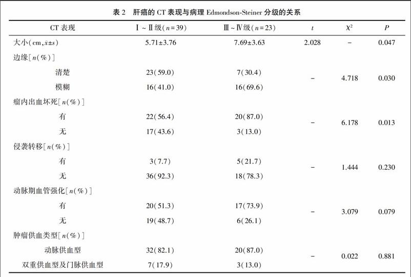
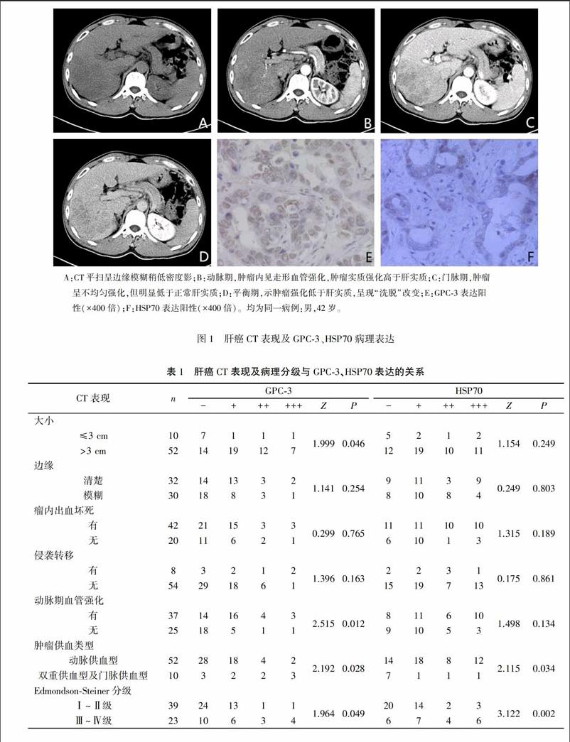
结果 本研究62例HCC患者中肝癌的大小、动脉期血管强化及肝癌的供血类型与GPC-3表达水平存在相关性,不同供血类型的肝癌HSP70的表达水平存在差异;肝癌的大小、边缘、瘤内出血坏死与其病理分化程度相关;肝癌的病理分化程度越低GPC-3及HSP70的表达水平越高。
结论 肝癌的CT征象可在一定程度上反映癌组织GPC-3及HSP70的表达情况及肝癌的病理分化程度,GPC-3和HSP70表达水平与肝癌的病理分级相关。
【关键词】 肝细胞肝癌; 病理分级; 磷脂酰肌醇蛋白聚糖-3; 热休克蛋白70
中图分类号:R735.7 文献标识码:A DOI:10.3969/j.issn.1003-1383.2017.03.005
【Abstract】 Objective To investigate the correlation of preoperative CT findings between expressions and pathological grade of postoperative glypican-3(GPC-3) and heat shock proteins 70(HSP70) protein in hepatocellular carcinoma(HCC).
Methods A total of 62 cases who were pathologically proved with HCC from July,2013 to August,2015 were enrolled in this study.All cases underwent CT plain scan and 3 phase enhanced scanning before operation.And by comparing preoperative CT findings and the expression levels of postoperative GPC-3,HSP70 and pathological grade,whether there was a correlation between them was counted and analyzed.
Results In this study,the size of liver cancer,arterial blood vessels and blood supply type of liver cancer were correlated with GPC-3 expression levels in 62 cases of HCC patients,and the expression level of HSP70 in HCC with different blood supply types was different.The size,margin,intratumoral hemorrhage and necrosis of HCC were correlated with the degree of pathological differentiation.In addition,the lower the degree of pathological differentiation,the higher the expression level of GPC-3 and HSP70.
Conclusion CT findings of HCC can reflect the expression of GPC-3,HSP70 and liver pathological differentiation in a certain extent,and the expression level of GPC-3 and HSP70 were correlated with HCC pathological grading.
【Key words】 HCC;pathological grade;GPC-3;HSP70
肝細胞癌(Hepatocellular carcinoma,HCC)在我国发病率较高,最新统计资料显示,肝癌发病率位于肺癌和胃癌后排第三位,而其所致的男性恶性肿瘤死亡率仅次于肺癌[1]。CT是目前肝癌诊断的重要影像学方法,不仅可以观察肝癌的形态、大小及其对周围邻近组织的关系,更能通过增强扫描的方法来观察肝癌的血供状况,对肝癌的检出、定性、分期及治疗后复查具有重要意义[2]。病理学检查是诊断肝癌的金标准,而免疫组织化学是诊断肝癌的重要辅助手段。磷脂酰肌醇蛋白聚糖-3 (Glypican-3, GPC-3)及热休克蛋白70(Heat shock proteins 70, HSP70)是肝癌诊断的代表性免疫组织化学标志物之一,近些年来,多个研究结果都表明GPC-3及HSP70在肝癌的发生、发展中都扮演了重要的角色[3~5]。笔者拟通过研究HCC的CT表现与癌组织中GPC-3、HSP70表达及病理分级的关系,为肝癌的诊断、治疗及预后评估提供更多信息。
1 资料与方法
1.1 一般资料
搜集右江民族医学院附属医院2013年7月至2015年8月病理科确诊为HCC的患者62例,其中男性52例,女性10例,年龄22~76岁,平均年龄49.74岁。所有病例术前行CT平扫及3期增强扫描,未接受放化疗、肿瘤靶向治疗等非手术治疗,粒子植入、碘油栓塞等介入治疗。术后用免疫组织化学法分析癌组织GPC-3及HSP70表达情况。本研究所有病例均有完整的随访资料。

