Effects of Labisia pumila on oxidative stress in rat model of post-menopausal osteoporosis
Nurdiana Nurdiana, Nelly Mariati, Noorhamdani Noorhamdani, Bambang Setiawan, Nicolaas Budhiparama, Zairin Noor
1Department of Pharmacology, Faculty of Medicine, University of Brawijaya, Malang, East Java, Indonesia
2Midwifery Master Study Programme, Faculty of Medicine, Brawijaya University, Malang, East Java, Indonesia
3Department of Microbiology, Faculty of Medicine, University of Brawijaya, Malang, East Java, Indonesia
4Research Center for Toxicology,Cancer, and Regenerative Medicine, Department of Medical Chemistry and Biochemistry, Faculty of Medicine, University of LambungMangkurat, Banjarmasin, South Kalimantan, Indonesia
5Budhiparama Institute of Hip and Knee Research and Education Foundation for Arthroplasty, Sports Medicine and Osteoporosis, Jakarta, Indonesia
6Research Center for Osteoporosis, Department of Orthopaedic and Traumatology, Ulin General Hospital, Faculty of Medicine, University of LambungMangkurat, Banjarmasin, South Kalimantan, Indonesia
Effects of Labisia pumila on oxidative stress in rat model of post-menopausal osteoporosis
Nurdiana Nurdiana1*, Nelly Mariati2, Noorhamdani Noorhamdani3, Bambang Setiawan4, Nicolaas Budhiparama5, Zairin Noor6
1Department of Pharmacology, Faculty of Medicine, University of Brawijaya, Malang, East Java, Indonesia
2Midwifery Master Study Programme, Faculty of Medicine, Brawijaya University, Malang, East Java, Indonesia
3Department of Microbiology, Faculty of Medicine, University of Brawijaya, Malang, East Java, Indonesia
4Research Center for Toxicology,Cancer, and Regenerative Medicine, Department of Medical Chemistry and Biochemistry, Faculty of Medicine, University of LambungMangkurat, Banjarmasin, South Kalimantan, Indonesia
5Budhiparama Institute of Hip and Knee Research and Education Foundation for Arthroplasty, Sports Medicine and Osteoporosis, Jakarta, Indonesia
6Research Center for Osteoporosis, Department of Orthopaedic and Traumatology, Ulin General Hospital, Faculty of Medicine, University of LambungMangkurat, Banjarmasin, South Kalimantan, Indonesia
ARTICLE INFO
Article history:
Received 2016
Received in revised form 2016
Accepted 2016
Available online 2016
Ovariectomy
Oxidative stress
Oxidative damage
Lipid peroxidation
Antioxidants
Objective:To analyze the effects ofLabisia pumila(L. pumila) extract on the markers of oxidative stress in ovariectomized rats.Methods:Twenty-five female Wistar rats were divided into five treatment groups (n= 5): the control group, the ovariectomized group, the ovariectomized group treated withL. pumilaextract of various doses (10 mg/kg; 20 mg/kg, and 40 mg/kg).L. pumilaextract was administered daily for 8 weeks.Results:The levels of malondialdehyde and superoxide dismutase (SOD) were subjected to spectrophotometric analysis. Serum levels of malondialdehyde and SOD were significantly higher in the group of rat model of post-menopausal osteoporosis than that of the control group (P<0.05). All doses ofL. pumilaadministered significantly decreased the serum levels of malondialdehyde relative to the group of rat model of post-menopausal osteoporosis, reaching the levels comparable to those of the control group (P<0.05). The second dose ofL. pumilasignificantly decreased the levels of SOD relative to those of the group of rat model of post-menopausal osteoporosis, although it did not reach the levels of SOD of the control group (P<0.05). Conclusions:The ethanol extract ofL. pumilanormalizes lipid peroxidation in the rat model of post-menopausal osteoporosis via the mechanism of endogenous antioxidantreplacement.
1. Introduction
Osteoporosis is characterized by the loss of bone mass and microarchitectural deterioration of bones, leading to bone fragility and increased risk of fracture[1]. Pathophysiology of the ovary-related loss of bone mass is complex; it cannot be explained simply by the increase in bone resorption or decreased bone formation[2]. Bone homeostasis is affected by a reduction in antioxidant defenses against oxidative stress along with increased reactive oxygen species. At the cellular level, a defect in bone remodeling caused by oxidative stress is associated with reduction in osteoblasts and osteoclasts as well as reduction in bone formation accompanied by increased apoptosis of osteoblasts and osteocytes[3,4]. The model of bilateral ovariectomy resembles accelerated bone loss in postmenopausal women, which is underlain by estrogen deficiency [5]. Bilaterally ovariectomized animals showed an increase in osteoclastic bone resorption and reactive osteoblastic bone formation with the net result of a loss of bone mass[6]. Previous studies demonstrated that the ovariectomized rats had an increase in lipid peroxidation and hydrogen peroxide and a decrease in antioxidants and antioxidant cofactors[7-9].
L. pumila(L. pumila) is a plant commonly found in Southeast Asia. In Southeast Asia communities this plant decoction is used as a health supplement by boiling the roots, leaves, or whole parts of the plant for irregular menstruation, menstrual pain, inducing labor and a tonic for the vagina walls after childbirth[10,11]. Other studies have evaluated the antioxidant contents and benefits ofL. pumilain rat model of ovariectomy[12-14], but no studies on the control of oxidative stress in the rat model of osteoporosis due to estrogen deficiency. Hence, the purpose of the present study was to analyze the effects ofL. pumilaextract on oxidative stress in ovariectomized rats.
2. Material and methods
2.1. Subjects
Twenty-five female Wistar rats, aged 3–4 months and body weight of 150 g–200 g, were used as the subjects of this study. After acclimatization to the laboratory conditions, they were randomly divided into five treatment groups: the control group, the ovariectomized group and the ovariectomized group administered withL. pumilaextract of various doses (10 mg/kg; 20 mg/kg and 40 mg/kg).L. pumilaextract was administered for 8 weeks. Dosage was determined on the basis of previous studies stating that the dose with no adverse effects forL. pumilawas 50 mg/kg [15]. In the course of the study rats were reared under conditions of 12 h of light and 12 h of dark at room temperature of 24 ℃. In addition, rats were given standard laboratory feed and access to water ad libitum. All research procedures were performed under the ethical guidelines for experimental animals and passed the ethical review of the Research Ethics Committee, Medicine Faculty of Brawijaya University Malang, East Java, Indonesia.
2.2. Procedures for ovariectomy
After anesthesia with ketamine (50 mg/kg) and xylacine (8 mg/kg), twenty rats of the ovariectomized group were subjected to bilateral ovariectomy through a ventral incision. In addition, five rats were subjected to sham surgery[16]. After ovariectomy, wound care was carried out for 10 d and then treatment was given.
2.3. Extract preparation
L. pumilain dry conditions was cut into small pieces and then crushed in a blender and sieved to separate the rough from fine particles. The extract ofL. pumilawas prepared by using the maceration method in which 100 g ofL. pumilapowder is soaked in 900 mL of 96% ethanol for 5–7 d while stirring occasionally. The soaked extract ofL. pumilawas put into the rotavapor until the ethanol solution separates from the active substance. The concentrated extract was then weighed and diluted with distilled water[17]. The ethanol extract ofL. pumilawas administered in accordance with the dosage using an oral feeding tube to the ovariectomized rats.
2.4. Serum collection
At the end of the study, all the rats were anesthetized and their blood was collected from their hearts by cardiac puncture. Furthermore, the blood was centrifuged to obtain serum.
2.5. Analysis of malondialdehyde (MDA) and superoxide dismutase (SOD)
Analysis of serum levels of MDA and SOD was performed according to the protocol in previous studies[18].
2.6. Ethics
This study passed the ethics review of the Ethics Committee of the Faculty of Medicine, Brawijaya University Malang of East Java, Indonesia.
2.7. Statistical analysis
All data was presented in mean ± SD. Differences in the levels among treatment groups were analyzed by ANOVA using SPSS 16.0 statistical package. APvalue of <0.05 was considered as statistically significant difference.
3. Results
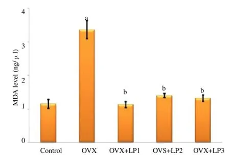
Figure 1 presents the serum levels ofMDA in each experimental group. Serum levels of MDA were significantly higher in the group of rat model of post-menopausal osteoporosis compared to those of the control group (P<0.05). All doses ofL. pumilasignificantly reduced the serum levels MDA relative to those of the group of rat model of post-menopausal osteoporosis, reaching the levels comparable to those of the control group (P<0.05).
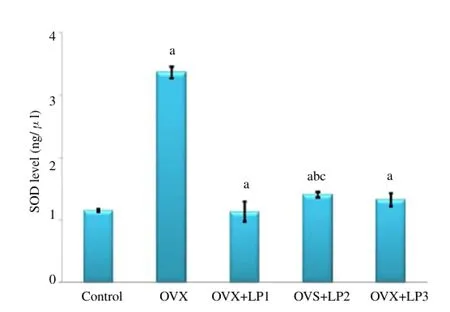
Figure 2 presents the serum levels of SOD in each experimental group. SOD levels were significantly higher in the group of rat model of post-menopausal osteoporosis than those of the control group (P<0.05). Of the three doses, the second dose ofL. pumilasignificantly lowered SOD levels relative to those of the group of rat model of post-menopausal osteoporosis, although it did not reach the levels of SOD of the control group (P<0.05).

Figure 1.Malondialdhyde level in serum of control and experimental groups.Note:aP<0.05 in comparison with control (untreated) group;bP<0.05 in comparison with ovariectomized rats (OVX) group; OVX + LP: ovariectomized rats treated byL. pumila(ng/μL).

Figure 2.Superoxide dismutase level in serum of control and experimental groups.aP<0.05 in comparison with control (untreated) group;bP <0.05; in comparison with OVX group; OVX + LP : ovariectomized rats treated byL. pumila(U/mL).
4. Discussion
Under normal conditions, reactive oxygen species (ROS) will be neutralized efficiently by cellular antioxidant defense mechanisms. In many conditions, there is an imbalance of ROS production and antioxidant defenses, leading to cellular destruction and dysfunction [19]. The present study showed that the levels of MDA, the marker of oxidative damage, increased significantly in the groups of rat model of post-menopausal osteoporosis relative to those of the control group (P<0.05). This suggests that ovariectomy induces lipid peroxidation caused by a systemic increase in oxidative stress. All doses ofL. pumilaadministered significantly decreased the serum levels of MDA relative to the group of rat model of post-menopausal osteoporosis, reaching the levels comparable to those of the control group (P<0.05). This shows thatL. pumilais capable of repairing oxidative damage[19,20].
The levels of SOD were significantly higher in the group of rat model of post-menopausal osteoporosis than those of the control group (P<0.05). These finding indicates that under conditions of osteoporosis-induced oxidative stress, there is endogenous antioxidant compensation in the form of increased levels of SOD. Depending on the levels of ROS, a variety of transcription factors sensitive to changes in the redox status will be activated and will coordinate certain biological responses. Low levels of oxidative stress induce Nrf2, a transcription factor implicated in transactivation of genes that encode enzymatic antioxidant activity[21]. Of the three doses, the second dose ofL. pumilasignificantly lowered SOD levels relative to those of the group of rat model of post-menopausal osteoporosis, although it did not reach the levels of SOD of the control group (P<0.05). We speculated that the antioxidant levels ofL. pumilawill replace SOD to reduce oxidative stress. This mechanism will restore SOD levels to the basal levels.Previous studies showed thatL. pumilaacts as an antioxidant, which is played by flavonoids, ascorbic acid, beta-carotene, anthocyanin and phenolic compounds[19,20]. Additionally, previous studies also demonstrated the activity of SOD inL. pumila[22].
In conclusion, the ethanol extract ofL. pumilanormalizes lipid peroxidation in the rat model of post-menopausal osteoporosis through the mechanism of endogenous antioxidantreplacement.
Conflict of interest statement
The authors declare that they have no conflict of interest.
[1] Zhao X, Wu Z, Zhang Y, Gao M, Yan Y, Cao P, et al. Locally administrated perindopril improves healing in an ovariectomized rat tibial osteotomy model.PLosONE2012;7(3): e33228. doi:10.1371/journal. pone.0033228.
[2] Zhang J, Lazarenko OP, Blackburn ML, Shankar K, Badger TM. Feeding blueberry diets in early live prevent senescence of osteoblast and bone loss in ovariectomized adult female rats.PLos ONE2011;6(9): e24486. doi:10.1371/journal.pone.0024486.
[3] Wauquier F, Leotoing L, Coxam V, Guicheux J, Wittrant Y. Oxidative stress in bone remodelling and disease.Trends Mol Med2009;15: 468-477.
[4] Kular J, TicknerJ, Chim SM, Xu J. An overview of the regulation of bone remodelling at the cellular level,Clin Biochem 2012;45: 863-873.
[5] Folwarczna J, Zych M, Trzeciak HI. Effects of curcumin on the skeletal system in rats.Pharmacol Rep2010;62: 900-909.
[6] de Freitas PHL, Hasegawa T, Takeda S, Sasaki M, Tabata C, Oda K, et al. Eldecalcitol, a second-generation vitamin D analog, drives bone minimodeling and reduces osteoclastic number in trabecular bone of ovariectomized rats.Bone2011;49: 335-342.
[7] Lean JM, Davies JT, Fuller K, Jagger CJ, Kirstein B, Partington GA, et al. A crucial role for thiol antioxidants in estrogen-deficiency bone loss.JClinInvestig2003;112: 915-923.
[8] Ha BJ. Oxidative stress in ovariectomy menopause and role of chondroitin sulfate.Arch Pharm Res2004;27: 867-872.
[9] Karimi E, Jaafar HZ, Ahmad S. Phytochemical analysis and antimicrobial activities of methanolic extracts of leaf, stem and root from different varieties ofLabisa pumilaBenth.Molecules2011;16: 4438-4450.
[10] Fazliana, M, Nazaimoon WMW, Gu HF.Labisia pumilaextract regulates body weight and adipokines in ovariectomized rats. Maturitas 2009; 62: 91-97.
[11] Ali Z, Khan IA. Chemical constituents ofLabisia pumila(Kacip Fatimah).Planta Med2009;75: 40-45.
[12] Fathilah SN, Shuid AN, Mohamed N, Muhammad N, Soelaiman IN.Labisia pumilaprotects the bone of estrogen-deficient rat model: A histomorphometric study.J Ethnopharmacol2012;142(1): 294-299.
[13] Shuid AN, Ping LL, Muhammad N, Mohamed N, Soelaiman IN. The effects ofLabisia pumilavar. alata on bone markers and bone calcium in a rat model of post-menopausal osteoporosis.J Ethnopharmacol2011;133(2): 538-542.
[14] Sing GD, Ganjoo M, Youssouf MS, Koul A, Sharma R, Singh S, et al. Sub-acute toxicity evaluation of an aqueous extract ofLabisia pumila, a Malaysian herb.Food &Chem Toxicol2009;47(10): 2661-2665.
[15] Yalin S, Sagir O, Comelekoglu U, Berkoz M, Eroglu P. Strontium ranelate treatment improves oxidative damage in osteoporotic rat model.Pharmacol Rep2011;63: 396-402.
[16] Fard SG, Shamsabadi FT, Ernadi M, Meng GY, Muhammad K, Mohamed S. Ethanolic extract of Eucheuma cottonii promotesin vivohair growth and wound healing.J Anim& Vet Adv2011;10(5): 601-605.
[17] Draper H, Hadley M. Malondialdehyde determination as index of lipid peroxidation.Methods Enzymol1990;186: 421-431.
[18] Halliwell B, Gutteridge JMC. Free radical in biology and medicine. 3rd Edition. Oxford: University Press, 1999.
[19] Mohamad N, Mahmood M, Mansor H. Antioxidative properties of leaf extracts of a popular Malaysian herb,L. pumila.J Med Plants Res2009;3: 217-223.
[20] Huang J, Zhang H, Shimizu N, Takeda T. Triterpenoid saponins from Arsidia mamillata.Phytochemistry2000;54:817-822.
[21] Glorie G, Legrand-Poels S, Piette J. NF-kB activation by reactive oxygen species: fifteen years later.Biochem Pharmacol2006;72: 1493-1505.
ment heading
10.1016/j.apjr.2016.07.015
*Corresponding author: Nurdiana Nurdiana, Department of Pharmacology, Faculty of Medicine, University of Brawijaya, Malang, East Java, Indonesia.
Tel: +628123388428
E-mail: farmakodes@gmail.com
 Asian Pacific Journal of Reproduction2016年5期
- Asian Pacific Journal of Reproduction的其它文章
- Heparin binding proteins and their relationship with vital sperm function tests visà-vis fertility of buffalo bull semen
- Conception rate in Holstein dairy cows having both normal sized follicles and cystic follicles at estrus
- Polymyxin B changes the plasma membrane integrity of cryopreserved bull semen
- Freezability of buffalo semen with TRIS extender enriched with disaccharides (trehalose or sucrose) and different glycerol concentrations
- Gum arabic improves semen quality and oxidative stress capacity in alloxan induced diabetes rats
- Ovarian hyperstimulation syndrome followed by ovarian torsion in premenopausal patient using adjuvant tamoxifen treatment for breast cancer
